Download presentation
Presentation is loading. Please wait.

1
Blood Human Anatomy Chapter 17

2
I. Overview: Composition of Blood
Blood is considered a connective tissue. It carries all substances that must travel throughout the body. These include nutrients, waste, hormones, electrolytes, antibodies, etc. Clinical examiners study blood of patients, more often than other tissues, to help in making a diagnosis. Blood looks red when it is oxygenated and blue when it is not (although it is not truly ever blue). It travels from the heart in arteries, arterioles, and capillaries, and returns to the heart through venuoles, and veins.
. It travels from the heart in arteries, arterioles, and capillaries, and returns to the heart through venuoles, and veins.")
3
Blood is composed of cells called formed elements and liquid called plasma. The formed elements are erythrocytes (red blood cells), leukocytes (white blood cells), and platelets. The plasma is composed of a high amount of water and all components that would be dissolved in blood. The components are separated when spun in a centrifuge. The heavier more dense red blood cells accumulate at the bottom and the yellowish liquid (plasma) float at the top. In between these two layers lie the white blood cells.
, leukocytes (white blood cells), and platelets. The plasma is composed of a high amount of water and all components that would be dissolved in blood. The components are separated when spun in a centrifuge. The heavier more dense red blood cells accumulate at the bottom and the yellowish liquid (plasma) float at the top. In between these two layers lie the white blood cells..")
4
II. Blood Plasma This fluid is about 90% water and contains over 100 different chemicals. Aside from nutrients, waste, and electrolytes, plasma also holds three important proteins: Albumin- prevents water from diffusing out into the tissue Globulin- antibodies and proteins that carry lipids, iron, and copper Fibrinogen- this is involved in forming blood clots.

5
II. Formed elements Two of these components are not considered true cells: erythrocytes and platelets. These components do not undergo mitosis. ***White blood cells are true cells because they have all organelles and they can multiply in the blood stream

6
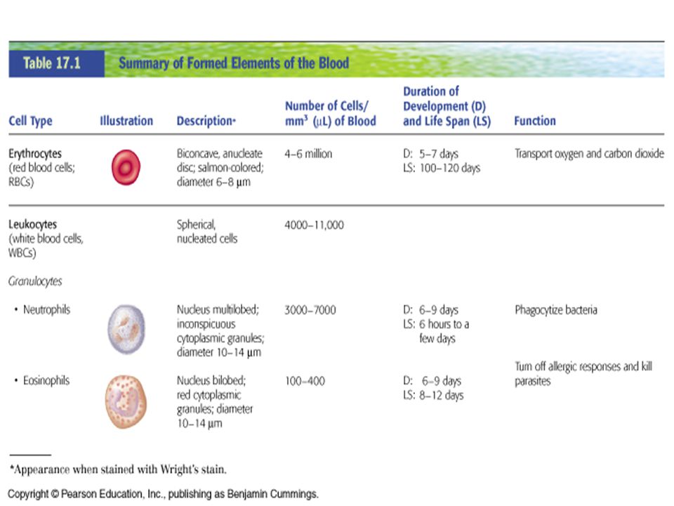
A. Erythrocytes- The specialty of these cells is to transport oxygen and carbon dioxide. Red blood cells originate in bone marrow and as they mature they expel their organelles before entering the blood stream. They are shaped like a disc with a depressed center. They are the most numerous component of formed elements. These cells contain no nucleus or organelles, instead they are packed with hemoglobin. Hemoglobin is a combination of proteins and iron molecules. Erythrocytes collect oxygen at the lungs and deliver it to the tissues then carry carbon dioxide back to the lungs. There are three important characteristics of red blood cells: 1. Their concave shape allows for 30% more surface area for carrying oxygen. 2. 97% of their content is hemoglobin. It is used for binding both oxygen and CO2 3. They depend on anaerobic respiration thus they do not consume any oxygen

7
B. Leukocytes- The specialty of these cells is to fight disease
B. Leukocytes- The specialty of these cells is to fight disease. These are true cells containing organelles and have the ability to divide. The have the ability to travel through the blood to a body region that is infected, exit the blood stream, and enter the site of infection. Site of the body that are infected release certain chemicals to attract white-blood cells. These cells also originate in the bone marrow and once matured entered the blood stream. During times of infections the amount WBC increases, thus a higher WBC count indicates infection. Some groups of WBC may contain large amount of granules that hold hydrolytic enzymes and some do not.

8
Neutrophil Eosinophil
1. Granulocytes- These are called neurtophils, eosinophils, and basophils. Larger than RBCs they are short lived. All granulocytes are phagocytic. Neutrophil Eosinophil Basophil Lymphocyte Monocyte

9
a. Neutrophil- Most common, makes up 60% of WBC count
a. Neutrophil- Most common, makes up 60% of WBC count. These cells have 2-6 lobed nucleus, they contain vesicles filled with digestive enzymes specifically designed to destroy the cell walls of bacteria. They are in the first line of defense of an inflammatory response. They can destroy the bacteria by phagocytosis or by releases chemical substances. These chemicals can even cause damage to the surrounding tissues. Pus is a collection of dead neutrophils, WBCs, and bacterial debri.

10
b. Eosinophils- Account for 1-4% of WBC is blood. It has a bi-
b. Eosinophils- Account for 1-4% of WBC is blood. It has a bi- lobed nucleus. Contain large vesicles that stain red. They are involved in ending allergic reactions by degrading histamine and parasitic infections by exposing the parasite to digestive enzymes.

11
c. Basophils-Most rare of all types, makes up 0. 5% of WBC count
c. Basophils-Most rare of all types, makes up 0.5% of WBC count. Has a bi-lobed nucleus. They release histamine and other chemicals that signal inflammation. They are present in the later stages of infection

12
Agranulocytes- These include lymphocytes and monocytes, cells that do not contain granules of digestive enzymes. a. Lymphocytes- Most important types, makes up about 20-45% of WBC count. When viewed under the microscope they appear to have a large purple nucleus. Most are embedded in lymphatic tissue. They specialized in attacking specific foreign molecules recognized as an antigen. The two main types of lymphocytes are B-cells and T-cells. They produce antibodies or attack a foreign cell directly by destroying it. Lymphocyte Monocyte

13
B-lymphocytes produce antibodies and respond to bacterial cells.
T lymphocytes respond to antigens presented by the membranes of eukaryotic cells. These are the ones responsible for rejection of transplanted tissue. They also destroy self cells that are infected. Natural killer cells spontaneously attack tumor cells and infected cells.

15
b. Monocytes- Contain a large nucleus that resembles a kidney
b. Monocytes- Contain a large nucleus that resembles a kidney. Its cytoplasm may have some granules. These cells travel through the blood stream and transform into macrophages once they enter the tissues Lymphocyte Monocyte

16
Part of a blood clot C. Platelets- These small cell fragments that broke off larger cells called megakaryocytes, are also called thrombocytes. Their specialty is to release chemicals that cause blood clots. They plug tears in the walls of blood vessels to reduce bleeding. Their secretions may induce more platelets to accumulation, or an inflammatory response, or constriction of blood vessels.

17
Platelets contract pulling the edges of a blood clot closer together in order to assist in healing of torn tissue. If a healthy tissue is roughened by scaring, inflammation, or atherosclerosis, platelets will attach and form a blood clot called a thrombus

20
IV. Blood Cell Formation
Hematopoiesis is the process by which blood cells form. After birth, about 1000 billion blood cells are created in the bone marrow per day. A. Bone marrow as the site of hematopoiesis- Red bone marrow generates blood cells. In adults it is located in bones of the axial skeleton and in the epiphysis of the humerus and femur. Newborns have only red bone marrow that is replaced by yellow bone marrow during the ages of Bone marrow contains reticular fibers and macrophages.

21
B. Cell lines in blood cell formation- blood cells arise from blood stem cells or pluripotential hematopoietic stem cell. They divide consistently and produces lines of progenitor cells called lymphoid stem cells and myeloid stem cells. These cells will divided until they become committed cells and thus differentiate into a specific type of blood cell. 1. Genesis of erythrocytes- See diagram on page Blood stem cell becomes a myeloid stem cell, these give rise to proerythroblast that eventually becomes a Nomoblast, then a Reticulocyte, then an Erythrocyte. 2. Formation of leukocytes and platelets- See diagram on page Blood stem cell becomes a myeloid stem cell or lymphoid stem cell. The Lymphoid stem cell directly becomes a B or T lymphocytes. The Myeloid stem cell becomes a Myeloblast, Monoblast, or Megakaryoblast, then specialize to for the granular leukocytes, macrophates, or platelets.

23
V. Blood Types Every person has a certain blood type. It is determined by the markers on the surface of the red blood cells. Immune system cells learn to identify those markers as “self” and if they encounter any RBCs with different marker they will attack them. Thus it is important to know one’s blood type and to be aware of which type one can receive if a blood transfusion is needed. Complete the chart below Blood Type Surface Marker Genotype Plasma antibodies (fights against) Can receive blood from Can donate blood to
 Can receive blood from. Can donate blood to.")
24
gslc.genetics.utah.edu/.../ABObloodsystem.gif

25
V. Blood Disorders- A. Erythrocytes- Polycythemia- abnormal excess of RBCs that can be caused by bone marrow cancer. This increase blood viscosity slowing or blocking blood flow. It can be treated by diluting or removing blood. Anemia- abnormally low RBCs or low hemoglobin. May be caused by excessive bleeding, iron deficiency, deficiencies in folic acid or B12 vitamin, excessive destruction of RBCs, or abnormal structure of hemoglobin.

26
Normal RBC Sickle Cell RBC
Sickle cell disease- it is an inherited abnormality of the hemoglobin. The RBC are shaped like a crescent and are fragile. They may block blood vessels and causing pain, strokes, or infections. There are several forms of treatment so the person lives into adult hood. 1 in 400 African Americans have sickle cell disease. Normal RBC Sickle Cell RBC

27
B. Leukocytes Leukemia- this is a form of cancer that causes an increase production of WBCs. There are several forms of leukemia but in all forms immature WBCs enter the blood stream and also take over the bone marrow crowding out the normal WBCs. Infections and hemorrhaging are the causes of death in patients with leukemia. C. Platelets Thrombocytopenia- abnormally low platelet concentration. Diminished clot formation and internal bleeding.

Similar presentations


 RBCs WBCs Platelets color ? volume ?>")


