Download presentation
Presentation is loading. Please wait.

1
Tracheostomy Dr. Vishal Sharma

2
Jackson’s metallic tube

3
Jackson’s metallic tube

4
Jackson’s metallic tube
Made of German silver (alloy of Ag + Cu + P) Has obturator (pilot), inner tube & outer tube Inner tube is longer than outer tube for its removal & cleaning. Outer tube maintains patency. Pilot is inserted into outer tube for smooth & non-traumatic insertion of tube Lock prevents expulsion of tube during cough
 Has obturator (pilot), inner tube & outer tube. Inner tube is longer than outer tube for its removal & cleaning. Outer tube maintains patency. Pilot is inserted into outer tube for smooth & non-traumatic insertion of tube. Lock prevents expulsion of tube during cough.")
5
Fuller’s bivalved metallic tube
O

6
Fuller’s metallic tube
Outer tube bi-valved. The 2 blades when pressed together, help in smooth entry of tube. Inner tube is longer & has a vent for phonation Pt phonates by closing main tube opening Vent also helps in decannulation of tube

7
Phonation via vent

8
Portex cuffed tube

9
Portex cuffed tube Made of siliconized PolyVinylChloride. It is thermolabile & prevents crusting. Low pressure high volume cuff maintains an air-tight seal required for: Prevention of aspiration of secretions Positive pressure ventilation

10
Cuffed double lumen tube

11
Cuffed fenestrated tube

12
Portex uncuffed tube For tracheostomy patient receiving radiation

13
Uncuffed double lumen fenestrated tube

14
Hands free speaking valve

15
Mechanism of speaking valve

16
Adjustable flange tube
Used in obese neck, oedema neck

17
Salpekar double cuff tube
Prevents ischemic necrosis of tracheal cartilage

18
Cold & hot water humidifiers

19
Heat & moisture exchanger

20
Nebulization attachment

21
Metallic Tubes Plastic Tubes
Easily cleaned without suction Cleaning requires suction Cuff is absent Cuff is present Cannot be connected to ventilator Can be connected Rigid & less comfortable to patient Soft & more comfortable Concomitant radio-therapy is to be avoided Can be given

22
Tracheostomy tube size
Age of pt Tracheostomy tube size Portex (I.D. in mm) Metallic (Fg) 1 – 3 yrs 4.0 – 4.5 16 4 – 6 yrs 5.0 18 7 – 9 yrs 5.5 20, 22 10 – 12 yrs 6.0 24, 26 13 – 18 yrs 7.0 – 7.5 28, 30 Adult 8.0 – 9.0 32, 34, 36
 Metallic (Fg) 1 – 3 yrs. 4.0 – – 6 yrs – 9 yrs , – 12 yrs , – 18 yrs. 7.0 – , 30. Adult. 8.0 – , 34, 36.")
23
Functions of Tracheostomy
1. Relieves upper airway obstruction 2. Improves alveolar ventilation by ing dead space by 30-50% & ing airflow resistance 3. Prevention of aspiration of blood & secretions 4. Removal of airway secretions in patient with inability to cough or with painful cough 5. Administration of anesthesia

24
Indications for Tracheostomy

25
A. Respiratory obstruction
Trauma to airway : external, endoscopic Infection: epiglottitis, croup, Ludwig’s angina, para-pharyngeal /retro-pharyngeal abscess Neoplasm: laryngo-tracheal, pharyngeal Foreign body in airway Oedema of larynx: irritant, allergic, irradiation Paralysis of larynx: B/L abductor palsy Congenital: laryngeal web, cyst, choanal atresia

26
B. Retained airway secretions
Inability to cough: coma, respiratory muscle palsy or spasm, laryngectomy Painful cough: chest injuries, pneumonia Excessive secretions: pulmonary oedema C. Respiratory insufficiency Chronic bronchitis, bronchiectasis, atelectasis, reatined airway secretions

27
D. Anesthesia administration in:
Laryngo-pharyngeal growths Maxillo-facial trauma Trismus Severe Ludwig’s angina Positive pressure ventilation for > 72 hrs

28
Types of Tracheostomy Emergency Elective Temporary Permanent
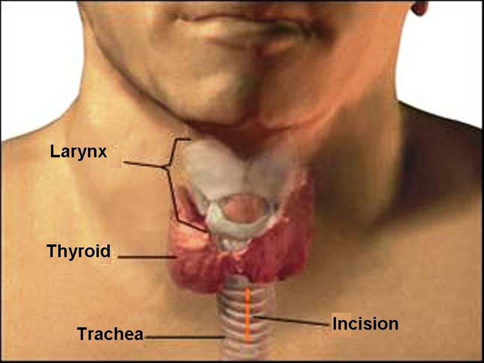
Therapeutic Prophylactic High (1st ring): above thyroid isthmus Mid (2nd – 4th ring): behind thyroid isthmus Low (below 4th ring): below thyroid isthmus
: above thyroid isthmus. Mid (2nd – 4th ring): behind thyroid isthmus. Low (below 4th ring): below thyroid isthmus.")
29
Mid tracheostomy preferred
High tracheostomy leads to subglottic stenosis Low tracheostomy is avoided as: Trachea is deeper Displacement of tracheostomy tube is common Proximity to great vessels Surgical emphysema is common Tracheostoma is close to tracheal bifurcation

30
Steps of Tracheostomy

32
Positioning Supine position with extension of neck. General anesthesia with endotracheal intubation.

33
Infiltration Cricoid palpated & a 5 cm horizontal incision marked 2 cm below it 2 % lignocaine & 1 in 2 lakh adrenaline injected into incision line

34
Horizontal Incision A 5 cm horizontal incision made with # 15 blade &
deepened below subcutaneous tissue

35
Vertical Incision A 5 cm midline vertical incision can be made below cricoid in emergency tracheostomy. This avoids injury to blood vessels.

38
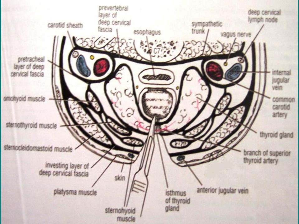
Exposure of strap muscles
Investing layer of deep cervical fascia opened vertically with artery forceps. Palpation for tracheal rings done regularly during the dissection.

40
Retraction of strap muscles

41
Exposure of thyroid isthmus
Strap muscles retracted laterally with Langenbeck retractors to expose the trachea & thyroid isthmus

42
Isthmus separation from trachea
Thyroid isthmus detached from tracheal surface & retracted with blunt tracheal hook.

43
Isthmus retraction to expose pre-tracheal fascia

44
Division of thyroid isthmus
If required, thyroid isthmus is divided between clamps. Transfixion sutures applied at the ends.

45
Confirmation of trachea
5 ml syringe containing 4 % Lignocaine taken, its needle inserted into trachea & aspirated. Air bubbles confirm presence of needle in trachea. 2 ml of solution injected into trachea & needle removed quickly to avoid breaking of needle during violent cough movements.

46
Creation of tracheal window
Sharp cricoid hook inserted below cricoid to steady trachea. Tracheal window created by excising anterior 1/3rd of 2nd & 3rd tracheal ring with No. 11 blade & Allis tissue forceps.

47
Cautery assisted window

48
Holding cartilage with Allis forceps

49
Tracheal window

50
Other options

51
Inferiorly based tracheal flap made & sutured
Bjork flap Inferiorly based tracheal flap made & sutured to lower skin edge

52
Insertion of tracheostomy tube
Endotracheal tube withdrawn into larynx Lubricated tracheostomy tube inserted into trachea Confirm presence of tube in trachea with help of ambu bag & auscultation

55
Suturing of flanges Cuff inflated with 5 ml of air & anesthetic circuit connected to the tube Neck extension released & flanges of tube sutured to skin to avoid tube movement

56
Tying the tapes Tapes of tracheostomy tube tied around the neck keeping a space for 1 finger. Neck kept flexed. Skin incision closed loosely to avoid surgical emphysema.

57
Padded tapes

58
Insertion of medicated gauze
Betadine soaked gauze or Sofratulle put around the tracheostomy opening.

60
Shower collar

61
Shower guard

62
Tracheostomy locket

63
Immediate Complications
Occurs during operation Primary Haemorrhage Air embolism Cardiac Arrest Aspiration of blood CO2 withdrawal Apnoea Injury to: Apical pleura (pneumothorax), recurrent laryngeal nerve, oesophagus
, recurrent laryngeal nerve, oesophagus.")
64
Intermediate Complications
Occurs within first few days Reactionary & secondary haemorrhage Blocking or displacement of tube Subcutaneous emphysema, pneumothorax Tracheitis & crusting Atelectasis & lung abscess Wound infection & granulation tissue

65
Surgical emphysema

66
Causes of surgical emphysema after tracheostomy
Dissection into many tissue planes in neck Use of smaller tracheostomy tube Tight closing of skin incision Excessive struggling & coughing of pt during extubation

67
Tracheostomy site granulation

68
Late Complications Occurs after weeks / months
Subglottic stenosis, tracheal stenosis Tracheo-arterial or Tracheo-venous fistula Tracheo-oesophageal fistula Persistent tracheo-cutaneous fistula Decannulation difficulty Tracheostomy wound scar / keloid Metallic tube corrosion & fragment aspiration

69
Anatomy of tracheal fistulae

70
Tube fragment aspiration

71
Tracheostomy care Pt given 100 % oxygen. Deflate the tube cuff.
Suction catheter with negative suction pressure ( mmHg) used Catheter diameter should be < 1/3rd of internal diameter of tracheostomy tube Catheter length introduced just enough to go beyond inner tube (10 cm)
 used. Catheter diameter should be < 1/3rd of internal diameter of tracheostomy tube. Catheter length introduced just enough to go beyond inner tube (10 cm)")
72
Tracheostomy care Multiple-eyed catheters produce less trauma than whistle tip catheters Lubricated catheter tip inserted (with suction off) as pt is inspiring. At end inspiration, suction put on & catheter withdrawn in rotating motion. Each suction procedure should last for seconds. Instill 0.5 ml NaHCO3 to liquefy crusts.
 as pt is inspiring. At end inspiration, suction put on & catheter withdrawn in rotating motion. Each suction procedure should last for seconds. Instill 0.5 ml NaHCO3 to liquefy crusts.")
73
Tracheostomy care Chest auscultated for confirmation of adequate suctioning. Re-inflate cuff to a pressure of 25 mmHg. Patient oxygenated again. Tracheostomy wound dressing done BID Steam inhalation TID. Moist gauze piece placed over tracheostomy tube opening. Regular chest physiotherapy, expectorants & mucolytics given.

75
Wall suction

76
Portable suction

77
Closed-system Multiple-use Suction Unit (CMSU)
")
78
Communication chart for pt

79
Electronic communication

80
Hand bells

81
Tracheostomy tube changing
Inner tube is removed & cleaned when blocked Outer tube never removed before 72 hrs to allow formation of tracheo-cutaneous tract Cuff of Portex tube deflated for 10 minutes every 2 hours to prevent pressure necrosis & dilatation of trachea

82
Pt position in tube changing

83
Cleaning of inner tube

84
Tube removal over bougie

85
Obturator guide wire insertion

86
Decannulation Adult: plug or seal tube opening & if tolerated for 24 hrs, remove tube. Child: Sequentially reduce size of tube. After tube removal close wound. Healing occurs within 1 week. Secondary closure after freshening the wound margin is required rarely.

87
Capping of tube opening

88
Decannulation difficulty
Organic causes: Persistence of cause requiring tracheostomy Obstructing tracheal granulations Tracheal oedema Subglottic stenosis Collapse of tracheal wall (tracheomalacia)
")
89
Decannulation difficulty
Non-organic causes: Emotional dependence in children Inability to tolerate upper airway resistance In-coordination of laryngeal opening reflex Long-standing tube leads to impaired laryngeal development

90
Tracheostomy Intubation
Invasive Non-invasive Complications are more Less Can be kept for > 7 days Should not be kept Pt can speak Cannot speak Tracheo-bronchial toilet is easy Difficult Decreases dead space by 30-50% Does not

91
Disadvantages of Tracheostomy
Anosmia: no nasal air entry Aphonia: avoided by phonatory vent Aspiration: avoided by cuffed tube Inability to lift heavy weight Inability to perform strenuous exercise Inability to swim

92
Percutaneous Tracheostomy

93
Insertion of cannula

94
Insertion of guide wire

95
Tracheal dilator over guide wire

96
Insertion of tracheal dilator

97
Tracheostomy tube

98
Insertion of tracheostomy tube

100
Percutaneous Tracheostomy
Trachea punctured with needle & cannula Needle removed & a guide wire passed into trachea via cannula Cannula removed & graded dilators passed over guide wire till the opening can admit a tracheostomy tube

101
Cricothyrotomy

102
Cricothyrotomy 1. Midline vertical skin incision made to identify cricothyroid notch. 2. Cricothyroid membrane incised horizontally, with # 11 blade, close to cricoid. 3. Knife handle inserted & rotated by 900, to widen the horizontal opening or tracheostomy tube is inserted. 4. Elective tracheostomy done as soon as possible to avoid subglottic stenosis.

103
Tracheal fenestration

104
Tracheal fenestration

105
Tracheal fenestration

106
Tracheal fenestration
Indicated for C.O.P.D. where tracheal opening is required for mechanical cleaning. Bilateral medial based skin flaps elevated & tracheal opening made. Distal edges of flaps sutured to margins of tracheal window. Lateral edges of 2 flaps sutured to each other to create watertight skin buttons.

107
Thank You

Similar presentations


 – Oral-tracheal – Naso-tracheal Tracheostomy (trach) 1.>")


 Amended 2012.>")


