Download presentation
Presentation is loading. Please wait.

1
David Kachlík and Petr Zach
Cerebellum David Kachlík and Petr Zach

2
Small brain = Cerebellum
10 % weight of whole brain More than ½ neurons of whole brain ¼ - ¾ area of whole brain

3
Mesencephalon Pons Medulla oblongata Medulla spinalis Cerebellum

4
Cerebellum

5
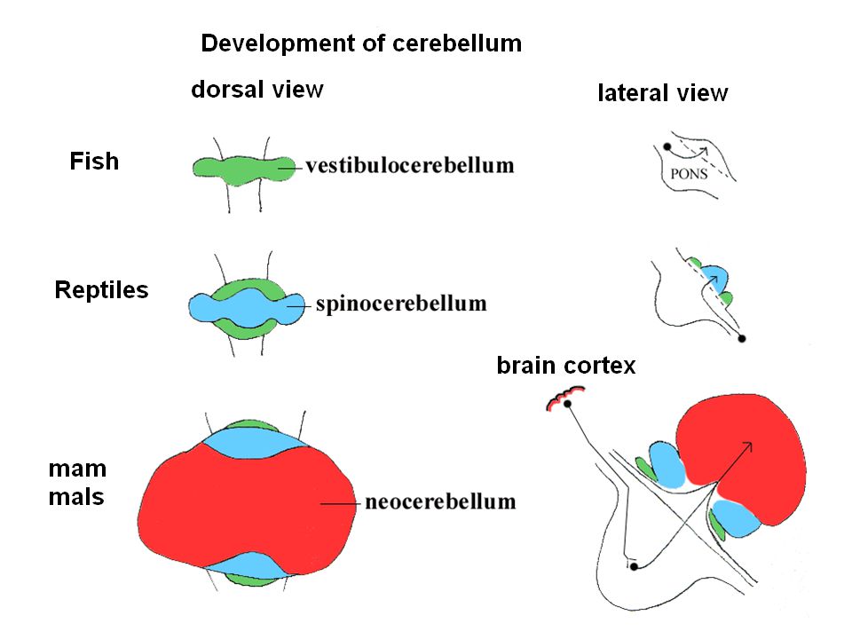
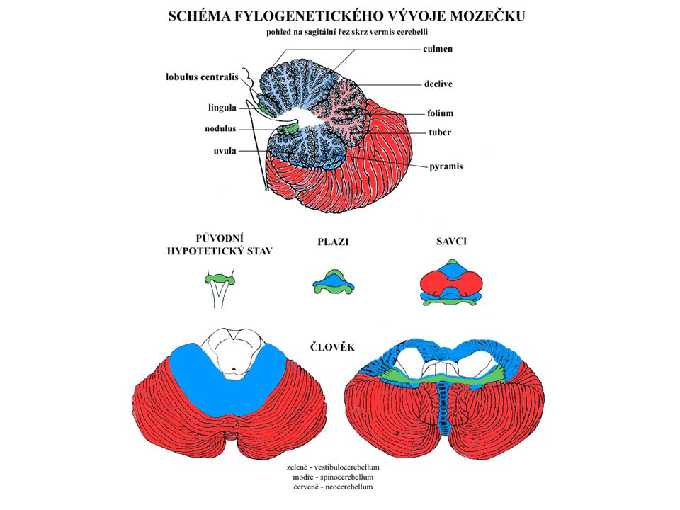
Cerebellum – parcellation
Functional placement: vermis a l. floculonodularis paravermální (intermediální) zóna hemisféry (laterální zóna) External structure: lobus anterior lobus posterior lobus flocculonodularis developmentally: archicerebellum paleocerebellum neocerebellum function: vestibulocerebellum spinocerebellum cerebrocerebellum (= pontocerebellum) 5
 zóna. hemisféry (laterální zóna) External structure: lobus anterior. lobus posterior. lobus flocculonodularis. developmentally: archicerebellum. paleocerebellum. neocerebellum. function: vestibulocerebellum. spinocerebellum. cerebrocerebellum (= pontocerebellum) 5.")
6
Cerebellum – description
folia cerebelli (leaves) fissurae cerebelli (crack) vermis (červ) – non paired in middle hemispheria – paired 3 lobi (lobes) Small parts 10 in vermis [I - X] – expample nodulus 9 in hemispheres [H II - H X] tonsilla – when edema it goes into foramen magnum and it compresses stem flocculus
 fissurae cerebelli (crack) vermis (červ) – non paired in middle. hemispheria – paired. 3 lobi (lobes) Small parts. 10 in vermis [I - X] – expample nodulus. 9 in hemispheres [H II - H X] tonsilla – when edema it goes into foramen magnum and it compresses stem. flocculus.")
7
Cerebellum – posterior view
VERMIS

8
Cerebellum – ventral view
NODULUS FLOCCULUS TONSILLA

9
Cerebellum – inferior view
VERMIS TONSILLA

10
Blood supply

11
Cerebellum – developmental parts
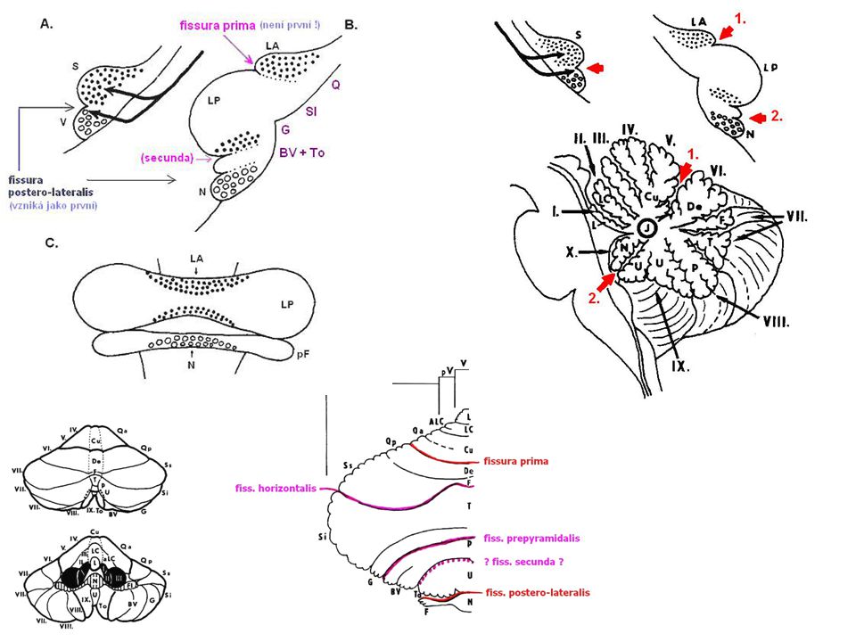
lobus anterior [I-V + H II - H V] = spinocerebellum = paleocerebellum fissura prima lobus posterior [VI-IX + H VI - H IX] = pontocerebellum = neocerebellum fissura posterolateralis lobus flocculonodularis [X + H X] = vestibulocerebellum = archicerebellum

15
Medial zone of anterior and posterior lobe =paleocerebellum=spinocerebellum=fine tune body and limb movement Lateral zone of anterior and posterior lobe = cerebrocerebellum=neocerebellum = ?? Plannning movement, cognitive functions Flocculonodular lobe =vestibulocerebellum=archicerebellum=balance and gait

17
Cerebellum – funkční části
3 podélné zóny vermis + lobus flocculonodularis paravermální kůra hemisféry

18
Cerebellum Anterior Lobe Posterior Lobe
Primary fissure Anterior Lobe Regulation of muscle tone, coordination of skilled voluntary movement Posterior Lobe Planning and initiation of voluntary activity Flocculo-Nodular Lobe (FN lobe) Maintenance of balance, control of eye movements Vestibulocerebellum Spinocerebellum Cerebrocerebelum Folia
 Maintenance of. balance, control. of eye movements. Vestibulocerebellum. Spinocerebellum. Cerebrocerebelum. Folia.")
19
Cerebellar homunkulus

20
Flocullonodular lobe Medial Lateral (Deiters)
Inferior (Bechterew) Superior Does not exit cerebellum via deep cerebellar nuclei!
 Superior. Does not exit cerebellum via deep cerebellar nuclei!")
21
Anterior lobe, spinocerebellum
Interposed nuclei=nc. globosi and nc. emboliformis Superior cerebellar peduncule Red nucleus Distal muscle group Vermis Fastigial nucleus

22
Cerebellum – pedicles pedunculus cerebellaris inferior
corpus restiforme corpus juxtarestiforme pedunculus cerebellaris medius (= brachium pontis) AF: tractus cortico-ponto-cerebellaris pedunculus cerebellaris superior (= brachium conjunctivum)
 AF: tractus cortico-ponto-cerebellaris. pedunculus cerebellaris superior (= brachium conjunctivum)")
23
Cerebellum – ventral view
P.C.SUPERIOR P.C.MEDIUS P.C.INFERIOR

24
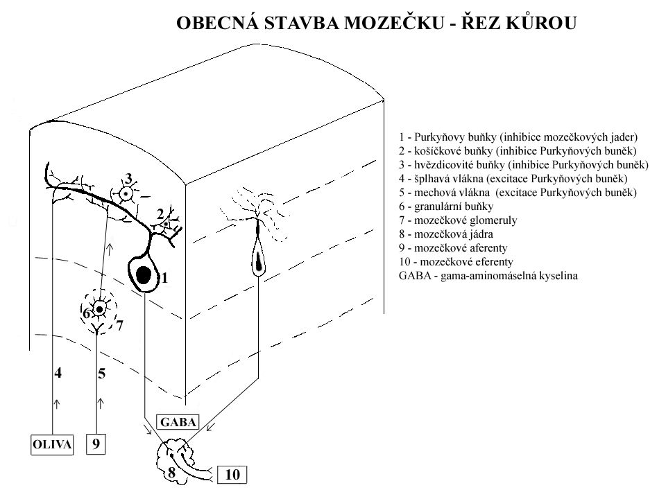
Cerebellum – internal structure
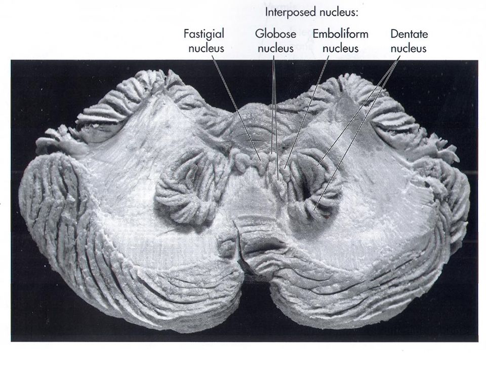
cortex cerebelli: strata (3 layers) – arbor vitae stratum moleculare stratum purkinjese stratum granulosum corpus medullare cerebelli: nuclei cerebelli 4 paired nuclei („Don't Eat Greasy Food“) nucleus dentatus (= lateralis cerebelli) nucleus emboliformis (= interpositus anterior) nucleus globosus (= interpositus medialis) nucleus fastigii (= medialis cerebelli)
 – arbor vitae. stratum moleculare. stratum purkinjese. stratum granulosum. corpus medullare cerebelli: nuclei cerebelli 4 paired nuclei („Don t Eat Greasy Food ) nucleus dentatus (= lateralis cerebelli) nucleus emboliformis (= interpositus anterior) nucleus globosus (= interpositus medialis) nucleus fastigii (= medialis cerebelli)")
25
Cerebellum - nuclei

26
Deep cerebellar nuclei
Via inferior peduncle Via superior peduncle

29
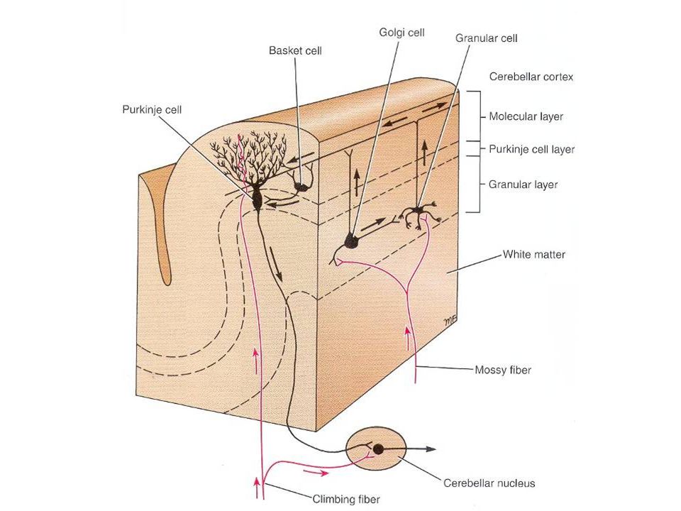
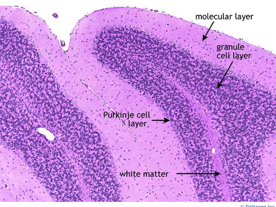
Cerebellum – layers of cortex
stratum moleculare neuron stellatum (stellate cells) neuron corbiforme (basket cells) neurofibra parellela (parallel fiber) – axons of granular cells stratum purkinjese = stratum neurium piriformium; formerly stratum gaglionicum neuron purkinjese (Purkynje cells) corbis neurofibrarum (rich branching to stratum moleculare) Axons to cerebellar nuclei stratum granulare neuron granulosum (granulr cell) neuron stellatum magnum Golghi (Golgi cell) Next 3 types of cells glomerulus cerebelli Afferent fibers: neurofibra muscosa (mossy fiber - Glu) + ascendens (climbing fiber - Asp)
 neuron corbiforme (basket cells) neurofibra parellela (parallel fiber) – axons of granular cells. stratum purkinjese. = stratum neurium piriformium; formerly stratum gaglionicum. neuron purkinjese (Purkynje cells) corbis neurofibrarum (rich branching to stratum moleculare) Axons to cerebellar nuclei. stratum granulare. neuron granulosum (granulr cell) neuron stellatum magnum Golghi (Golgi cell) Next 3 types of cells. glomerulus cerebelli. Afferent fibers: neurofibra muscosa (mossy fiber - Glu) + ascendens (climbing fiber - Asp)")
32
Cellular connection

33
Cerebellum: 3 layered cortex
Climbing fibers: excite the Purkinje cells Mossy fibers: excite the granule cells Granule cells: make excitatory contact with the Purkinje cells Purkinje cells: Tonic inhibition on the activity of the neurons of the cerebellar nuclei => All excitatory inputs will be converted to the inhibition => Removing the excitatory influence of the cerebellar inputs (erasing)
")
34
Cerebellar glomerulus

37
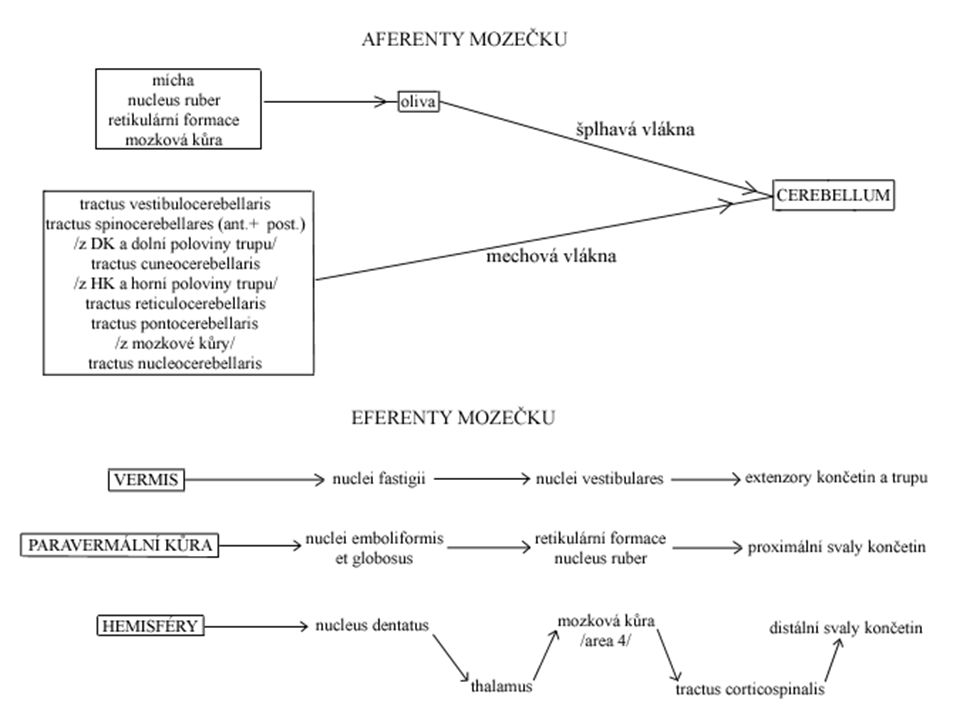
Cerebellum – afferentation balance
tractus vestibulocerebellaris directus vestibulum corpus juxtarestiforme (v PCI) nodulus + uvula (ipsilat.) tractus vestibulocerebellaris indirectus vestibulum ncl. vestibulares corpus juxtarestiforme (v PCI) lobus flocculonodularis + vermis (bilat.) tractus trigeminocerebellaris Information from head
 nodulus + uvula (ipsilat.) tractus vestibulocerebellaris indirectus. vestibulum ncl. vestibulares corpus juxtarestiforme (v PCI) lobus flocculonodularis + vermis (bilat.) tractus trigeminocerebellaris. Information from head.")
38
Cerebellum – afferentation passive proprioception
tractus spinocerebellaris posterior ncl. thoracicus post. Stilling-Clarke medulla oblongata pedunculus cer. inf. vermis + paravermal cortex (ipsilateral) proprioception from trunk and LL tractus cuneocerebellaris Posterior fascicle tract nucleus cuneatus accessorius Proprioception from UL and thorax
 proprioception from trunk and LL. tractus cuneocerebellaris. Posterior fascicle tract nucleus cuneatus accessorius. Proprioception from UL and thorax.")
39
Cerebellum – afferentation active proprioception
tractus spinocerebellaris anterior ncl. thoracicus post. Stilling-Clarke crossing at spinal lvl mesencephalon pedunculus cer. superior crossing in crbl cortex vermis + paravermal cortex (ipsilateral) – LL tractus spinocerebellaris rostralis ncl. thoracicus post. Stilling-Clarke pedunculus cer. inferior vermis + paravermal cortex (ipsilateral) – UL tractus spinoolivaris – motoric learning for example walking steep steps
 – LL. tractus spinocerebellaris rostralis. ncl. thoracicus post. Stilling-Clarke pedunculus cer. inferior vermis + paravermal cortex (ipsilateral) – UL. tractus spinoolivaris. – motoric learning. for example walking steep steps.")
40
Cerebellum – afferentation form cortex
tractus cortico-ponto-cerebellaris ( fibers) lobus f,p,o,t capsula interna ncll. pontis fibrae pontis transversae crossing pedunculus cer. medius crbl cortex (contralat.) tractus cortico-olivo-cerebellaris lobus f,p,o,t capsula interna complexus olivaris inf. (bilat.) crossing pedunculus cer. inferior crbl cortex tractus cortico-reticulo-cerebellaris lobus f,p,o,t (mostly sensorimotor cortex) capsula interna RF (bilat.) crossing pedunculus cer. medius + inf. crbl cortex Will motoric, movement preparation, setting of proper muscle tonus
 lobus f,p,o,t capsula interna ncll. pontis fibrae pontis transversae crossing pedunculus cer. medius crbl cortex (contralat.) tractus cortico-olivo-cerebellaris. lobus f,p,o,t capsula interna complexus olivaris inf. (bilat.) crossing pedunculus cer. inferior crbl cortex. tractus cortico-reticulo-cerebellaris. lobus f,p,o,t (mostly sensorimotor cortex) capsula interna RF (bilat.) crossing pedunculus cer. medius + inf. crbl cortex. Will motoric, movement preparation, setting of proper muscle tonus.")
41
Cerebellum – efferentation
ncl. fastigii 1. PCI RF (bilat.) tr. reticulospinalis 2. PCI ncl. vestibularis lat. Deitersi (bilat.) tr. vestibulospinalis 3. cranial nerves, neck muscles ncll. interpositi (globosus + emboliformis) PCS crossing ncl. ruber (pars magoncellularis) tractus rubrospinalis crossing spine (ipsilat.) ncl. dentatus PCS crossing ncl. VA+VL thalami area 4 tr. pyramidalis crossing spine (ipsilat.)
 tr. reticulospinalis. 2. PCI ncl. vestibularis lat. Deitersi (bilat.) tr. vestibulospinalis. 3. cranial nerves, neck muscles. ncll. interpositi (globosus + emboliformis) PCS crossing ncl. ruber (pars magoncellularis) tractus rubrospinalis crossing spine (ipsilat.) ncl. dentatus. PCS crossing ncl. VA+VL thalami area 4 tr. pyramidalis crossing spine (ipsilat.)")
42
Cerebellum – inferior pedicles pedunculus cerebellaris inferior
corpus restiforme AF↑: tr. spinocerebellaris posterior + rostralis, tr. cuneocerebellaris, tr. spinoolivaris AF↑: tr. spino-reticulo-cerebellaris AF↓: tr. cortico-reticulo-cerebellaris, cortico-olivo-cerebellaris, cortico-arcuato-cerebellaris corpus juxtarestiforme AF↑ tr. vestibulocerebellerais directus + indirectus EF↓: tr. cerebello-reticulospinalis, -cerebellovestibularis, cerebelospinalis, cerebellonuclearis (all from z ncl. fastigii)
")
43
Cerebellum – middle and upper pedicles pedunculus cerebellaris medius et superior
pedunculus cerebellaris medius AF↓: tractus cortico-ponto-cerebellaris pedunculus cerebellaris superior AF↑: tr. spinocerebellaris anterior + tectocerebellaris EF↓: tr. cerebello-rubro-thalamo-corticalis + tr. cerebello-rubro-spinalis EFcircuit: tr. cerebello-rubro-olivo-cerebellaris (Papezův control circuit)
")
46
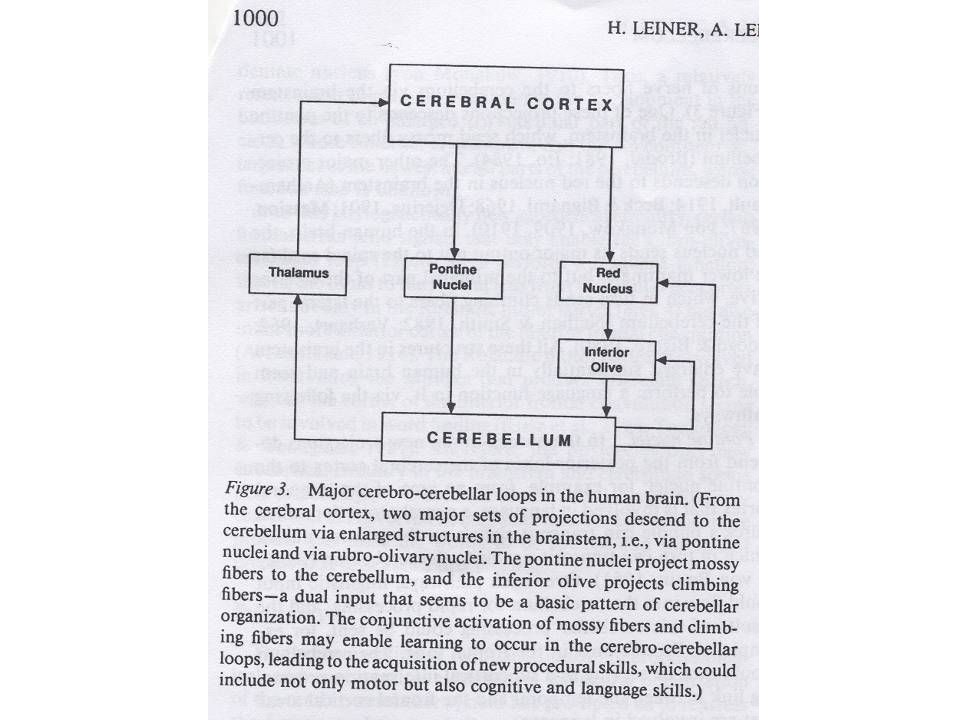
Dentate nuclei: project contralaterally through the superior cerebellar peduncle to neurons in the contralateral thalamus & from thalamus to motor cortex Func.: influence planning and initiation of voluntary movement Emboliform & Globose nuclei: project mainly to the contralateral red nuclei & a small group is projected to the motor cortex Red Nuclei Rubrospinal Tract control of proximal limb muscles Fastigial nuclei: project to the vestibular nuclei & to the pontine and medullary reticular formation Vestibulospinal & Reticulospinal tracts

47
Inputs to cerebellum from spinocerebellar tracts have a somatotopic organization.
2 maps of body Primary fissure Signals from the motor cortex, which is also arranged somatotopically, project to corresponding points in the sensory maps of the cerebellum.

48
Inputs and outputs of the Cerebellum

51
Archicerebellum (vestibulocerebellum)
lobus flocculonodularis + vermis balance nystagmus Connection with ncll. vestibulares (inferior + medialis) Common reason: meduloblastoma
 Common reason: meduloblastoma.")
53
Paleocerebellum (spinocerebellum)
lobus anterior AF: tractus spinocerebellaris anterior + posterior Proprioception (passive and active) (information about reflexes) Collaterals to cerebellar nuclei EF: action of anti gravitatory muscles, coordination of agonists/antagonists gait (walk)
 (information about reflexes) Collaterals to cerebellar nuclei. EF: action of anti gravitatory muscles, coordination of agonists/antagonists. gait (walk)")
54
Neocerebellum (cerebrocerebellum)
lobus posterior AF: tractus cortico-ponto-cerebellaris Collaterals to cerebellar nuclei EF: motoric control Coordination of subtle limbs movements Loop sided trim of motor activity Together with cortex plans movements

55
Cerebellar syndrome Muscular hypotonia (increased interval and pasivity of movements) ataxia (loss of coordination) hypermetria – dysmetria makrografia, saccadic speech, megafonia, bradylalia adiadochokinesis asynergia („drunken sailor walk“) Intention tremor nystagmus and vertigo (hyporeflexia of elementar postural reflexes)
 Intention tremor. nystagmus and vertigo. (hyporeflexia of elementar postural reflexes)")
56
Cerebellar cognitive-affective syndrome
Deficit of executive functions Impairment of spatial tasks Personality changes Flattening, desinhibition, non adequate behavior Language problems dysprosodia (melody, temp, rhytm), agrammatismus, light anomia (wrong social behavior) (Schmahmann a Sherman, 1998)
, agrammatismus, light anomia (wrong social behavior) (Schmahmann a Sherman, 1998)")
57
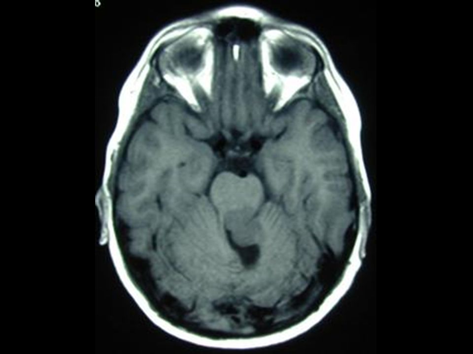
Cerebellar ataxia Ataxic gait and position: Left cerebellar tumor
a. Sways to the right in standing position b. Steady on the right leg c. Unsteady on the left leg d. ataxic gait

58
Clinical Findings and Localization of Cerebellar Lesions
Ataxia refers to disordered contractions of agonist and antagonist muscles and lack of coordination between movements at different joints typically seen in patients with cerebellar lesions. Normal movements require coordination of agonist and antagonist muscles at different joints in order for movement to have smooth trajectory. In ataxia movements have irregular, wavering course consisting of continuous overshooting, overcorrecting and then overshooting again around the intended trajectory. Dysmetria = abnormal undershoot or overshoot during movements toward a target finger-nose-finger test).
.")
59
Cerebellar lesions

60
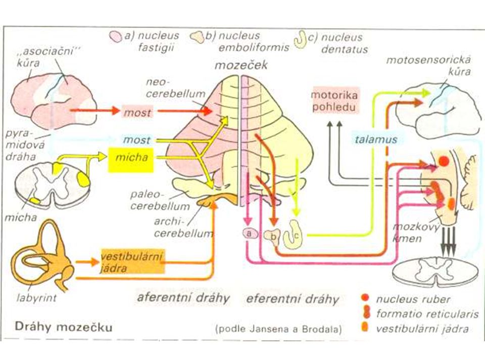
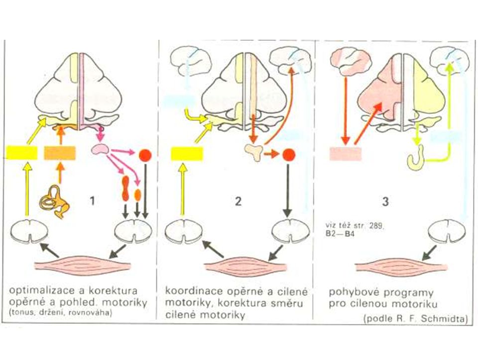
Cerebellum – principal circuits
circuit: cortex-cerebellum cortex → pons / oliva / RF (oliva) – ncll. pontis / complexus olivaris inferior → crossing → cortex → ncl. dentatus → crossing → thalamus (ncl. VL) → cortex Papez cerebellar control circuit: ncl. dentatus → ncl. ruber (pars parvocellularis) → oliva → ncl. dentatus Learning of motor, cognitive and language skills cortex – cerebellum: always contralaterally cerebellum – body: always ipsilaterally
 – ncll. pontis / complexus olivaris inferior → crossing → cortex → ncl. dentatus → crossing → thalamus (ncl. VL) → cortex. Papez cerebellar control circuit: ncl. dentatus → ncl. ruber (pars parvocellularis) → oliva → ncl. dentatus. Learning of motor, cognitive and language skills. cortex – cerebellum: always contralaterally. cerebellum – body: always ipsilaterally.")
62
Cerebellum - summary balance Mostly motoric functions „comparator“
Creation, support and maintenance of muscle tonus Planning of movement with cortex Complicated and subtle movements: dance, speech, writing „comparator“ Other functions – cognition, sensory perception

63
Lack of thiamin (B1) causes degeneration of lobus anterior cerebelli
 causes degeneration of lobus anterior cerebelli")
64
Optional reading: Cerebellar lesions

65
Cerebellar symptomes Dysmetria (hypermetria) – invalid targeting and finishing of movements due to delayed or insufficient contraction of antagonists, which normally end movement Spontaneous movements are incorrect (cerebellar macrography of Henner – increasing size of letters during writing as opposed to parkinson micrography) Bradytelokinesis – ending of movement before reaching target, compensated by cortical atactic spasms
 – invalid targeting and finishing of movements due to delayed or insufficient contraction of antagonists, which normally end movement. Spontaneous movements are incorrect (cerebellar macrography of Henner – increasing size of letters during writing as opposed to parkinson micrography) Bradytelokinesis – ending of movement before reaching target, compensated by cortical atactic spasms.")
66
Cerebellar symptomes Dyssynergia (asynergia) – individual muscle groups work independently and complex movement patterns split into particular movements, movement fragments are usually performed with too much/less strength. Small asynergia – lesion of cerebellar hemispheres, targeting limb coordination Great asynergia – palleocerebellar lesion, deficit of trunk axial muscle – standing, sitting from laying, erecting etc.
 – individual muscle groups work independently and complex movement patterns split into particular movements, movement fragments are usually performed with too much/less strength. Small asynergia – lesion of cerebellar hemispheres, targeting limb coordination. Great asynergia – palleocerebellar lesion, deficit of trunk axial muscle – standing, sitting from laying, erecting etc.")
67
Cerebellar symptomes Hypotonia (pasivity) – decrease of muscle tonus, increase in movement range in joints, more pronounced in acute then chronic cerebellar lesion
 – decrease of muscle tonus, increase in movement range in joints, more pronounced in acute then chronic cerebellar lesion.")
68
Cerebellar symptomes I. Ataxia -uncoordinated voluntary mvmt.
II. Hypotonia III. Cerebellar Gait -wide base -may veer towards side of lesion -will sway standing with feet together eyes open or closed(not a sign of Rhomberg b/c because none of those three senses are causing the patient to loose balance) IV. Intention Tremor -present when moving, not at rest V. Dysdiadochokinesia -inability to move rapidly VI. Dysmetria -can’t measure distance, so there is a loss of control of range mvmts. (pastpointing), cant reach out to perform tasks VII. Dysarthria -slurred (scanning) speech
 IV. Intention Tremor. -present when moving, not at rest. V. Dysdiadochokinesia. -inability to move rapidly. VI. Dysmetria. -can’t measure distance, so there is a loss of control of range mvmts. (pastpointing), cant reach out to perform tasks. VII. Dysarthria. -slurred (scanning) speech.")
69
Causes of cerebellar lesions
I. Multiple Sclerosis II. Cerebellar Strokes III.Tumors IV. Degeneration V. Wernicke-Korsakoff Syndrome -caused by Thyamine Deficiency, mostly from alcohol abuse -Wernicke’s encephalopathy symptoms are gait ataxia, nystagmus, diplopia, strabismus -Korsakoff syndrome- sever anterograde and retrograde amnesia -treatment with glucose and no thiamine can result in death VI. Alcoholic Cerebellar Degeneration -gait ataxia without limb ataxia -different pathology than Wernicke’s VII. Cerebellar Hemorrhage -vomiting -ataxia VIII. Fredrick’s Ataxia -Genetic (triple repeat GAA on Chrm.9) -gradual onset in first 3 decades of life -gait disturbances,dysarthria, sensory loss to extremities
 -gradual onset in first 3 decades of life. -gait disturbances,dysarthria, sensory loss to extremities.")
70
Cerebellar symptomes Tremor
A) intention tremor during intended movements, worse at the beginning and end of movement, lesion of dentate nc. or mesencephalic pedunculus B) Gordon-Holmes tremor when mesencephalic pedunculus without lesion of nc. ruber, rough irregular tremor even in rest (wing-beating tremor) C) titubation – tremor of head (3-4 Hz) or upper trunk in ventrodorsal direction, medial cerebellar lesions
 intention tremor during intended movements, worse at the beginning and end of movement, lesion of dentate nc. or mesencephalic pedunculus. B) Gordon-Holmes tremor when mesencephalic pedunculus without lesion of nc. ruber, rough irregular tremor even in rest (wing-beating tremor) C) titubation – tremor of head (3-4 Hz) or upper trunk in ventrodorsal direction, medial cerebellar lesions.")
71
Cerebellar symptomes Slurred speech – caused by dyssynergia and dysdiadochokinesis of speech and respiratory muscles, speech tempo slowing down, changes of articulation, words expressed with first syllable accentation (similar to limbs hypermetria) Cerebellar dysarthria – blurred pronunciation, slow speech (like drunkard speech)
 Cerebellar dysarthria – blurred pronunciation, slow speech (like drunkard speech)")
72
Cerebellar symptomes Eyeball problems – usually when vestibulocerebellum (archicerebellum) is damaged or connections with vestibular nuclei, nystagmus (saccadic dysmetria) Astasia – damage of standing, nonstabile standing on wide basis with fall tendency without direction Abasia – „drunkard walking“ when vermis damaged (also paleocerebellar syndrome)
 is damaged or connections with vestibular nuclei, nystagmus (saccadic dysmetria) Astasia – damage of standing, nonstabile standing on wide basis with fall tendency without direction. Abasia – „drunkard walking when vermis damaged (also paleocerebellar syndrome)")
73
Cerebellar syndromes Paleocerebellar syndrome – astasia, abasia (flocculonodular lobe), rough (big) dyssynergia, axial ataxia (does not get worse with closed eyes – as opposed to posterior fasciculi damage), spontaneous falls Neocerebellar syndrome – hypermetria, adiadochokinesis, small asynergia, intention tremor, pasivity, neocerebellar ataxia
, rough (big) dyssynergia, axial ataxia (does not get worse with closed eyes – as opposed to posterior fasciculi damage), spontaneous falls. Neocerebellar syndrome – hypermetria, adiadochokinesis, small asynergia, intention tremor, pasivity, neocerebellar ataxia.")
74
Cerebellar syndromes Global cerebellar syndrome – mixed up together other syndromes Cerebellar cognitive-affective syndrome – after tumor operation (best described in children Levinson et al., 2000), perseveration, personality changes, memory deficits, prosody, agramatismus, decrease of intellect
, perseveration, personality changes, memory deficits, prosody, agramatismus, decrease of intellect.")
Similar presentations


 ◦ Mossy (Not Olive) ◦ Parallel Output ◦ Purkinje Cells M P G W Climbing.>")


