Download presentation
Presentation is loading. Please wait.

1
Methods in Molecular Biology and Genetic Engineering
Chapter 3 Methods in Molecular Biology and Genetic Engineering

2
1953. Double helix structure, 1962. Noble Prize
for their discoveries concerning the molecular structure of nucleic acids and its significance for information transfer in living material".

3
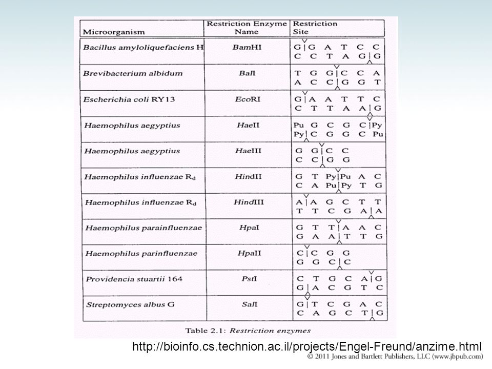
3.1 Introduction restriction endonuclease – An enzyme that recognizes specific short sequences of DNA and cleaves the duplex (sometimes at the target site, sometimes elsewhere, depending on type). Type II: recognition site and cleavage site are the same Type I: cleavage site can be up to 1000 bp away from recognition site Type III: have closer cleavage sites, usually 20 to 30 bp
. Type II: recognition site and cleavage site are the same. Type I: cleavage site can be up to 1000 bp away from recognition site. Type III: have closer cleavage sites, usually 20 to 30 bp.")
4
3.1 Introduction cloning vector – DNA (often derived from a plasmid or a bacteriophage genome) that can be used to propagate an incorporated DNA sequence in a host cell. Vectors contain selectable markers and replication origins to allow identification and maintenance of the vector in the host.
 that can be used to propagate an incorporated DNA sequence in a host cell. Vectors contain selectable markers and replication origins to allow identification and maintenance of the vector in the host.")
5
nucleases hydrolyze an ester bond within a phosphodiester bond.
phosphatases hydrolyze the ester bond in a phosphomonoester bond. FIGURE 01: The target of a phosphatase (a) and a nuclease (b). An endonuclease (c) and an exonuclease (d)
 and a nuclease (b). An endonuclease (c) and an exonuclease (d)")
6
3.2 Nucleases endonuclease – Nucleases that cleave phosphoester bonds within a nucleic acid chain. They may be specific for RNA or for single-stranded or double-stranded DNA. exonuclease – Nucleases that cleave phosphoester bonds one at a time from the end of a polynucleotide chain. They may be specific for either the 5′ or 3′ end of DNA or RNA.

7
FIGURE 02: Restriction endonuclease
3.2 Nucleases sticky end blunt end Restriction endonucleases can be used to cleave DNA into defined fragments. Recognize a specific sequence Cut, or restrict, that sequence FIGURE 02: Restriction endonuclease

9
The Nobel Prize in Medicine 1978
Biozentrum der Universität, Basel, Switzerland Johns Hopkins University School of Medicine, Baltimore, MD, USA Johns Hopkins University School of Medicine, Baltimore, MD, USA Discovery of restriction enzymes and their application to problems of molecular genetics

10
3.2 Nucleases A map can be generated by using the overlaps between the fragments generated by different restriction enzymes. FIGURE 03: A restriction map is a linear sequence of sites separated by defined distances on DNA

11
3.3 Cloning Cloning a fragment of DNA requires a specially engineered vector. recombinant DNA – A DNA molecule that has been created by joining together two or more molecules from different sources. ligating (or ligation) – The process of joining together two DNA fragments.
 – The process of joining together two DNA fragments.")
12
3.3 Cloning subclone – The process of breaking a cloned fragment into smaller fragments for further cloning. MCS (multiple cloning site) – A sequence of DNA containing a series of tandem restriction endonuclease sites used in cloning vectors for creating recombinant molecules.
 – A sequence of DNA containing a series of tandem restriction endonuclease sites used in cloning vectors for creating recombinant molecules.")
13
FIGURE 04: Plasmid transformation
3.3 Cloning Three site in plasmid: ori ampr lacZ with MCS FIGURE 04: Plasmid transformation

14
Action of DNA ligase 5’ Base Base O O O OH _ Restriction Enzyme O P O
CH2 Base O O CH2 Base O 3’

15
Enhance of the cloning efficiency:
Dephosphorylation of the 5’-phophate in the vector by alkaline phosphatase to prevent self-ligation From Dr. Yu

16
3.3 Cloning transformation – The acquisition of new genetic material by incorporation of added exogenous, nonviral DNA. Blue/white selection allows the identification of bacteria that contain the vector plasmid and vector plasmids that contain an insert. FIGURE 05: E. coli colonies on agar plates with ampicillin, IPTG, and the color indicator X-gal Transformation: CaCl2, electroporation LacZ gene encodes β-galactoside (β-gal) β-gal can cleave X-gal into blue compound IPTG as β-gal inducer
 β-gal can cleave X-gal into blue compound. IPTG as β-gal inducer.")
17
3.4 Cloning Vectors Can Be Specialized for Different Purposes
FIGURE 06: Several types of cloning vectors are available

18
3.4 Cloning Vectors Can Be Specialized for Different Purposes
Shuttle vectors can be propagated in more than one type of host cell. Expression vectors contain promoters that allow transcription of any cloned gene.

19
FIGURE 07: A vector can be used in both yeast and bacteria
Shuttle Vector FIGURE 07: A vector can be used in both yeast and bacteria (shuttle vector)
")
20
Expression vector RBS: Ribosome Binding Site

21
FIGURE 08: Luciferase graph/Firefly
Expression Vector FIGURE 08: Luciferase graph/Firefly Photo © Cathy Keifer/Dreamstime.com

22
3.4 Cloning Vectors Can Be Specialized for Different Purposes
Reporter genes can be used to measure promoter activity or tissue-specific expression. FIGURE 10: Fluorescent proteins are powerful research tools Courtesy of Joachim Goedhart, Molecular Cytology, SILS, University of Amsterdam. FIGURE 09: A mouse promoter controls tissue-specific expression of lacZ Photo courtesy of Robb Krumlauf, Stowers Institute for Medical Research

23
Fluorescent proteins are powerful research tools
GFP(Green), YFP(Yellow), CFP(Cyan), BFP(Blue) Reprinted from Vision Res., vol. 45, T. G. Wensel, et al., Rhodopsin-EGFP knock-ins..., pp Copyright 2005, with permission from Elsevier [ Photo courtesy of Theodore G. Wensel, Baylor Col
, YFP(Yellow), CFP(Cyan), BFP(Blue) Reprinted from Vision Res., vol. 45, T. G. Wensel, et al., Rhodopsin-EGFP knock-ins..., pp Copyright 2005, with permission from Elsevier [ Photo courtesy of Theodore G. Wensel, Baylor Col.")
24
The Fly brain

25
Targeted gene expression in fly
GAL4/UAS Binary System Brand & Perrimon 1993

26
GFP expression in subset of fly mushroom body
Shih & Wu unpublished

27
GFP express in single neuron in fly brain
Wu et al., 2011

28
The Nobel Prize in Chemistry 2008
Woods Hole & Boston University Medical School Columbia University Howard Hughes Medical Institute For the discovery and development of the green fluorescent protein, GFP

29
3.4 Cloning Vectors Can Be Specialized for Different Purposes
Numerous methods exist to introduce DNA into different target cells. FIGURE 11: DNA can be introduced into cells in several ways

30
3.5 Nucleic Acid Detection
DNA, RNA nucleic acids absorb light at 260 nm Protein absorb light at 280 nm 260/280 ratios to quantify the amount of nucleic acid DNA, RNA can be nonspecifically stained with ethidium bromide(EtBr) or SYBR
 or SYBR.")
31
3.5 Nucleic Acid Detection
Hybridization of a labeled nucleic acid to complementary sequences can identify specific nucleic acids. probe – A radioactive nucleic acid, DNA or RNA, used to identify a complementary fragment.

32
3.5 Nucleic Acid Detection
autoradiography – A method of capturing an image of radioactive materials on film. FIGURE 12: An autoradiogram of a gel prepared from the colonies described in Figure 3.5

33
3.5 Nucleic Acid Detection
in situ hybridization – Hybridization of a probe to intact tissue to locate its complementary strand by autoradiography. FIGURE 13: The fluorescent in situ hybridization (FISH) technique Adapted from an illustration by Darryl Leja, National Human Genome Research Institute (
 technique. Adapted from an illustration by Darryl Leja, National Human Genome Research Institute (")
34
In situ Hybridization: Locating genes in chromosomes
FISH: Fluorescence in situ hybridization From Dr. Yu

35
3.6 DNA Separation Techniques
Gel electrophoresis separates DNA fragments by size, using an electric current to cause the DNA to migrate toward a positive charge. FIGURE 14: DNA sizes can be determined by gel electrophoresis Adapted from an illustration by Michael Blaber, Florida State University.

36
FIGURE 15: Agarose gel electrophoresis pattern of SV40 DNA
Type I topoisomerase: Relaxes negative supercoiled DNA to relaxed or linear DNA Reproduced from W. Keller, Proc. Natl. Acad. Sci. USA 72 (1975): Photo courtesy of Walter Keller, University of Basel.
: Photo courtesy of Walter Keller, University of Basel.")
37
3.6 DNA Separation Techniques
DNA can also be isolated using density gradient centrifugation. DNA density dependents on G-C content An AT base pair has a lower molecular weight than a GC base pair

38
3.7 DNA Sequencing Chain termination sequencing uses dideoxynucleotides (ddNTPs) to terminate DNA synthesis at particular nucleotides. Primer - A single stranded nucleic acid molecule with a 3′ –OH used to initiate DNA polymerase replication of a paired template strand.

39
3.7 DNA Sequencing Chain termination sequencing uses ddNTPs to terminate DNA synthesis at particular nucleotide. Fluorescently tagged ddNTPs and capillary gel electrophoresis allow automated, high-throughput DNA sequencing.

40
FIGURE 17: DideoxyNTP sequencing using fluorescent tags
Different fluorescents label for each ddNTP Hit with a laser and pass by an optical sensor Glass capillary tube dissipate heat rapidly FIGURE 17: DideoxyNTP sequencing using fluorescent tags Inset photo courtesy of Jan Kieleczawa

41
3.8 PCR and RT-PCR Thermus aquaticus (Taq DNA polymerase)
")
42
3.8 PCR and RT-PCR Polymerase chain reaction (PCR) permits the exponential amplification of a desired sequence, using primers that anneal to the sequence of interest. Tm: annealing temperature for primer/template pair *Kary Mullis in the 1980s *Taq polymerase from Thermus aquaticus *Thermal cycler 95oC oC oC 20x
 permits the exponential amplification of a desired sequence, using primers that anneal to the sequence of interest. Tm: annealing temperature for primer/template pair. *Kary Mullis in the 1980s. *Taq polymerase from Thermus aquaticus. *Thermal cycler 95oC 40oC 72oC. 20x.")
43
Exponential produce the primer to primer-defined sequence

44
The Nobel Prize in Chemistry 1993
Press Release 13 October 1993 The Royal Swedish Academy of Sciences has decided to award the 1993 Nobel Prize in Chemistry for contributions to the development of methods within DNA-based chemistry, with half to Dr Kary B. Mullis, La Jolla, California, U.S.A., for his invention of the polymerase chain reaction (PCR) method, and half to Professor Michael Smith, University of British Columbia, Vancouver, Canada, for his fundamental contributions to the establishment of oligonucleotide-based, site-directed mutagenesis and its development for protein studies. Decisive progress in gene technology through two new methods: the polymerase chain reaction (PCR) method and site-directed mutagenesis Cetus生物科技公司以三億三千五百萬美金專利金賣出(史上最貴的發明專利) Kary B. Mullis Michael Smith
 method, and half to Professor Michael Smith, University of British. Columbia, Vancouver, Canada, for his fundamental. contributions to the establishment of. oligonucleotide-based, site-directed mutagenesis. and its development for protein studies. Decisive progress in gene technology through two new methods: the polymerase chain reaction (PCR) method and site-directed mutagenesis. Cetus生物科技公司以三億三千五百萬美金專利金賣出(史上最貴的發明專利) Kary B. Mullis. Michael Smith.")
45
3.8 PCR and RT-PCR RT-PCR uses reverse transcriptase to convert RNA to cDNA for use in a PCR reaction. Real-time, or quantitative, PCR detects the products of PCR amplification during their synthesis, and is more sensitive and quantitative than conventional PCR.

46
3.8 PCR and RT-PCR fluorescence resonant energy transfer (FRET) – A process whereby the emission from an excited fluorophore is captured and reemitted at a longer wavelength by a nearby second fluorophore whose excitation spectrum matches the emission frequency of the first fluorophore.
 – A process whereby the emission from an excited fluorophore is captured and reemitted at a longer wavelength by a nearby second fluorophore whose excitation spectrum matches the emission frequency of the first fluorophore.")
47
FIGURE 20: Fluorescence Resonant Energy Transfer (FRET)
")
48
Fluorescent Tags in Real-Time PCR
This fluorescent-tagged oligonucleotide serves as a reporter probe Fluorescent tag at 5’-end Fluorescence quenching tag at 3’-end With PCR rounds the 5’ tag is separated from the 3’ tag Fluorescence increases with incorporation into DNA product

49
TeqMan probe Primer binding Probe hybridization PCR conditions
wikipedia

50
SYBR Green I Dye Primer binding PCR conditions
SYBR Green I Dye binds to the minor groove of ds DNA Reporter dye are not sequence specific, spurious products produced by the reaction may lead to false positive signals.

Similar presentations


 Analysis of DNA (Sequencing) Chemical Synthesis of DNA.>")


>")