Download presentation
Presentation is loading. Please wait.

1
Chapter 24: The Forearm, Wrist, Hand and Finger
Jennifer Doherty-Restrepo, MS, LAT, ATC Academic Program Director, Entry-Level ATEP Florida International University Acute Care and Injury Prevention

2
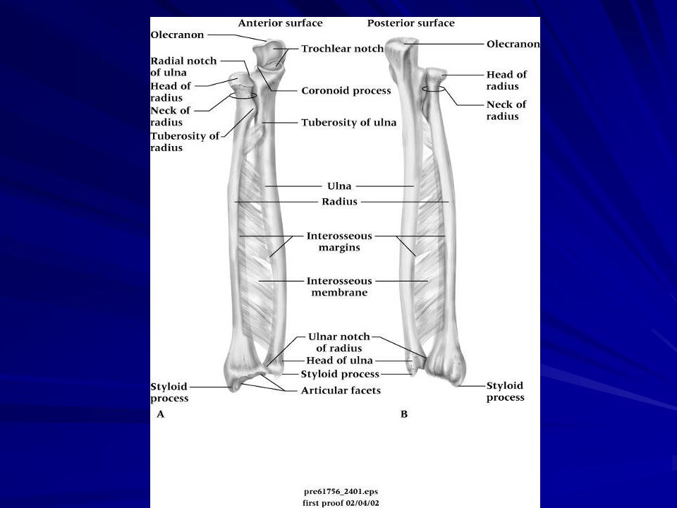
Anatomy of the Forearm

6
Recognition and Management of Injuries to the Forearm
Contusion Etiology Ulnar side receives majority of blows due to arm blocks Can be acute or chronic Result of direct contact or blow Signs and Symptoms Pain, swelling and hematoma If repeated blows occur, heavy fibrosis and possibly bony callus could form w/in hematoma

7
Contusion (continued)
Management Proper care in acute stage involves RICE for at least one hour and followed up w/ additional cryotherapy Protection is critical - full-length sponge rubber pad can be used to provide protective covering

8
Forearm Splints Etiology Signs and Symptoms Management
Forearm strain - most come from severe static contraction Cause of splints - repeated static contractions Difficult to manage Signs and Symptoms Dull ache between extensors which cross posterior aspect of forearm Weakness and pain w/ contraction Point tenderness in interosseus membrane Management Treat symptomatically If occurs early in season, strengthen forearm; when it occurs late in season treat w/ cryotherapy, wraps, or heat Can develop compartment syndrome in forearm as well and should be treated like lower extremity

9
Forearm Fractures Etiology Signs and Symptoms
Common in youth due to falls and direct blows Ulna and radius generally fracture individually Fracture in upper third may result in abduction deformity Fracture in lower portion will remain relatively neutral Older athlete may experience greater soft tissue damage and greater chance of paralysis due to Volkman’s contracture Signs and Symptoms Audible pop or crack followed by moderate to severe pain, swelling, and disability Edema, ecchymosis w/ possible crepitus

10
Management Initially RICE followed by splinting until definitive care is available Long term casting followed by rehab plan

11
Colles’ Fracture Etiology Occurs in lower end of radius or ulna
MOI is fall on outstretched hand, forcing radius and ulna into hyperextension Less common is the reverse Colles’ fracture

12
Signs and Symptoms Management
Forward displacement of radius causing visible deformity (silver fork deformity) When no deformity is present, injury can be passed off as bad sprain Extensive bleeding and swelling Tendons may be torn/avulsed and there may be median nerve damage Management Cold compress, splint wrist and refer to physician X-ray and immobilization Severe sprains should be treated as fractures Without complications a Colles’ fracture will keep an athlete out for 1-2 months In children, injury may cause lower epiphyseal separation
 When no deformity is present, injury can be passed off as bad sprain. Extensive bleeding and swelling. Tendons may be torn/avulsed and there may be median nerve damage. Management. Cold compress, splint wrist and refer to physician. X-ray and immobilization. Severe sprains should be treated as fractures. Without complications a Colles’ fracture will keep an athlete out for 1-2 months. In children, injury may cause lower epiphyseal separation.")
13
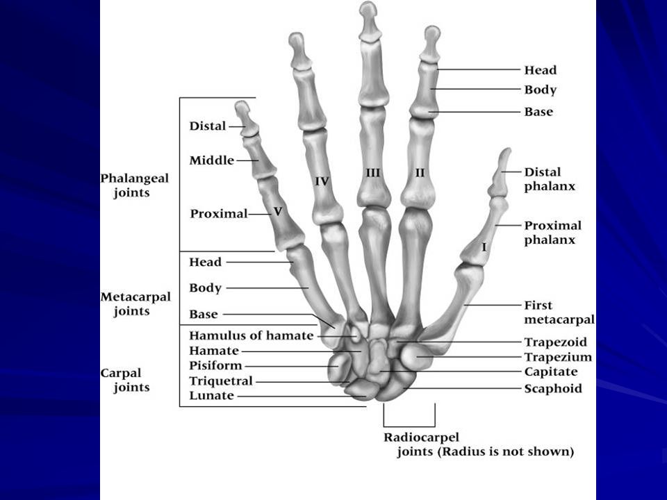
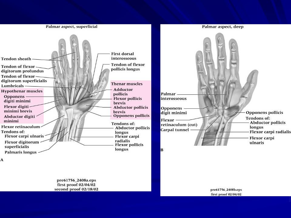
Anatomy of the Wrist, Hand and Fingers

20
Recognition and Management of Injuries to the Wrist, Hand and Fingers
Wrist Sprains Etiology Most common wrist injury Arises from any abnormal, forced movement Falling on hyperextended wrist, violent flexion or torsion Multiple incidents may disrupt blood supply Signs and Symptoms Pain, swelling and difficulty w/ movement

21
Management Refer to physician for X-ray if severe
RICE, splint and analgesics Have athlete begin strengthening soon after injury Tape for support can benefit healing and prevent further injury

22
Triangular Fibrocartilage Complex (TFCC) Injury
Etiology Occurs through forced hyperextension, falling on outstretched hand Violent twist or torque of the wrist Often associated w/ sprain of UCL Signs and Symptoms Pain along ulnar side of wrist, difficulty w/ wrist extension, possible clicking Swelling is possible, not much initially Athlete may not report injury immediately

23
Management Referred to physician for treatment
Treatment will require immobilization initially for 4 weeks Immobilization should be followed by period of strengthening and ROM activities Surgical intervention may be required if conservative treatments fail

24
Tenosynovitis Etiology Signs and Symptoms Management
Cause of repetitive wrist accelerations and decelerations Repetitive overuse of wrist tendons and sheaths Signs and Symptoms Pain w/ use or pain in passive stretching Tenderness and swelling over tendon Management Acute pain and inflammation treated w/ ice massage 4x daily for first hours, NSAID’s and rest When swelling has subsided, ROM is promoted w/ contrast bath Ultrasound and phonphoresis can be used PRE can be instituted once swelling and pain subsided

25
Tendinitis Etiology Signs and Symptoms Management
Repetitive pulling movements; repetitive pressure on palms (cycling) Primary cause is overuse of the wrist Signs and Symptoms Pain on active use or passive stretching Isometric resistance to involved tendon produces pain, weakness or both Management Acute pain and inflammation treated w/ ice massage 4x daily for first hours, NSAID’s and rest When swelling has subsided, ROM is promoted w/ contrast bath PRE can be instituted once swelling and pain subsided (high rep, low resistance)
 Primary cause is overuse of the wrist. Signs and Symptoms. Pain on active use or passive stretching. Isometric resistance to involved tendon produces pain, weakness or both. Management. Acute pain and inflammation treated w/ ice massage 4x daily for first hours, NSAID’s and rest. When swelling has subsided, ROM is promoted w/ contrast bath. PRE can be instituted once swelling and pain subsided (high rep, low resistance)")
26
Nerve Compression, Entrapment, Palsy
Etiology Median and ulnar nerve compression Result of direct trauma to nerves Signs and Symptoms Sharp or burning pain associated w/ skin sensitivity or paresthesia May result in benediction/ bishop’s deformity (damage to the ulnar nerve) or claw hand deformity (damage to both nerves) Palsy of radial nerve produces drop wrist deformity caused by paralysis of extensor muscles Palsy of median nerve can cause ape hand (thumb pulled back in line w/ other fingers) Management Chronic entrapment may cause irreversible damage Surgical decompression may be necessary
 or claw hand deformity (damage to both nerves) Palsy of radial nerve produces drop wrist deformity caused by paralysis of extensor muscles. Palsy of median nerve can cause ape hand (thumb pulled back in line w/ other fingers) Management. Chronic entrapment may cause irreversible damage. Surgical decompression may be necessary.")
27
Carpal Tunnel Syndrome
Etiology Compression of median nerve due to inflammation of tendons and sheaths of carpal tunnel Result of repeated wrist flexion or direct trauma to anterior aspect of wrist Signs and Symptoms Sensory and motor deficits (tingling, numbness and paresthesia); weakness in thumb Management Conservative treatment - rest, immobilization, NSAID’s If symptoms persist, corticosteroid injection may be necessary or surgical decompression of transverse carpal ligament
; weakness in thumb. Management. Conservative treatment - rest, immobilization, NSAID’s. If symptoms persist, corticosteroid injection may be necessary or surgical decompression of transverse carpal ligament.")
28
Dislocation of Lunate Bone
Etiology Forceful hyperextension or fall on outstretched hand Signs and Symptoms Pain, swelling, and difficulty executing wrist and finger flexion Numbness/paralysis of flexor muscles due to pressure on median nerve Management Treat as acute, and sent to physician for reduction If not recognized, bone deterioration could occur, requiring surgical removal Usual recovery is 1-2 months

29
Scaphoid Fracture Etiology Signs and Symptoms Management
Caused by force on outstretched hand, compressing scaphoid between radius and second row of carpal bones Often fails to heal due to poor blood supply Signs and Symptoms Swelling, severe pain in anatomical snuff box Presents like wrist sprain Pain w/ radial flexion Management Must be splinted and referred for X-ray prior to casting Immobilization lasts 6 weeks and is followed by strengthening and protective tape Wrist requires protection against impact loading for 3 additional months

30
Hamate Fracture Etiology Signs and Symptoms Management
Occurs as a result of a fall or more commonly from contact while athlete is holding an implement Signs and Symptoms Wrist pain and weakness, along w/ point tenderness Pull of muscular attachment can cause non-union Management Casting wrist and thumb is treatment of choice Hook of hamate can be protected w/ doughnut pad to take pressure off area

31
Wrist Ganglion Etiology Signs and Symptoms Management
Synovial cyst (herniation of joint capsule or synovial sheath of tendon) Generally appears following wrist strain Signs and Symptoms Appear on back of wrist generally Occasional pain w/ lump at site Pain increases w/ use May feel soft, rubbery or very hard Management Old method was to first break down the swelling through distal pressure and then apply pressure pad to encourage healing New approach includes aspiration, chemical cauterization w/ subsequent pressure from pad Ultrasound can be used to reduce size Surgical removal is most effective treatment method
 Generally appears following wrist strain. Signs and Symptoms. Appear on back of wrist generally. Occasional pain w/ lump at site. Pain increases w/ use. May feel soft, rubbery or very hard. Management. Old method was to first break down the swelling through distal pressure and then apply pressure pad to encourage healing. New approach includes aspiration, chemical cauterization w/ subsequent pressure from pad. Ultrasound can be used to reduce size. Surgical removal is most effective treatment method.")
32
Bowler’s Thumb Etiology Signs and Symptoms Management
Perineural fibrosis of subcutaneous ulnar digital nerve of thumb Pressure from bowling ball on thumb Signs and Symptoms Pain, tingling during pressure on irritated area and numbness Management Padding, decrease amount of bowling If condition continues, surgery may be required

33
Mallet Finger Etiology Signs and Symptoms Management
Caused by a blow that contacts tip of finger avulsing extensor tendon from insertion Signs and Symptoms Pain at DIP; X-ray shows avulsed bone on dorsal proximal distal phalanx Unable to extend distal end of finger (carrying at 30 degree angle) Point tenderness at sight of injury Management RICE and splinting for 6-8 weeks
 Point tenderness at sight of injury. Management. RICE and splinting for 6-8 weeks.")
34
Boutonniere Deformity
Etiology Rupture of extensor tendon dorsal to the middle phalanx Forces DIP joint into extension and PIP into flexion Signs and Symptoms Severe pain, obvious deformity and inability to extend DIP joint Swelling, point tenderness Management Cold application, followed by splinting Splinting must be continued for 5-8 weeks Athlete is encouraged to flex distal phalanx

35
Jersey Finger Etiology Signs and Symptoms Management
Rupture of flexor digitorum profundus tendon from insertion on distal phalanx Often occurs w/ ring finger when athlete tries to grab a jersey Signs and Symptoms DIP can not be flexed, finger remains extended Pain and point tenderness over distal phalanx Management Must be surgically repaired Rehab requires 12 weeks and there is often poor gliding of tendon, w/ possibility of re-rupture

36
Sprains, Dislocations and Fractures of Phalanges
Etiology Phalanges are prone to sprains caused by direct blows or twisting MOI is also similar to that which causes fractures and dislocations Signs and Symptoms Recognition primarily occurs through history Sprain symptoms - pain, severe swelling and hematoma

37
Gamekeeper’s Thumb Etiology Signs and Symptoms Management
Sprain of UCL of MCP joint of the thumb Mechanism is forceful abduction of proximal phalanx occasionally combined w/ hyperextension Signs and Symptoms Pain over UCL in addition to weak and painful pinch Management Immediate follow-up must occur If instability exists, athlete should be referred to orthopedist If stable, X-ray should be performed to rule out fracture Thumb splint should be applied for protection for 3 weeks or until pain free

Similar presentations


![What am I?. What am I? Articulations of the humerus, radius, and ulna Articulations of the humerus, radius, and ulna. [ olecranon process ] Medial.](/14/4241906/big_thumb.jpg "What am I?. What am I? Articulations of the humerus, radius, and ulna Articulations of the humerus, radius, and ulna. [ olecranon process ] Medial.>")