Download presentation
Presentation is loading. Please wait.

1
Margaret Elaine J. Villamayor, RMT, MD
MEDICINE GRAND ROUNDS Hypertrophic Osteoarthropathy: a pretender, a cheater Margaret Elaine J. Villamayor, RMT, MD Makati Medical Center April 8, 2010

2
Objective To present a case of a patient presenting with joint pains as a symptom of a distant pathology And I’m here to present a case of a patient presenting with joint pains as a symptom of an underlying pathology

3
GENERAL DATA EB, 52/male, married, Filipino, Engineer, admitted last January 13, 2010 Our px is EB, a 52 yr old man, who was admitted in our institution last January 13, 2010

4
Joint pains CHIEF COMPLAINT
Patient came in w/ a chief complaint of joint pains

5
HISTORY OF PRESENT ILLNESS
2 months Intermittent pain on tibial & ankles, sharp, deep-seated, 5/10, not related to physical activity, no specific timing and lasting more than 30 minutes Self-medicated : Ibuprofen and Meloxicam

6
HISTORY OF PRESENT ILLNESS
1 month Consult: Normal Blood tests Impression: Gouty Arthritis Unrecalled pain meds Diet modification incldu

7
HISTORY OF PRESENT ILLNESS
2 weeks (+) pain on wrists, toes (+) enlargement & pain on fingertips (+) difficulty in ambulating and with fine movements (+) sore throat & fever Consult at MMC
 pain on wrists, toes (+) enlargement & pain on fingertips (+) difficulty in ambulating and with fine movements (+) sore throat & fever Consult at MMC")
8
HISTORY OF PRESENT ILLNESS
1 week On follow-up: (+) increase in pain severity Advised admission for further workup
 increase in pain severity Advised admission for further workup")
9
REVIEW OF SYSTEMS General: (-) pallor, anorexia, weight loss HEENT: (-) signs of head injury, (-) redness, itchiness, lacrimation of eyes, (-) deafness, tinnitus, and ear discharge, (-) colds, nasal stuffiness, epistaxis, (-) mouth sores, sore throat, gum bleeding, (-) stiffness, lumps, tenderness, adenopathy Respiratory: (-) cough, wheezing, shortness of breath, hemoptysis Cardiovascular: (-) chest pain, palpitation, syncope, cyanosis Gastro-intestinal: (-) nausea, vomiting, hematemesis, dysphagia, diarrhea, constipation Renal: (-) hematuria, no dysuria Neurologic: (-) fainting Psychiatric: (-) history of depression
 pallor, anorexia, weight loss HEENT: (-) signs of head injury, (-) redness, itchiness, lacrimation of eyes, (-) deafness, tinnitus, and ear discharge, (-) colds, nasal stuffiness, epistaxis, (-) mouth sores, sore throat, gum bleeding, (-) stiffness, lumps, tenderness, adenopathy Respiratory: (-) cough, wheezing, shortness of breath, hemoptysis Cardiovascular: (-) chest pain, palpitation, syncope, cyanosis Gastro-intestinal: (-) nausea, vomiting, hematemesis, dysphagia, diarrhea, constipation Renal: (-) hematuria, no dysuria Neurologic: (-) fainting Psychiatric: (-) history of depression")
10
PAST MEDICAL HISTORY No serious illness during childhood.
No known co-morbidities Diagnosed with Cholelithiasis in 2006 No history of previous surgeries, blood transfusions, trauma, allergies

11
FAMILY HISTORY (+) Gastric CA – father (+) Colon CA - paternal grandfather
 Gastric CA – father (+) Colon CA - paternal grandfather")
12
PERSONAL AND SOCIAL HISTORY
36 pack years smoking history Occasional alcoholic beverage drinker Denies illicit drug use & multiple sexual partners Patient works as an engineer in Saudi Arabia for the past 20 years Plays golf every week.

13
PHYSICAL EXAMINATION General Survey: conscious, coherent, oriented to person, place and time, ambulates with assistance BP 120/80, HR 72 bpm, RR 18 cpm, T 36.9C, Height: 162cm Weight: 68.18kg BMI: kg/m2 HEENT: anicteric sclerae, pink conjunctivae, no naso-aural discharge, no cervical lymphadenopathies, no tonsilopharyngeal congestion, no neck vein distention, no carotid bruits Chest and Lungs: No gross lesions, no retractions, no point tenderness, equal chest expansion, equal fremiti, lungs resonant, clear breath sounds

14
PHYSICAL EXAMINATION Heart: adynamic precordium, AB at 5th LICS MCL, regular rate & rhythm, no friction rub, no murmurs Abdomen: flabby abdomen, non-distended, normoactive bowel sounds, soft, tympanitic, nontender, no hepatosplenomegaly, no Murphy’s sign, no costovertebral angle tenderness, no peritoneal signs

15
PHYSICAL EXAMINATION Extremities:
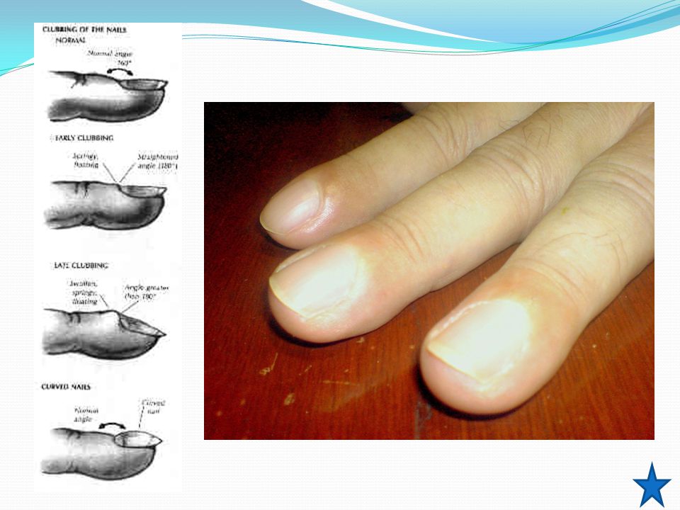
(+) digital clubbing on all extremities (+)bilateral tibial tenderness, non erythematous, not warm to touch (+) limitation of movement on wrist & ankle flexion & finger flexion and opposition (-) cyanosis, crepitus Shamroth’s test/The apposition sign: The distal interphalangeal (DIP) joints and the nail bed of the right and left index finger are placed in apposition. Normally, an oblong diamond-shaped aperture is visible between the 2 juxtaposed nail beds. With clubbing, this space is obliterated. Shamroth’s test
 digital clubbing on all extremities. (+)bilateral tibial tenderness, non erythematous, not warm to touch. (+) limitation of movement on wrist & ankle flexion & finger flexion and opposition. (-) cyanosis, crepitus. Shamroth’s test/The apposition sign: The distal interphalangeal (DIP) joints and the nail bed of the right and left index finger are placed in apposition. Normally, an oblong diamond-shaped aperture is visible between the 2 juxtaposed nail beds. With clubbing, this space is obliterated. Shamroth’s test.")
16
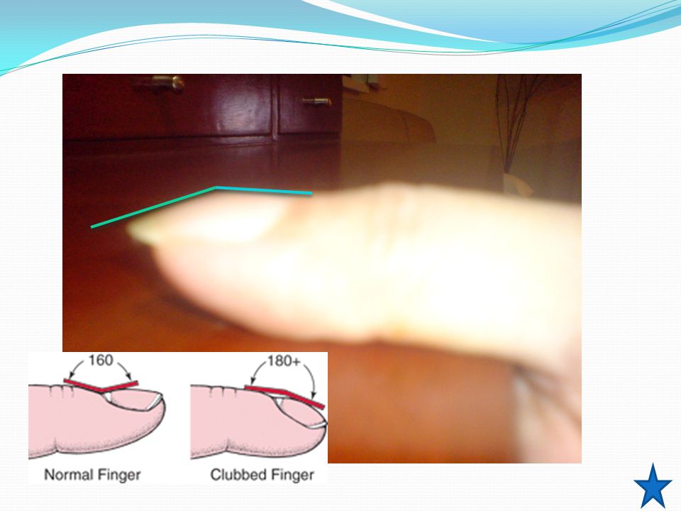
Normal Angle = less than or equal to 160°.
Lovibond angle the angle made by the proximal nail fold and nail plate (Lovibond angle) typically is less than or equal to 160°. In clubbing, the angle flattens out and increases as the severity of the clubbing increases. If the angle is greater than 180°, definitive clubbing exists. An angle between ° falls in a gray area and may indicate early stages of clubbing Normal Angle = less than or equal to 160°. Flat/ >180 = clubbing Swartz MH. Textbook of Physical Diagnosis: History and Examination. 2nd ed. Philadelphia, Pa: WB Saunders; 1994:76-8.
 typically is less than or equal to 160°. In clubbing, the angle flattens out and increases as the severity of the clubbing increases. If the angle is greater than 180°, definitive clubbing exists. An angle between ° falls in a gray area and may indicate early stages of clubbing. Normal Angle = less than or equal to 160°. Flat/ >180 = clubbing. Swartz MH. Textbook of Physical Diagnosis: History and Examination. 2nd ed. Philadelphia, Pa: WB Saunders; 1994:76-8.")
17
NEUROLOGIC EXAMINATION:
Mental status: awake, alert, oriented Cranial Nerve Exam: CN I – not assessed CN II, III – pupils 3mm ERTL, (+) ROR, clear media, no visual field defects CN III, IV, VI – full EOMs CN V – intact V1-V3 CN VII – no facial asymmetry CN VIII – no gross hearing defects CN IX, X – tongue and uvula at midline CN XI – good SCM tone CN XII – tongue at midline
 ROR, clear media, no visual field defects CN III, IV, VI – full EOMs CN V – intact V1-V3 CN VII – no facial asymmetry CN VIII – no gross hearing defects CN IX, X – tongue and uvula at midline CN XI – good SCM tone CN XII – tongue at midline")
18
NEUROLOGIC EXAMINATION
Cerebellar: no dysdiadochokinesia, no dysmetria Reflexes: +2 on all extremities, no Babinski Meninges: supple neck, no Brudzinski, no Kernig’s Motor : 5/5 on all extremities Sensory: no sensory deficits

19
SALIENT FEATURES OBJECTIVE
SUBJECTIVE 36 pack year smoking history (+) Family History of Cancer (+) fever, (-) weight loss, cardiac, gastrointestinal or respiratory symptoms Joint & tibial pain x 2 months - symmetrical, no specific timing, not relieved by rest/ analgesics, >30 mins OBJECTIVE (+) digital clubbing both upper and lower extremities (+)bilateral tibial tenderness
 Family History of Cancer. (+) fever, (-) weight loss, cardiac, gastrointestinal. or respiratory symptoms. Joint & tibial pain x 2 months. - symmetrical, no specific timing, not relieved by rest/ analgesics, >30 mins. OBJECTIVE. (+) digital clubbing both upper and lower extremities. (+)bilateral tibial tenderness.")
20
Admitting Impression t/c Hypertrophic Osteoarthropathy, etiology to be determined Obese Class I (BMI kg/m2)
 .")
21
Weakness, easy fatigability, weight loss, ±fever, None ±fever
Patient Rheumatic Fever Rheumatoid Arthritis Osteoarthritis HOA Systemic sxs Fever Fever, sore throat Weakness, easy fatigability, weight loss, ±fever, None ±fever PE symmetrical Tibial, wrists, toes digits, wrist, clubbing (-) crepitus Migratory, carditis, PIP, MCP/MTP, wrist, elbow, knee, ankle, nodules LS spine, hip, knee, 1st MTP DIP PIP spared are the wrist, elbow, and ankle (+) crepitus Tibia, wrists, ankles, digits, elbow, clubbing Timing Movement Movement, morning , >1 hr RA: Pain in affected joints, aggravated by movement, is the most common manifestation of established RA. Generalized stiffness is frequent and is usually greatest after periods of inactivity. The majority of patients will experience constitutional symptoms such as weakness, easy fatigability, anorexia, and weight loss. Although fever to 40°C occurs on occasion OA: OA is joint failure, a disease in which all structures of the joint have undergone pathologic change, often in concert. The pathologic sine qua non of disease is hyaline articular cartilage loss, present in a focal and, initially, nonuniform manner. This is accompanied by increasing thickness and sclerosis of the subchondral bony plate, by outgrowth of osteophytes at the joint margin, by stretching of the articular capsule, by mild synovitis in many affected joints, and by weakness of muscles bridging the joint. In knees, meniscal degeneration is part of the disease. There are numerous pathways that lead to joint failure, but the initial step is often joint injury in the setting of a failure of protective mechanisms
 crepitus. Migratory, carditis, PIP, MCP/MTP, wrist, elbow, knee, ankle, nodules. LS spine, hip, knee, 1st MTP DIP PIP. spared are the wrist, elbow, and ankle. (+) crepitus. Tibia, wrists, ankles, digits, elbow, clubbing. Timing. Movement. Movement, morning , >1 hr. RA: Pain in affected joints, aggravated by movement, is the most common manifestation of established RA. Generalized stiffness is frequent and is usually greatest after periods of inactivity. The majority of patients will experience constitutional symptoms such as weakness, easy fatigability, anorexia, and weight loss. Although fever to 40°C occurs on occasion. OA: OA is joint failure, a disease in which all structures of the joint have undergone pathologic change, often in concert. The pathologic sine qua non of disease is hyaline articular cartilage loss, present in a focal and, initially, nonuniform manner. This is accompanied by increasing thickness and sclerosis of the subchondral bony plate, by outgrowth of osteophytes at the joint margin, by stretching of the articular capsule, by mild synovitis in many affected joints, and by weakness of muscles bridging the joint. In knees, meniscal degeneration is part of the disease. There are numerous pathways that lead to joint failure, but the initial step is often joint injury in the setting of a failure of protective mechanisms.")
22
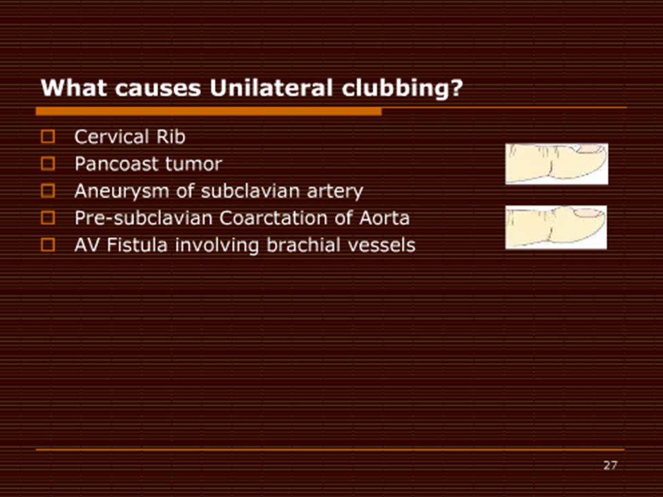
Approach to HOA Cervical rib, pancoast tumor, aneurysm of subclavian artery, pre-subclavian coarctation of aorta, AVF involving brachial vessels Pineda C, Fonseca C, Martinez-Lavin M. The spectrum of soft tissue and skeletal abnormalities of hypertrophic osteoarthropathy. J Rheumatol. May 1990;17(5):
:")
23
Course in the Wards Jan 13 Sodium 135 Potassium 4.6 BUN 10.12 Crea
0.72 Calcium 8.78 Total Protein 6.9 Albumin 3.6 Globulin 3.3 A/G 1.09 ALP 140 AST 25 Total Bili 7.8 Uric Acid 3.8 Trigly 56 Chole 185.57 HDL 53.78 LDL 101.76 Jan 13 Hb 12.7 Hct 37 RBC 4.42 WBC 15.25 Seg 76 Lympho 16 Mono 8 Plt 531,000 CRP positive up to 1:64 dilutions (384 mg/l) Jan 13 ESR 93 CRP (+)
 Jan 13. ESR. 93. CRP. (+)")
24
Xray of Both Hands No osseous nor joint abnormalities seen in both hands

25
Chest Xray A mass density measuring about 7.5x6.5cm in its widest dimension is seen in the right parahilar area. The mass has lobulated border with compression of the inferior bronchus. No cavitations, no evident calcification within the lesion. The rest of the lungs are unremarkable

26
CT-Scan of the Chest: January 13, 2010
CT scan of the chest showed 4.6 x 5.5 x 4.6 cm well-defined soft tissue mass in the right hilar region, accompanied by some ground glass opacity changes in the surrounding right upper lung segment (obstructive pneumonitis).
.")
27
CT-Scan of the Chest: January 13, 2010
A small 0.6cm nodule is noted along the peripheral margin of the right middle lobe, emphysematous changes are noted scattered throughout the right and left lungs.

28
CT-Scan of the Chest: January 13, 2010
At least 3 about 1.0 to 1.6 cm nodes are noted in the subcarinal region.

29
Course in the Wards Cranial MRI & CT of the Abdomen TCVS Referral
CT-guided Biopsy Plan: if NSCLC, mediastinoscopy and possible pneumonectomy of the right lung Cranial MRI & CT of the Abdomen

30
Cranial MRI

31
SMEAR, DIFF-QUIK STAIN, HPO
ON THIS slide, neoplastic cells are arranged singly, in small groups and in clusters with glandular formation seen in some areas. The cells are enlarged, pleomorphic and hyperchromatic with occasional prominent nucleoli, with scanty , angulated, eosinophilic cytoplasm. Few inflammatory cells and RBCs are seen in the background. SMEAR, DIFF-QUIK STAIN, HPO

32
CELLBLOCK: Right Lung Mass s/p CT-guided Biopsy
The cell block shows similar cells as previously described, seen in clusters & in small sheets.

33
CK7 (+) Immunohistochemical staining are done & disclose tumor cells are focally positive in CK 7 , & negative for CK20.
 Immunohistochemical staining are done & disclose tumor cells are focally positive in CK 7 , & negative for CK20.")
34
CK 20 (-) Positive for malignant cells, cytomorphologic features consistent with Non-small cell Cancer Focally positivity of CK7 favors an Adenocarcinoma.

35
TTF –1 indeterminate TTF-1 Thyroid Transcription Factor was indeterminate in tumor cells. (some tumor cells, while others doesn’t). The indeterminate result of TF1 cannot exclude with certainty a squamous and adenosquamous carcinoma. So a CKHMW.

36
CKHMW (-) Tumor cells was negative for high molecular weight cytokeratin
 Tumor cells was negative for high molecular weight cytokeratin")
37
FINAL DIAGNOSIS POSITIVE FOR MALIGNANT CELLS
CK 7 (+) POSITIVE, TTF1 INDETERMINATE, CK 20- NEGATIVE, CKHMW-FOCALLY POSITIVE SUPPORTS THE DIAGNOSIS OF NON-SMALL CELL ADENOCARCINOMA CYTOMORPHOLOGIC FEATURES CONSISTENT WITH NON-SMALL CELL CARCINOMA
 POSITIVE, TTF1 INDETERMINATE, CK 20- NEGATIVE, CKHMW-FOCALLY POSITIVE. SUPPORTS THE DIAGNOSIS OF NON-SMALL CELL ADENOCARCINOMA. CYTOMORPHOLOGIC FEATURES CONSISTENT WITH NON-SMALL CELL CARCINOMA.")
38
Course in the Wards Plan:
LN biopsy (+) for malignancy: defer Thoracotomy & proceed with Radiotherapy LN biopsy (-): Right lung Pneumonectomy and Mediastinal lymph node dissection IIB T2 N1 M0 large tumor, <?> mediastinal & subcarinal ipsi, no mets
 for malignancy: defer Thoracotomy & proceed with Radiotherapy. LN biopsy (-): Right lung Pneumonectomy and Mediastinal lymph node dissection. IIB T2 N1 M0 large tumor, < > mediastinal & subcarinal ipsi, no mets.")
39
PET Scan The accurate assessment of the extent of disease is critical to determine whether the patient is treated by surgical resection, chemotherapy, radiation therapy, or combination of these therapies. PET imaging and integrated PET/CT (positron emission tomography+CT) imaging play an important role in the evaluation of pxs with lung CA. PET scans use glucose (a form of sugar) that contains a radioactive atom. A special camera can detect the radioactivity. Cancer cells absorb high amounts of the radioactive sugar because of their high rate of metabolism
 imaging play an important role in the evaluation of pxs with lung CA. PET scans use glucose (a form of sugar) that contains a radioactive atom. A special camera can detect the radioactivity. Cancer cells absorb high amounts of the radioactive sugar because of their high rate of metabolism.")
40
PET Scan standardized uptake value (SUV) of >2.5 is highly suspicious for malignancy. Hypermetabolic right hilar mass compatible with the known tumor of the patient. Right hilar lymphadenopathy with low-grade uptake is suspicious for metastasis, Low grade subcarinal nodal uptake is also suspicious but not specific for metastasis Enlarged lymph node is present in the right hilum measuring 1.3 cm with mild FDG uptake (SUV 2.0); Mild FDG uptake is also noted in at least 2 unenlarged nodes in the subcarinal area (SUV=2.0, 2.1) An FDG-avid (SUV 11.0) mass lesion is present in the right hilum measuring 6.5x6.4cm.
 of >2.5 is highly suspicious for malignancy. Hypermetabolic right hilar mass compatible with the known tumor of the patient. Right hilar lymphadenopathy with low-grade uptake is suspicious for metastasis, Low grade subcarinal nodal uptake is also suspicious but not specific for metastasis. Enlarged lymph node is present in the right hilum measuring 1.3 cm with mild FDG uptake (SUV 2.0); Mild FDG uptake is also noted in at least 2 unenlarged nodes in the subcarinal area (SUV=2.0, 2.1) An FDG-avid (SUV 11.0) mass lesion is present in the right hilum measuring 6.5x6.4cm.")
41
Quantitative Perfusion Imaging
This is the Quantitative perfusion scan of our patient done on January 27, Intravenous injection of Technetium 99m-macroaggregated albumin was done prior to the scan. Film shows a Right/Left Lung ratio of 47/53 in anterior view (0.9) ; and 46/54 in posterior view (0.84). Values fall below the acceptable right /left lung ratio (N= 52.5/47.5 +/- 2.1% or 1.1) . Preoperative analysis of relative right to left lung ratio reveals essentially asymmetric function with the left lung contributing more than the right lung.
 ; and 46/54 in posterior view (0.84). Values fall below the acceptable right /left lung ratio (N= 52.5/47.5 +/- 2.1% or 1.1) . Preoperative analysis of relative right to left lung ratio reveals essentially asymmetric function with the left lung contributing more than the right lung.")
42
Perfusion Scan Static images show non-segmental perfusion defects at the hilar and upper regions of the right lung, the rest of the lungs show satisfactory and fairly uniform distribution of radioactivity. Non-segmental defects may suggest tumors, trauma, hemorrhage, bullae, mediastinal and hilar adenopathy, atelectasis, penumonia, and aortic ectasia or aneurysm. Other causes which is most likely not present in our patient are pacemaker artifact, pleural effusion, cardiomegaly.

43
Course in the wards February 6, 2010
Mediastinoscopy with Frozen Section and possible right pneumonectomy with mediastinal lymph node dissection.

44
OR Findings Bronchoscopic findings: carina & main bronchi free of tumor, slight narrowing of right upper lung bronchus, Frozen section of the Right tracheobronchial mediastinal lymph node was negative for metastasis. Frozen Section shows fibrocollagenous tissue infiltrated with carbon-laden macrophages admixed with lymphoid cells in clusters in different maturation. No evidence of malignancy is seen in this submitted specimen.

45
OR Findings Exploration revealed a central tumor encompassing all 3 right lung lobes with some enlarged lymph nodes.

46
Intraoperative Status
Patient was noted to have desaturation 84% on 100% FiO2 Elevation of mean PA/Swan Ganz Consistently observed with single lung ventilation Pneumonectomy was aborted Section biopsy of the right hilar node done.

47
CT-Scan of Chest Jan. 14, 2010 Feb. 15.2010
: comparative study 7.3 x 7.3 x 7.0 cm appearing more cystic/necrotic than before. Accompanying right paratracheal – precarinal lymph node is noted measuring 2.2cm. the subcarinal/right hilar rods remain unchanged. There is again note of another mainly cystic “nodule” posterior to the above along the right lung fissure measuring 4 cm greatest axial diameter subcm pulmo nodule remain unchanged. Now with minimal right sided pleural effusion, pleural calcification seen. Jan. 14, 2010 Feb

48
Course in the Wards Induction chemotherapy with radiation therapy
Referral Oncology and Radio-Onco Induction chemotherapy with radiation therapy For unresectable stage II non-small cell lung cancer, combined thoracic radiation therapy and cisplatin-based chemotherapy reduces mortality by about 25% at 1 year. Unresectable non small cell cancer, radiation tx + addition of a cisplatin/taxane based chemo may reduce death risk by 13% at 2 yrs and improve quality of life SMALL CELL: combination chemois standard mode of tx, response after 6-12 wks predicts median and long-term survival. Addition of radio to chemo

49
STAGING T3 large lung mass w/ obstuctive pneumonitis, subcarinal suspicous of malignancy on PET scan, absence of distant metastasis

50
Non-Small Cell Lung Adenocarcinoma Stage IIIA
STAGING Non-Small Cell Lung Adenocarcinoma Stage IIIA

51
FINAL DIAGNOSIS Non-Small Adenosarcinoma Lung Carcinoma Stage IIIA
Hypertrophic Pulmonary Osteoarthropathy secondary to malignancy

52
Hypertrophic Osteoarthropathy
DISCUSSION Hypertrophic Osteoarthropathy

53
The incidence of HOA is so small that only a few cases have been reported in literature. Most of them have similar clnical presentation of arthritic-like pains on the joints & tibial area and clubbing.

54
Diagnostic Criteria Clubbing, Joint Pains, Periostosis of the tubular bones Three other forms of hypertrophic osteoarthropathy are described: clubbing alone periostosis without clubbing in the setting of an illness known to be associated with hypertrophic osteoarthropathy, and pachydermia associated with minor manifestations In an article written by the of Martinez-Lavin in the Journal of Rheumatology…. (eg, synovial effusion, seborrhea, folliculitis, hyperhidrosis, hypertrophic gastropathy, acroosteolysis). Martinez-Lavin M, Matucci-Cerinic M, Jajic I, Pineda C. Hypertrophic osteoarthropathy: consensus on its definition, classification, assessment and diagnostic criteria. J Rheumatol. Aug 1993;20(8):1386-7
. Martinez-Lavin M, Matucci-Cerinic M, Jajic I, Pineda C. Hypertrophic osteoarthropathy: consensus on its definition, classification, assessment and diagnostic criteria. J Rheumatol. Aug 1993;20(8):")
55
Hypertrophic Osteoarthropathy
Bilateral and symmetric It may occur as a primary condition, which is familial and affects mainly males 90% of secondary cases are associated with intra-thoracic pathology 90% of secondary cases are associated with intra-thoracic pathology so the syndrome is often called HPOA Karkuca Murat, Erturk Engin, Capkin Erhan, Akyazi Hikmet, Ozden, Gonca, Tosun Mehmet: Primary hypertrophic osteoarthropathy (pachydermoperiostosis): a case report. Rheumatol Int2007, 27:
: a case report. Rheumatol Int2007, 27:")
56
Hypertrophic Osteoarthropathy
HPOA has only been reported in 1–10% of cases of Lung carcinomas Among these, HPOA is most commonly found with Non–small cell lung carcinoma Sridhar KS, Lobo CF, Altman RD. Digital clubbing and lung cancer. Chest. Dec 1998;114(6):
:")
57
Clinical Presentation
Mild to severe arthralgias on metacarpal joints, wrists, elbows, knees, ankles. Range of motion of affected joints may be slightly decreased When effusions are present, they usually involve the large joints (eg, knees, ankles, wrists). Martinez-Lavin M, Matucci-Cerinic M, Jajic I, Pineda C. Hypertrophic osteoarthropathy: consensus on its definition, classification, assessment and diagnostic criteria. J Rheumatol. Aug 1993;20(8):1386-7
. Martinez-Lavin M, Matucci-Cerinic M, Jajic I, Pineda C. Hypertrophic osteoarthropathy: consensus on its definition, classification, assessment and diagnostic criteria. J Rheumatol. Aug 1993;20(8):")
58
Hypertrophic Pulmonary Osteoarthropathy
The occurrence of HPOA without clubbing of the digits is so rare as to be published only in as few as four case reports in the past 10 years. It is more common with large central tumors than peripheral tumors, with no predilection for cell type Clarke S, Barnsley L, Peters M, Morgan L, Van der WH. Hypertrophic pulmonary osteoarthropathy without clubbing of the digits. Skeletal Radiol 2001;30:652–5

59
Hypertrophic Pulmonary Osteoarthropathy
It is a general conception that the malignant neoplasms associated with HPO are primary in the lung and that metastatic tumors to the lungs are rarely associated with HPO. Hammarsten JF, O'Leary J: The features and significance of hypertrophic osteoarthropathy, Arch Intern Med 99: , 1956

60
Hypertrophic pulmonary osteoarthropathy (HPOA)
The pulmonary lesions were generally larger than 5 cm However, in some cases, the major mass lesion was located in the lower lobes of the lung & was in direct contact with the pleural surfaces, either anteriorly or diaphragmatically

61
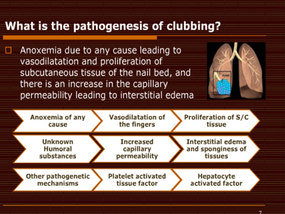
Pathophysiology Exact mechanism of the syndrome is still unclear.
This theory did not gain acceptance and does not feature in standard texts. It was suggested that megakaryocytes bypass the pulmonary circulation through arteriovenous shunting and escape fragmentation platelet derived growth factor (PDGF) & vascular endothelial growth factor (VEGF) by endothelial cells resulting in angiogenesis and endothelial hyperplasia clubbing of fingers and toes Marinez-Levin M, Pineda C: Hypertrophic osteoarthropathy. Rheumatology 3rd edition. Edited by: Hochberg MC, Silman AJ, Smolen JS, Weinblatt ME, Weisman MH. Mosby, Edinburgh; 2003:
 & vascular endothelial growth factor (VEGF) by endothelial cells resulting in angiogenesis and endothelial hyperplasia clubbing of fingers and toes. Marinez-Levin M, Pineda C: Hypertrophic osteoarthropathy. Rheumatology 3rd edition. Edited by: Hochberg MC, Silman AJ, Smolen JS, Weinblatt ME, Weisman MH. Mosby, Edinburgh; 2003:")
62
Other secondary causes
Pulmonary Tuberculosis Congenital Cyanotic Heart Disease Hepatic and Colorectal Carcinoma Inflammatory Bowel Disease Cirrhosis Pulmonary Fibrosis and Empyema These are other secondary causes of HOA Armstrong David, McCausland Elisabeth, Wright Gary: Hypertrophic pulmonary osteoarthropathy (HPOA) (Pierre Marie-Bamberger syndrome): two cases presenting as acuteinflammatory arthritis. Description and review of the literaliteratureRheumatol Int 2007, 27
 (Pierre Marie-Bamberger syndrome): two cases presenting as acuteinflammatory arthritis. Description and review of the literaliteratureRheumatol Int 2007, 27.")
63
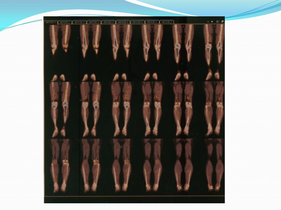
Whole-body bone scintigraphy
Preferred method for diagnosing the syndrome Increased tracer uptake along the cortical margins of the bilateral lower extremities, which is compatible with the “parallel stripe” sign or “tramline” sign Pericortical uptake involving the long bones of the lower limbs. Thicker, more extensive alterations are indicative of long-standing disease. The periostitis of HOA is not dependent on the primary or secondary nature of the disease but principally on its duration.

64
HPOA vs Metastasis on Bone Scan
HPOA: Diffuse increased uptake along the margins of the long bones. The cardinal features of skeletal metastatic disease are multiple focal areas of increased MDP uptake, randomly distributed but favoring the axial skeleton, with asymmetric involvement. Sharp et al, Practical Nuclear Medicine 3rd edition,2005

65
HPOA diagnosed by FDG PET-CT: a case report
The CT portion : extensive irregular bilateral periosteal new bone formation in the long bones. The PET images : diffuse moderately increased FDG uptake in the periostea of the long bones of the legs, with some focal sites of more intense FDG uptake in the thicker portions of the periosteum. Makis. Clin Nucl Med Sep;34(9):625-7
:")
66
Digital Clubbing : Demonstration With PET: A Case Report
The PET scan revealed increased glucose metabolism in all of the patient's fingertips An increased signal, indicating increased glucose metabolism, has been demonstrated in the distal part of the clubbed fingers. These changes are not seen in fingertips with normal morphology. The increase in signal supports the theory that clubbing is caused by the presence of a factor (eg, platelet-derived growth factor) that increases cellular metabolism Ward RW, Chin R Jr, Keyes JW Jr, Haponik EF. Digital clubbing. Demonstration with positron emission tomography. Chest. Apr 1995;107(4):
 that increases cellular metabolism. Ward RW, Chin R Jr, Keyes JW Jr, Haponik EF. Digital clubbing. Demonstration with positron emission tomography. Chest. Apr 1995;107(4):")
67
Treatment The only effective treatment for HPOA is treatment of the underlying condition Atropine, antitumor chemotherapy In patients with HPOA, tumor resection often results in improvement of symptoms within 2 to 4 weeks and, sometimes, complete resolution by 3 to 6 months Nakayama, Ann Thorac Surg, 2007;83:685–7

68
Effective Symptomatic Relief of HPOA by VATS Vagotomy
Relief of all joints and she regained full range of movements within 24 hours Discharged on the third post-op day and remained pain free and fully mobile 3 months after the surgery. Surgical vagotomy had been effective in providing symptomatic relief when the lung tumor proved unresectable. Clinical examination found classic clubbing with red, warm, and enlarged tender terminal phalanges of both hands and feet. She had painful and limited range of movement of both knees and ankles. normal except for a raised erythrocyte sedimentation rate. Relief of all joints, and she regained full range of movements within 24 hours. Discharged on the third post-op day and remained pain free and fully mobile 3 months after the surgery. Martinez-Lavin M: Hypertrophic osteoarthropathy. Curr Opin Rheumatol 1997;9:83– 6.

69
Current Status: At present, patient finished 33 sessions of radiotherapy and is on his 5th cycle of chemotherapy Patient still experiences intermittent joint pains but lesser in severity

70
Thank You!

83
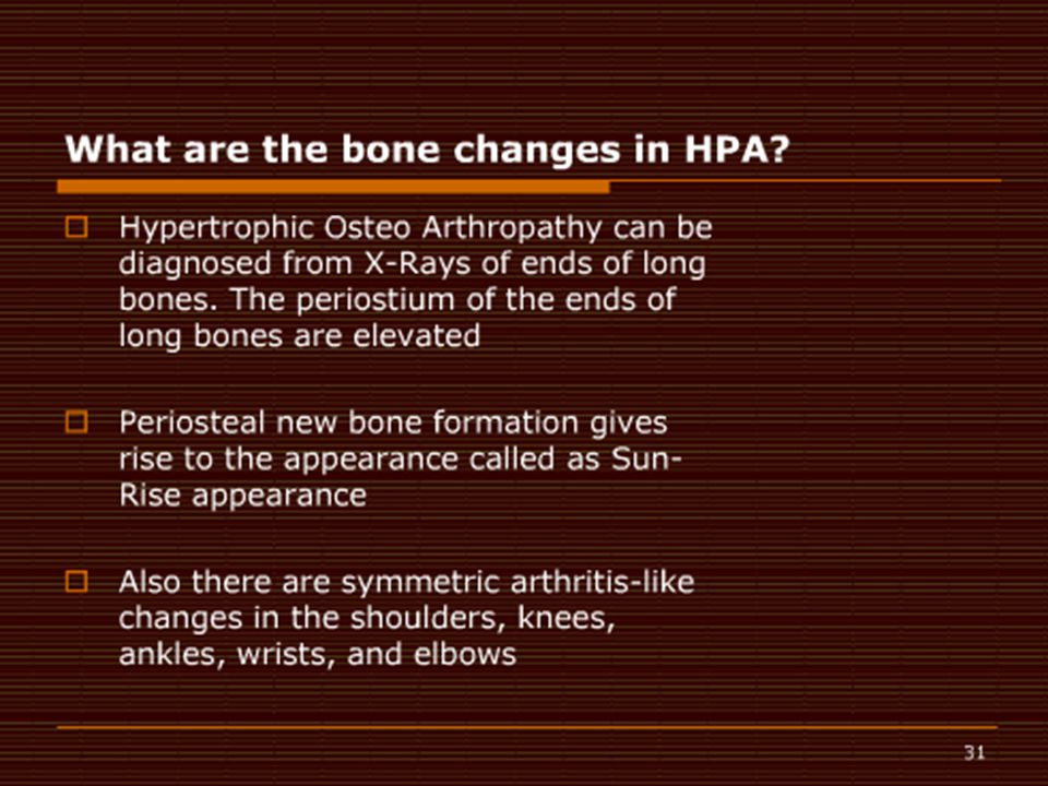
Hypertrophic Osteoarthropathy on Xray
The periostium of the ends of long bones are elevated Periosteal new bone formation gives rise to the appearance called as “Sun-Rise” appearance

84
PET vs Bone Scan in Detecting NSCLC Metastasis
Patients and methods: April May 2007, retrospectively reviewed to identify all patients with newly diagnosed NSCLC and who underwent staging with both PET/CT and bone scan prior to the initiation of therapy. Presence of bone metastases was confirmed by considering all available clinical information. This search identified 1000 patients, 265 women and 735 men (age range, 18–89 years; median age, 65 years). Song, et. al, Lung Cancer Journal, Volume 65, Issue 3, Pages , September 2009
. Song, et. al, Lung Cancer Journal, Volume 65, Issue 3, Pages , September")
85
PET vs Bone Scan in Detecting NSCLC Metastasis
Results Bone metastases were confirmed in 105 (10.5%) patients. PET/CT Bone Scan p value Accuracy 98.3% 95.1% <0.001 Sensitivity 94.3% 78.1% 0.001 Specificity 98.8% 97.4% 0.006 False (+) 1.2% 2.9% False (-) 5.7% 21.9% Song, et. al, Lung Cancer Journal, Volume 65, Issue 3, Pages , September 2009
 patients. PET/CT. Bone Scan. p value. Accuracy. 98.3% 95.1% < Sensitivity. 94.3% 78.1% Specificity. 98.8% 97.4% False (+) 1.2% 2.9% False (-) 5.7% 21.9% Song, et. al, Lung Cancer Journal, Volume 65, Issue 3, Pages , September")
86
Hypertrophic pulmonary osteoarthropathy diagnosed by FDG PET-CT: a case report
The CT portion : extensive irregular bilateral periosteal new bone formation in the long bones. The PET images : diffuse moderately increased FDG uptake in the periostea of the long bones of the legs, with some focal sites of more intense FDG uptake in the thicker portions of the periosteum. Makis. Clin Nucl Med Sep;34(9):625-7
:")
87
Patient Rheumatoid Arthritis Osteoarthritis Acute Rheumatic Fever HOA
Joints Tibial area, digits, wrist PIP, MCP/MTP, wrist, elbow, knee, ankle Knees, hips Migratory polyarthritis Tibia, wrists, digits, elbow Systemic symptoms fever Weakness, easy fatigability, anorexia, weight loss, ±fever, nodules None Fever, sore throat Timing Movement Movement, morning , >1 hr No specific timing Labs acute phase reactants (+) RF, elevated acute phase reactants Radio findings Normal Hand Xray erosions or unequivocal bony decalcification Normal/ RA: Pain in affected joints, aggravated by movement, is the most common manifestation of established RA. It corresponds in pattern to the joint involvement but does not always correlate with the degree of apparent inflammation. Generalized stiffness is frequent and is usually greatest after periods of inactivity. Morning stiffness of >1-h duration is an almost invariable feature of inflammatory arthritis. Notably, however, the presence of morning stiffness may not reliably distinguish between chronic inflammatory and noninflammatory arthritides, as it is also found frequently in the latter. The majority of patients will experience constitutional symptoms such as weakness, easy fatigability, anorexia, and weight loss. Although fever to 40°C occurs on occasion, temperature elevation in >38°C is unusual and suggests the presence of an intercurrent problem such as infection.
 RF, elevated acute phase reactants. Radio findings. Normal Hand Xray. erosions or unequivocal bony decalcification. Normal/ RA: Pain in affected joints, aggravated by movement, is the most common manifestation of established RA. It corresponds in pattern to the joint involvement but does not always correlate with the degree of apparent inflammation. Generalized stiffness is frequent and is usually greatest after periods of inactivity. Morning stiffness of >1-h duration is an almost invariable feature of inflammatory arthritis. Notably, however, the presence of morning stiffness may not reliably distinguish between chronic inflammatory and noninflammatory arthritides, as it is also found frequently in the latter. The majority of patients will experience constitutional symptoms such as weakness, easy fatigability, anorexia, and weight loss. Although fever to 40°C occurs on occasion, temperature elevation in >38°C is unusual and suggests the presence of an intercurrent problem such as infection.")
90
Articular manifestations of hypertrophic pulmonary osteoarthropathy in bronchogenic carcinoma. A clinical and pathologic study Eight patients with bronchogenic carcinoma presented with painful joint effusions as part of the syndrome of hypertrophic osteoarthropathy. Elevated sedimentation rates and symptomatic relief with aspirin were common, but synovial fluids all had leukocyte counts less than 500/mm3. .

91
Clubbing usually progresses through 4 phases.8
Fluctuation and softening of the nail bed, with a rocking sensation upon palpation due to increased edema and soft tissue Loss of the normal 15° angle (Lovibond angle27 ) between the nail and cuticle Accentuation of the convexity of the nails and clubbed appearance of the fingertips, with warmth and sweating Shiny or glossy change in the nail and adjacent skin, with disappearance of the normal creases and appearance of longitudinal striation of the nail Clubbing can be classified into 3 topographical groups.28,29 Symmetrical: All the fingers and toes are involved, as depicted in the image below. Clubbing associated with hypertrophic osteoarthropathy can be classified into 3 topographical groups (ie, symmetrical, unilateral, unidigital). This is symmetrical clubbing; it involves all the fingers and toes. [ CLOSE WINDOW ] Unilateral: The fingers or toes of 1 hand or foot are involved. Unidigital: Only 1 finger or toe is involved.
 between the nail and cuticle. Accentuation of the convexity of the nails and clubbed appearance of the fingertips, with warmth and sweating. Shiny or glossy change in the nail and adjacent skin, with disappearance of the normal creases and appearance of longitudinal striation of the nail. Clubbing can be classified into 3 topographical groups.28,29. Symmetrical: All the fingers and toes are involved, as depicted in the image below. Clubbing associated with hypertrophic osteoarthropathy can be classified into 3 topographical groups (ie, symmetrical, unilateral, unidigital). This is symmetrical clubbing; it involves all the fingers and toes. [ CLOSE WINDOW ] Unilateral: The fingers or toes of 1 hand or foot are involved. Unidigital: Only 1 finger or toe is involved.")
92
Immunohistochemistry
Braunwald, Fauci, Harrison’s Internal Medicine 17th edition, 2009

94
Pre-operative Testing
ABG Jan 26 pO2 96.2 pH 7.50 pCO2 34 HCO3 26.3 O2 Sat 97.9 Base Excess +3.6 Room air PRED Pre-Rx Post-Rx Best % Pred %Pred %Change FVC 3.05 3.25 106 3.15 103 -3 FEV1 2.41 2.49 2.55 2 FEV1/FVC% 82 77 93 81 98 5 FEF 25-75% 3.03 DLCO 14.3 66 MVV 130 74 57 55 42 -26 The FEV1/FVC ratio w/ a normal FVC. The FEV1 and & fev % are normal. The mvv is low. No signif response to bronchodilator. The tlc & rv are decreased. The dlco mildlt reduced. Spiromtery compatible w/ a restrictive ventilatory defect. The diffusing capacity is mildly is reduced.

95
Jones Criteria for Rheumatic Fever
MAJOR CRITERIA MINOR CRITERIA Carditis Migratory polyarthiritis Sydenham’s chorea Subcutaneous nodules Erythema marginatum Clinic Fever Arthalgia Laboratory Acute Phase Reactants Prolonged PR interval PLUS Supporting evidence of a recent Group A streptococcal fever Increased antistreptolysin O or other streptococcal antibodies Positive throat culture for Group A beta-hemolytic streptococci Positive rapid direct Group A strep carbohydrate antigen test Recent scarlet fever. \ GABHS + 2 major and 1 minor criteria, or 1 major and 2 minor criteria carditis or chorea exists with no other cause, previous history of rheumatic fever who have 1 major or 2 minor criteria in association with a recent streptococcal infection.

99
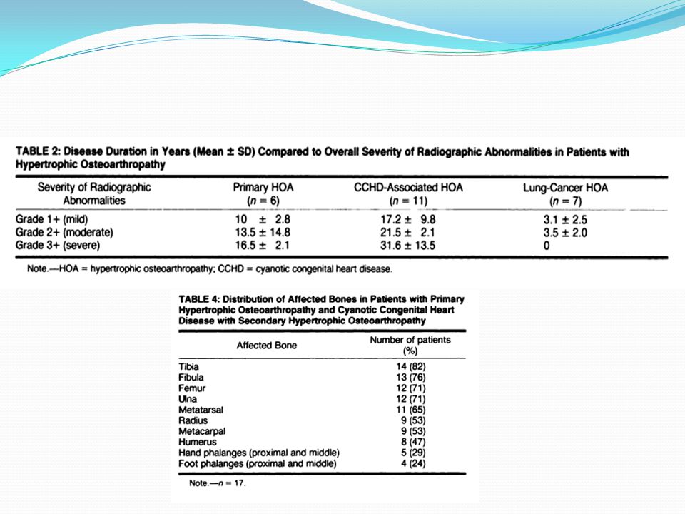
Periostitis in HOA Primary HOA and congenital heart disease-associated HOA had multilayered periostitis vs Lung-cancer HOA, there was only a single layer Scores indicating severity of periostitis showed that a severe degree was present in a significant percentage of patients with both primary HOA and CCHD-associated HOA compared to HPOA The most common type of periostitis observed in patients with primary HOA and congenital heart disease-associated HOA was multilayered; an irregular pattern of periostitis was present in one-third of these patients Pineda, Martinez, Periostitis in Hypertrophic Osteoarthropathy: relationship to disease duration

100
Periostitis in HOA In our seven patients with lung-cancer HOA, less prominent periosteal changes were noted in a more limited distribution. Malignancy-associated HOA, a disease process of much shorter duration, the natural evolution of the osseous changes cannot fully be expressed. When radiographs were analyzed according to disease duration, they found no significant differences between this group of patients and those with primary and CCHD-associated HOA; these observations suggest that the duration of the disease is most important in determining the distribution and morphology of the skeletal changes.

101
Periostitis in HOA

103
4 of 7 = our px was only to

104
Prediction of Hypoxemia during OLV
Side of Operation Lung Function Abnormalities “Paradoxical effect” Distribution of Perfusion Central vs Peripheral Gravity

Similar presentations


 Consult.>")

