Download presentation
Presentation is loading. Please wait.

1
Electron Spectroscopies of InN grown by HPCVD Department of Physics and Astronomy Georgia State University Atlanta, Georgia Rudra P. Bhatta Solid State Physics (Physics - 8510) Fall 2005
 Fall")
2
Motivation: InN and its application InN sample grown by HPCVD Auger Electron Spectroscopy Data Analysis to Determine Composition Composition vs. Treatment and Position Low energy electron diffraction High Resolution Electron Energy Loss Spectroscopy Surface Structure and Bonding Film Polarity Summary Future work Outline

3
High-efficient energy conversion system solid state lighting (high-efficient light emitting diodes) High speed opto-electronics for optical communication systems Solid state lasers operating in the blue and ultraviolet regions Terahertz device structures (emitters and detectors) Nonlinear optical switching elements. Spintronic device structures. Application of InN & In rich group III-Nitides

4
Motivation for studying indium nitride Research on indium nitride growth and characterizarion has increased tremendously in recent years. Controversy in the measurement of fundamental properties such as band gap, lattice constant, and effective mass. Difficulty of InN growth due to its low dissociation temperature and the high vapor pressure of nitrogen over InN. Potential of high pressure chemical vapor deposition (HPCVD): - stabilizes InN to higher temperature, and - allows growth of InN, GaN, and AlN at similar conditions.
: - stabilizes InN to higher temperature, and - allows growth of InN, GaN, and AlN at similar conditions..")
5
Flow Direction Reactor pressure 15 bar Gas flow velocity 41 cm /s Ammonia:TMI ratio 240 Substrate HPCVD GaN buffer on sapphire (0001) HPCVD grown Indium Nitride HPCVD Growth: N. Dietz and coworkers, JVST B 23, 1790 (2005) or phys. stat. sol. latest issue
 or phys. stat. sol. latest issue.")
6
Auger Electron Spectroscopy (AES) AES is a surface-sensitive spectroscopic technique used for elemental analysis of surfaces; it offers: High sensitivity (nearly 1% monolayer) for all elements except H and He. Quantitative compositional analysis of the surface region. A means of monitoring surface cleanliness of samples.

7
Auger electrons are the secondary ionized electrons

8
Nitrogen and Indium AES peaks (dN/dE) Indium Metal Nitrogen Si 0.54 N 0.46 Hand book of Auger Electron Spectroscopy, 2 nd Edition, L.E.Davis et al., Physical Electronics Division, 1978
 Indium Metal Nitrogen Si 0.54 N 0.46 Hand book of Auger Electron Spectroscopy, 2 nd Edition, L.E.Davis et al., Physical Electronics Division, 1978")
9
AES Lineshapes for InN and In

10
Peak fitting of InN Auger Spectra

11
Assumed linear background Integrated area under peaks carbon: 220 – 285 eV nitrogen: 358 – 392 eV indium: 392 – 418 eV oxygen: 500 – 522 eV O/In calibrated from native oxide of metallic indium (In 2 O 3 ) N/In calibrated from highest nitrogen content InN (assumed 1:1)
 N/In calibrated from highest nitrogen content InN (assumed 1:1)")
12
Atomic Fraction vs. Sample Treatment Argon Sputtered Region

13
Atomic Fraction vs. Sample Treatment Atomic Hydrogen Cleaning (AHC) 1000 L H 2 over 1800 K Tungsten filament with sample at 350 K + 1000 L H 2 over 1800 K Tungsten filament with sample at 600 K. 1 L= 1x10 -6 torr s McConville and coworkers, Univ. of Warwick Piper et al., JVST A 23, 617 (2005).
 1000 L H 2 over 1800 K Tungsten filament with sample at 350 K L H 2 over 1800 K Tungsten filament with sample at 600 K. 1 L= 1x10 -6 torr s McConville and coworkers, Univ. of Warwick Piper et al., JVST A 23, 617 (2005)..")
14
Atomic Fraction vs. Position After Atomic Hydrogen Cleaning Flow Direction

15
Schematic of LEED optics operated as RFA Sample sits at the center of the grids. Grid 1&4 are grounded. Grids 2 &3 are at potential slightly less than that of electron gun. Only elastically scattered electrons reach to the fluorescent screen. LEED: A technique used for the determination of surface structure

16
LEED image of InN Spot positions yield information on the size, symmetry and rotational alignments of surface unit cell with respect to substrate unit cell. Distance between the spots gives information about the distances between the atoms. Sharpness of the spots gives insight on how well ordered the surface atoms are arranged. E = 39.5 eV

17
e - E = 12.5 eV 60 o from normal e - E < 500 meV (4000 cm -1 ) specular collection InN HREELS Surface Vibrational Spectroscopy High Resolution Electron Energy Loss Spectroscopy
 specular collection InN HREELS Surface Vibrational Spectroscopy High Resolution Electron Energy Loss Spectroscopy")
18
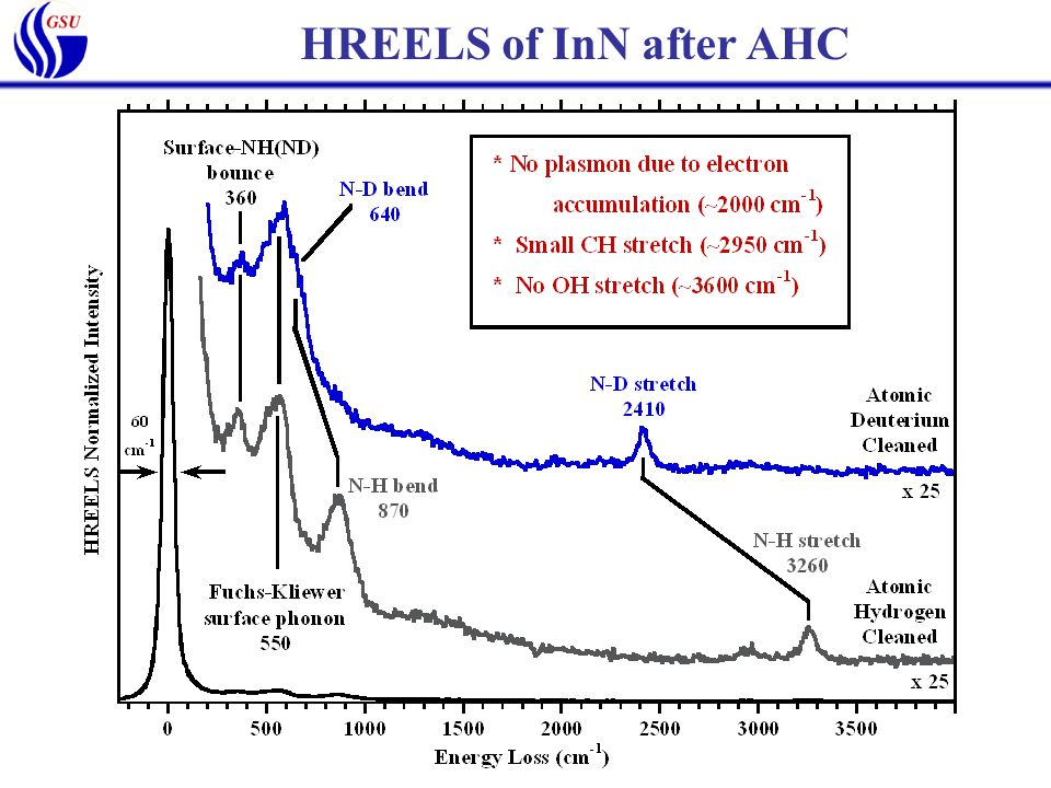
HREELS of InN after AHC

25
Surface Structure of InN after AHC N-polar surface consists of N atoms bonded to three In atoms in the second layer and one dangling bond normal to the surface. Atomic hydrogen saturates the dangling bonds to stabilize the surface. Growth Direction N In H

26
Summary Indium nitride sample grown by high pressure chemical vapor deposition was investigated by AES, LEED, and HREELS. The composition of the InN surface was determined by integrating areas under peaks in N(E) Auger Electron Spectra. Sputtering produces nitrogen deficient surface. Atomic hydrogen cleaning (AHC) produces a contaminant-free, well- ordered c-plane InN surface with a 1x1 LEED pattern. HREELS of InN after atomic hydrogen (deuterium) cleaning shows NH (ND) stretch, bend and bounce vibrational modes. No InH, NH 2, or OH vibrational modes are observed. InN surface is N-terminated and N-polar, i.e.
 Auger Electron Spectra. Sputtering produces nitrogen deficient surface. Atomic hydrogen cleaning (AHC) produces a contaminant-free, well- ordered c-plane InN surface with a 1x1 LEED pattern. HREELS of InN after atomic hydrogen (deuterium) cleaning shows NH (ND) stretch, bend and bounce vibrational modes. No InH, NH 2, or OH vibrational modes are observed. InN surface is N-terminated and N-polar, i.e..")
27
Future work To study the desorption rate of hydrogen from the surface at different temperature by the process of HREELS and temperature programmed desorption (TPD). To study the reaction of ammonia and trimethyl indium (TMI) on the indium nitride surface in order to understand the surface reaction during the growth. Thank you for your attention.
 on the indium nitride surface in order to understand the surface reaction during the growth. Thank you for your attention..")
Similar presentations


 and Al(110) Surfaces at Room Temperature* N.R. Shivaparan, M.A. Teter, and.>")


 2609-2613.>")
