Download presentation
Presentation is loading. Please wait.

1
Orthodontics Hospital of Stomatology,Xi’an Jiaotong University
Department of Orthodontics Professor ZhouHong

2
overview Orthodontics and Dentofacial orthopedics
Orthodontics is a branch of Clinical Stomatology , the mechanisms of major research and development of dentofacial deformities, diagnosis, prevention and treatment. Orthodontics Dentofacial Orthopedics Malocclusion Dentofacial Deformity Orthodontics and Dentofacial orthopedics

3
Orthodontics and Dentofacial orthopedics
The area and specialty of dentistry concerned with the supervision, guidance and correction of the growing or mature dentofacial structures, including those conditions that require movement of teeth or correction of malrelationships and malformations of their related structures and the adjustment of relationships between and among teeth and facial bones by the application of forces and/or the stimulation and redirection of functional forces within the craniofacial complex.

4
Orthodontics and Dentofacial orthopedics
Major responsibilities of orthodontic practice include the diagnosis, prevention, interception and treatment of all forms of malocclusion of the teeth and associated alterations of their surrounding structures; the design, application and control of functional and corrective appliances; and the guidance of the dentition and its supporting structures to attain and maintain optimal occlusal relations, physiologic function and esthetic harmony of facial and cranial structures.

5
What is Dentofacial Deformity ?

6
Dentofacial deformity
A malformation of the teeth, jaws and/or face characterized by disharmonies of size, form and/or function. The term encompasses problems such as malocclusion, cleft lip and palate and other skeletal or soft tissue anomalies, or syndromes that involve the face and the dentoalveolar complex.

7
一、The manifestation of Dentofacial Deformity
1 malposition of individual, abnormity of arch form ,tooth malalignment 2 maxillomandibular malrelationship 3 malrelationship between jaw and cranium

8
(Microdontia) (Anterior crossbite) (Spaces) (Suprenumerary tooth)
 (Anterior crossbite) (Spaces) (Suprenumerary tooth)")
9
(Congenital missing tooth)
(Ectopic eruption)
")
10
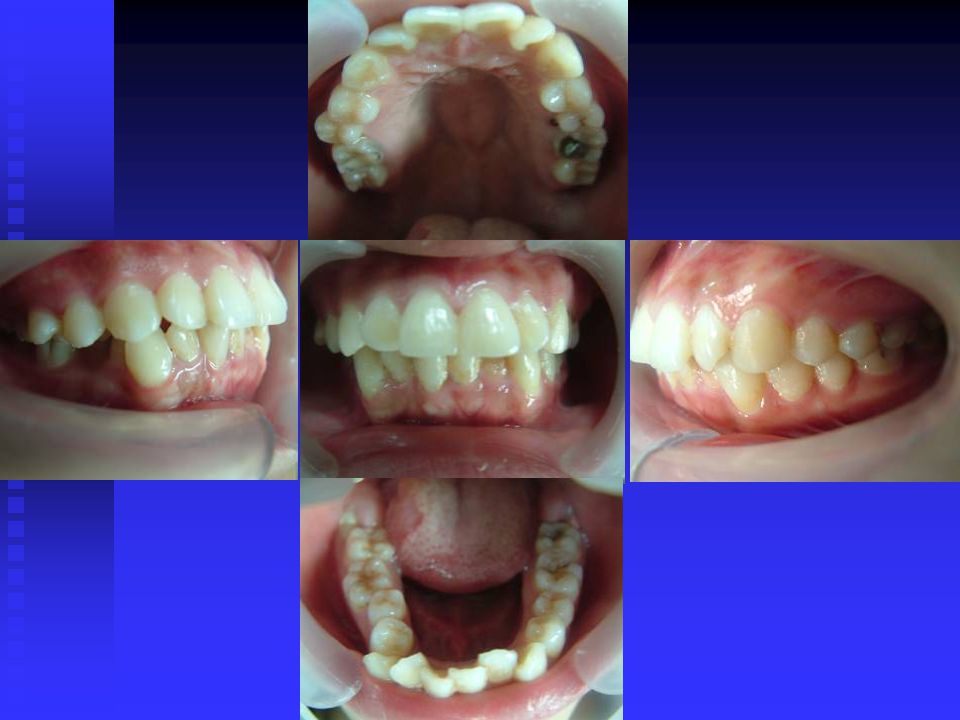
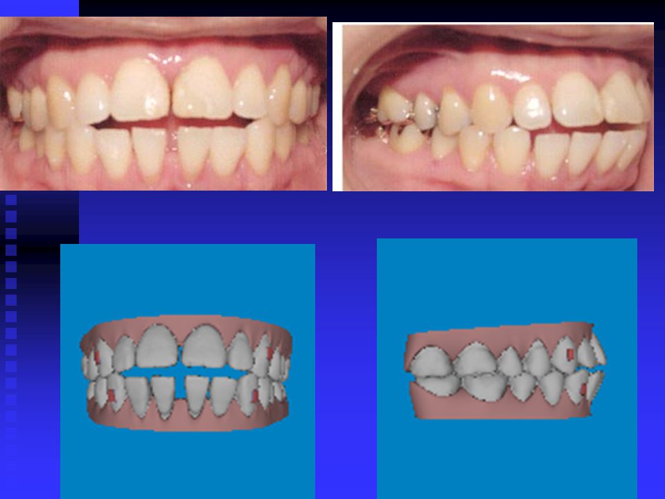
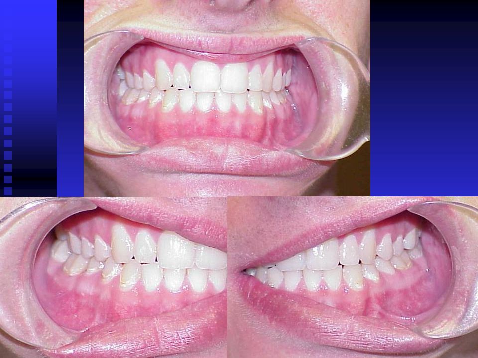
Bimaxillary Dentoalveolar Protrusion and Crowding

12
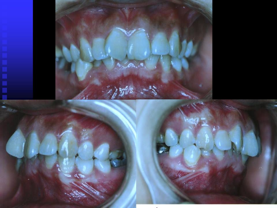
(Deep Overbite and Overjet)
")
13
Deep overjet 11.0 mm Deep overbite 90 %

14
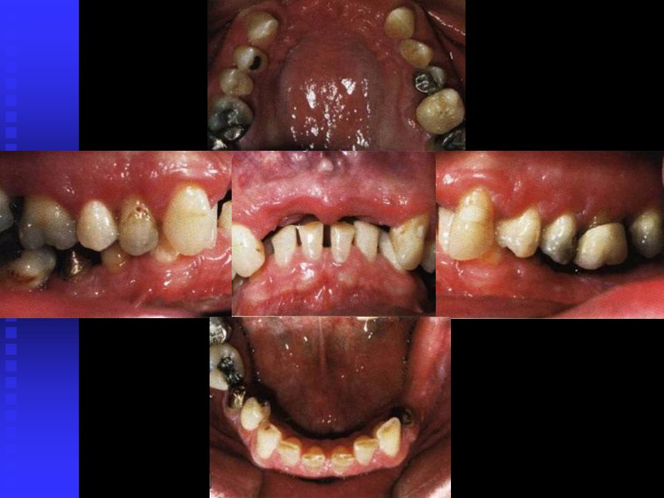
(Deep Overbite with Crowding)
")
16
Mandibular prognathism

17
Maxillar Retrusion with Mandibular Protrusion

18
Maxillary retrognathism
Narrow of upper arch Mandibular prognathism

19
(Edge-to-edge bite)
")
20
Crowding with edge-to edge bite

21
Bimaxillary dentoalveolar protrusion

22
(frontal view) (Lateral view )
 (Lateral view )")
23
Open bite

24
(frontal view) (Lateral view )
 (Lateral view )")
25
Mandibular Shift

26
(frontal view) (Lateral view )
 (Lateral view )")
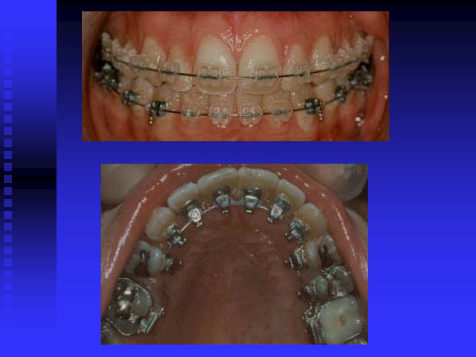
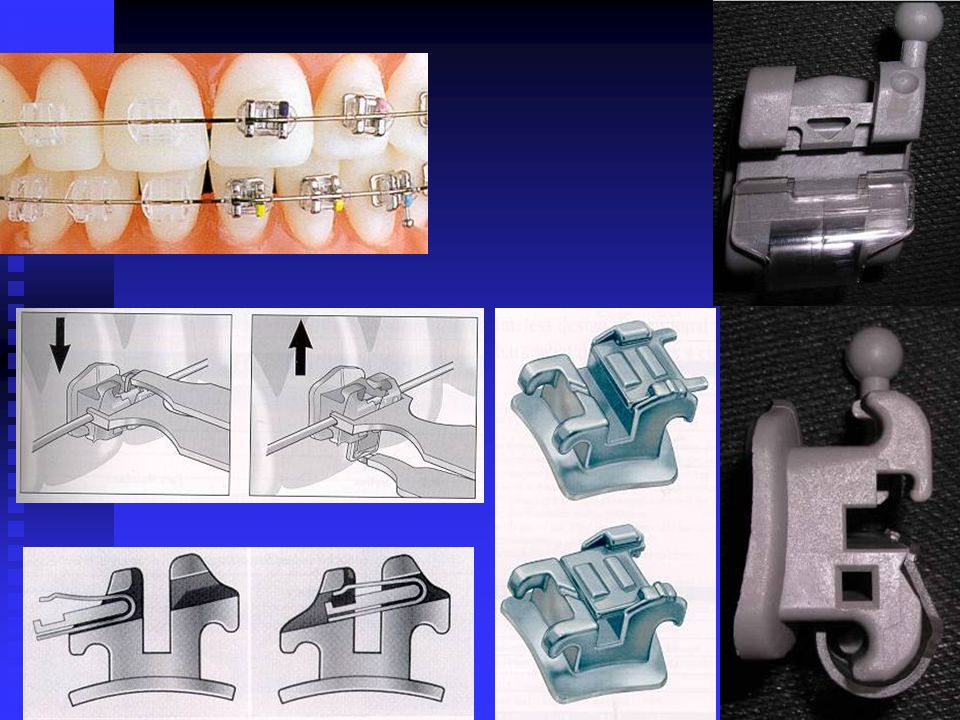
27
Introversion deep overbite

28
二、epidemiology prevalence rare : 60`s: 29.33% - 48.87%
Individual Normal Occlusion Ideal Normal Occlusion Incisor Irregularity Index

29
A+B+C+D+E= Anterior lower incisor crowding
ideal mild crowding moderate crowding severe crowding > extreme crowding

30
三、 Perniciousness 1 psychosocial influences 2 oral function
3 relation to dental disease 4 Aesthetic impact

31
Relation between size of overjet and
prevalence of traumatised anterior teeth Overjet (mm) Incidence % >
 Incidence % >9 44.")
32
Dr Sarver: Malocclusion of teeth is not disease , rather, it is a disability with a potential influence on physical and mental health. Orthodontics — current principles and techniques 2000 By Graber

33
Reason for orthodontics
1. To improve dentofacial appearance. 2. To correct the occlusal function of the teeth 3. To eliminate occlusion that could damage the long-term health of the teeth and periodontium

34
四、 standard and target 1、 changes of target
Crowed,irregular and protruding teeth have been a problem for some individuals since antiquity,and attempts to correct this disorder go back at least to 1000 BC.primitive orthodontic appliance have been found in both Greek and Etruscan matrials.

35
1850 the first texts that systematically described orthodontics appeared,the most notable being Norman Kingsley’s Oral Deformities.Kingsley who had a tremendous influence on American dentistry in the latter half of the nineteeth century,was among the first to use extroral force to correct protruding teeth.He was also a pioneer in the treatment of celft palate and related problems.

36
Their emphasis in orthodontics remaind the alignment of the teeth and the correction of facial proportions. Little attention was paid to the dental occlusion.In an era when an intact dentition was a rarity,the details of occlusal relationships were considered unimportant.

37
Edward H Angle can be credited with much of the development of a concept of occlusion in the natural dentition.His increasing interest in dental occlusion and in the treatment necessary to obtain normal occlusion led directly to his development of orthodontis as a specialty,with himself as the “father of modern orthodontics.”

38
The publication of Angle’s classification of malocclusion in the 1890s was an important step in the development of orthodontics because it not only subdivided major types of malocclusion but also included the first clear and simple definition of normal occlusion in the natural dentition.If this molar relationship existed and the teeth were arranged on a smoothly curving line of occlusion.

39
Angle`s classification of malocclusion

40
Orthodontics was no longer just the alignment of irregular teeth
Orthodontics was no longer just the alignment of irregular teeth.Angle and his followers strongly opposed extraction for orthodontic purpose.With the emphasis on dental occlusion that followed,however,less attention came to be paid to facial proportions and esthetics.

41
As time passed,it became clear that even an excellent occlusion was unsatisfactory if it was achieved at the expense of proper facial proportions.Not only were there esthetic problems,it often proved impossible to maintain an occlusal relationship.Extraction of teeth was reintroduced into orthodontics in the 1930s to enhance facial esthetics and achieve better stability of the occlusal relationships.

42
Cephalometric radiography enabled orthodontists to measure the changes in tooth and jaw positions produced by growth and treatment.These radiographs made it clear that many malocclusions resulted from faulty jaw relationships,not just malposed teeth.By use of cephalometrics,it also was possible to see that jaw growth could be altered by orthodontic treatment.

43
As the 21st century begins,orthodontics differs from what was done previously in three important ways: ⑴ there is more emphasis now on dental and facial esthetics, and less on details of dental occlusion.

44
⑵ patients now expect and are granted a greater degree of involvement in planning treament。No longer is it appropriate for the paternalistic doctor to simply tell patients what treament they should have.

45
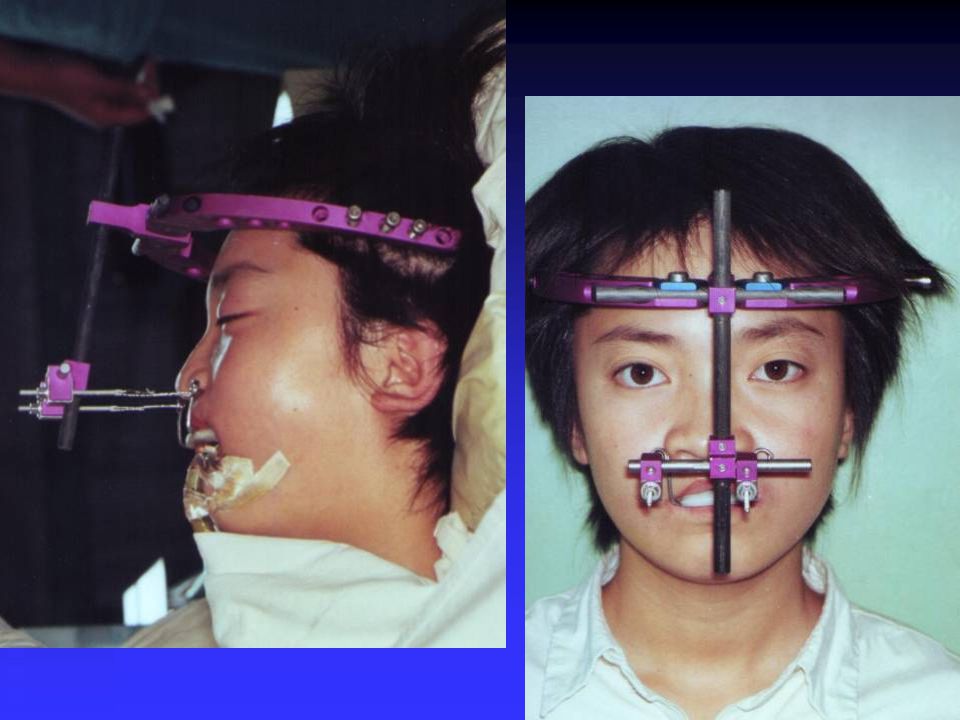
computer simulation post-treatment before treatment

46
⑶ orthodontics now is offered much frequently to older patients as part of a multidisciplinary treament plan involing other dental and medical specialties。 (Multidisciplinary Treatment ) (Interdisciplinary Treatment )
 (Interdisciplinary Treatment )")
49
The goal is not necessarily the best possible dental occlusion or facial esthetics but the best chance for long-term maintenance of the dention.This increased emphasis on treatment coordinated with other dentists has the effect of integrating orthodontics back into the main stream of dentistry,from which Angle’s teachings had tended to separate it.

50
⑴ target: Harmony Stable Aesthetic

51
⑵ Andrews ’s sixElements :
1. Molar relationship 2. Crown angulation (Mesiodistal “tip”) 3. Crown inclination 4. Rotations 5. Spaces 6. Occlusal plane
 3. Crown inclination. 4. Rotations. 5. Spaces. 6. Occlusal plane.")
52
五、The relationship between orthodontics and other subjects 1、Prosthodontics 2、implantodontics 3、 periodontics 4、Computer Technology 5、 Materialogy

55
Tooth Extrusion

57
significant development in stomatology are related to materials
enamel adhesive The super-elastic titanium alloy arch wire implant anchorage

59
不不锈钢丝 马氏体钛丝 应变 奥氏体钛丝丝 应力

60
(extraoral force )
")
62
1. Preventive Orthodontics regular oral examination
六、methods 1. Preventive Orthodontics antenatal care regular oral examination Get rid of bad habits space maintainer extractions of Supernumerary Teeth

63
2. Interceptive Orthodontics
serial extraction early treatment of crossbite

64
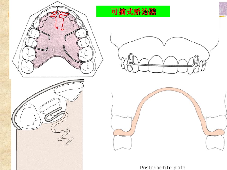
3. general Orthodontics Removable appliances fixed appliances function appliances lnvisalign appliances

66
Edgewise Appliance

71
Dental digital modeling and invisible appliance
牙颌光固化在牙轿器技术流程中的位置 Dental data laminar analysis Reverse correction appliance mold appliance

73
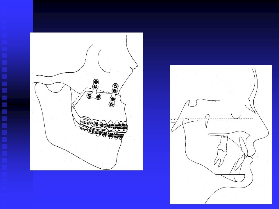
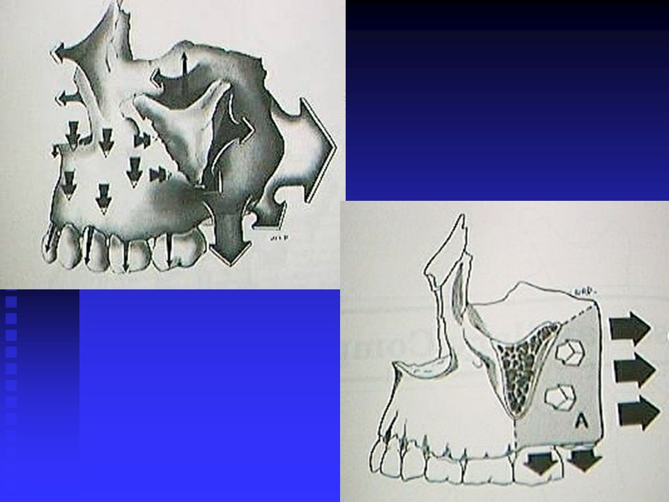
4. Orthodontics - Surgical correction
Orthognthic Surgery Surgical Orthodontics Distraction Osteogenesis

74
type of Orthognathic surgery

75
Le Fort I、II、III osteotomy

76
(Multijaw maxillary osteotomy )
")
78
Maxillar Impaction

79
sagittal split ramus osteotomy
oblique split ramus osteotomy

81
Preparation before traction

82
zone of ossification

83
Anterior crossbite III traction

89
Orthodontic Materials and Bio-mechanics
In the 20th century, major developments : Monobloc,1920,Pierre Edgewise,1928,Angle Begg、Straight-wire、Tipedge X-cephalometry,1931,Broadbent Eatraction,1941,Tweed Orthodontic Materials and Bio-mechanics Wire Materials, gold, stainless steel, O wire, hot-activated, nickel, titanium and titanium ß Tooth movement, Burstone, power systems, force size, force direction Bonding technology Orthognathic surgery and orthodontics Computer applications 80`s

90
In 21st century ,the direction of the development of orthodontics
Craniofacial growth and development Biology of tooth movement Biomechanics and BioMaterials Computer use in orthodontics Three Dimensional Diagnosis Interdisciplinary Treatment

92
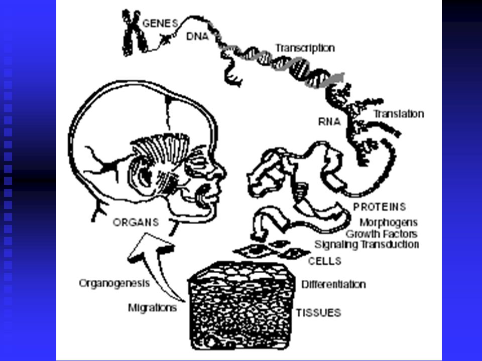
Craniofacial Growth and Development

93
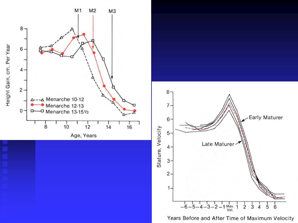
Why should we study the growth and development??
What is the craniofacial growth pattern ?

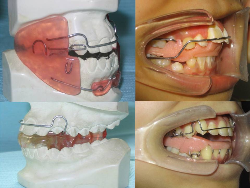
94
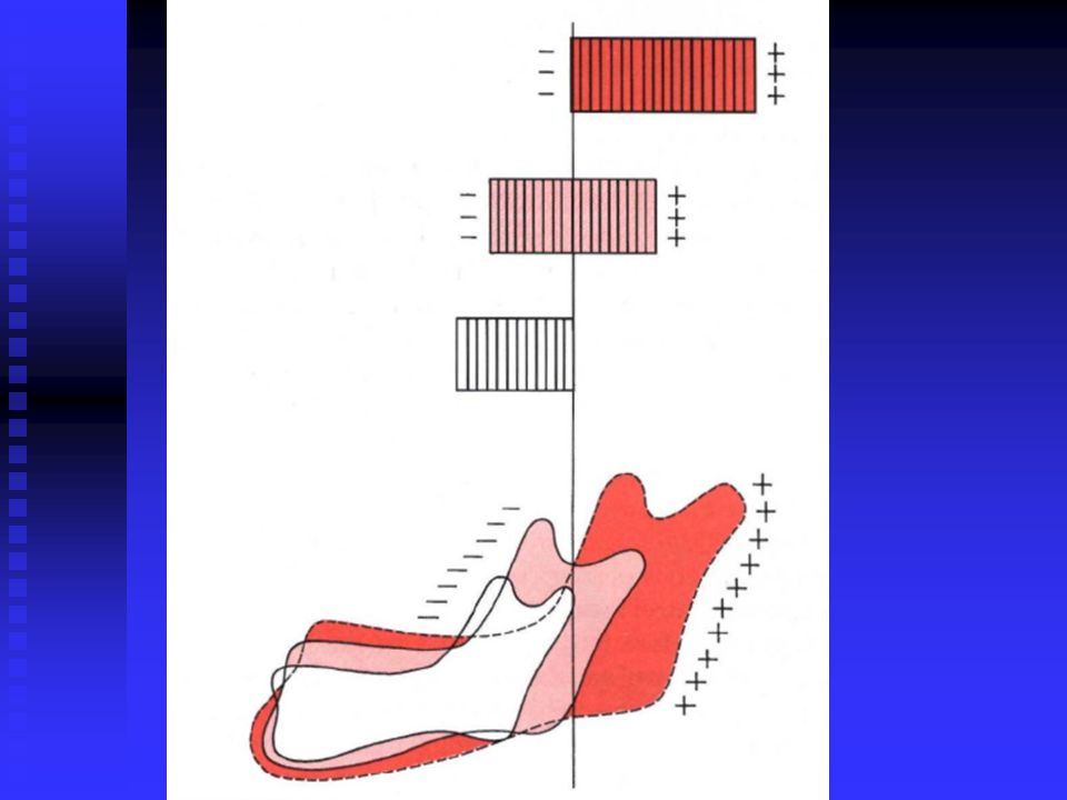
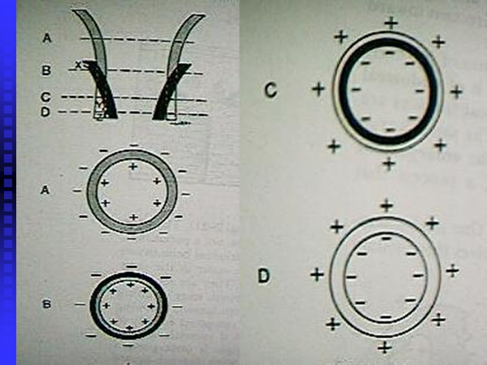
8 months, 6 y, 8 y and 20 y old

95
Craniofacial Growth and Development
一、methods of collecting information longitudinal study cross-sectional study mixed longitudinal study

96
二、 1. Measurement Study Craniometry
Anthropometry Cephalometry

97
Craniometry Cephalometry

98
Anthropometry

99
Three-dimensional structure
Anthropometry techniques for measuring living individuals Three-dimensional structure surface measurement Poor accuracy The stability of the measurement system and method ) The basis for evaluation of facial morphology studying the deep structure is impossible)
 The basis for evaluation of facial morphology. studying the deep structure is impossible)")
100
二、2 . experiment Vital staining Radioactive Tracer Implant radiography Molecular Genetics

101
Implant radiography (Radioactive Tracer)
")
102
三、 basic concept Pattern of facial growth : Average growth pattern
1. (Growth Patten) Pattern of facial growth : Average growth pattern Horizontal growth pattern Vertical growth pattern
 Pattern of facial growth : Average growth pattern. Horizontal growth pattern. Vertical growth pattern.")
103
Growth and Development
Terminology Growth Development

104
Pattern Normal growth pattern Changes in overall body proportions

105
Pattern Scammon’s Curve

106
Average growth pattern

107
Vertical growth pattern
Horizontal growth pattern Vertical growth pattern

108
2. Variability Everyone is not alike in the way that they grow as in everyting else.It can be difficult but clinically very important to decide whether an individual is merely at the extreme of the normal variation or falls outside the normal range.

111
Variability Racial and ethnic differences Gender Sickness nutrition
Timing factor -Late/early maturers Problems with growth (hormones or genetics)
")
112
3. Timing Variability in growth arises in several ways:from normal variation,from timing effects.Variation in timing arises because the same event happens for different individuals at different times. developmental age and chronologic age

113
Timing Variation Early, average, and late matuerers
Chronological age vs. Developmental age

116
4. Rapid phase of growth and development
Rapid and slow phase of craniofacial growth and development is close to rapid and slow phase of body growth and development .

120
Why do we assess growth? To determine optimum time for treatment (growth modification and surgery) to determine the amount of growth left to determine type of growth

121
5. Growth site and Growth center
A site of growth is merely a location at which growth occurs,whereas a center is a location at which independent (genetically controlled) growth occurs.All growth centers also are growth sites, whereas the reverse is not true.
 growth occurs.All growth centers also are growth sites, whereas the reverse is not true.")
122
Growth Center and Growth Site
For example, it is now known that the sutures between the membranous bones of the cranium and the maxilla that previously were considered as primary growth centers, actually are mere sites of growth.

125
Questions Do you know the hazards of Dentofacial deformities? Orthodontic treatment goal? What is ideal normal occlusion , what is individual normal occlusion? What is the growth pattern? What is the growth site and growth center?

126
四、postnatal Craniofacial Growth and Development
1. Craniofacial dividing line Bolton – nasion plane Frankfort plane Ba-N plane

127
Bolton - 鼻根平面, A line connecting points Bolton and Nasion; an alternate representation of the cranial base. Frankfort平面

128
前颅底平面(S-N)Representing the anterior cranial base
前颅底平面(S-N)Representing the anterior cranial base. A line joining points S and Na. 全颅底平面(N – Ba) To represent the cranial base more accurately than the SN line or the Bolton plane.
Representing the anterior cranial base. A line joining points S and Na. 全颅底平面(N – Ba) To represent the cranial base more accurately than the SN line or the Bolton plane.")
129
2. ways of Bone growth and development
⑴ surface apposition of bone periosteum osteoblast osseous tissue ⑵ interstitial growth Connective tissue cells Fibroblast Collagen fibers and matrix calcification

131
⑶ central cartilage cell proliferate hypertrophy
⑶ central cartilage cell proliferate hypertrophy Peripheral cartilage calcification Cells of deep Connective tissue membrane differentiate into cartilage cells and matrix form hyaline cartilage, that calcifiy into new bone

132
Reserve zones (RZ) Proliferating zones (PZ) Prehypertrophic zones (PHZ) Hypertrophic zones (HZ)
 Proliferating zones (PZ) Prehypertrophic zones (PHZ) Hypertrophic zones (HZ)")
133
Reserve zones (RZ) Proliferating zones (PZ) Prehypertrophic zones (PHZ) Hypertrophic zones (HZ) Articular cartilage (AC) Growth cartilage (GC)
")
134
3. Cranial growth and development A. cranial cavity
function:protecting the brain structure:flat bone Site and mechanism of the growth :suture and Surface hyperplasia

137
Clinical Significance : Aperts Syndrome
timing:(6-7 years old reach 90% of people ) Clinical Significance : Aperts Syndrome
 Clinical Significance : Aperts Syndrome.")
138
Major Features of Apert Syndrome
Prematurely fused cranial sutures A retruded midface Fused fingers Fused toes

140
B. cranial base function:stability
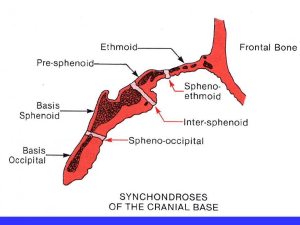
growth site and timing :The growth of cartilage (intersphenoid synchondrosis、spheno-occipital synchondrosis、spheno-ethmoidal synchondrosis)
")
144
Growth characteristics : depth >Height > Width
Clinical Significance : Hypoplasia cause deficiency of middle 1 / 3 face

145
4. Facial Growth and Development
A. Nasomaxillary Complex main Maxillary growth,but septal cartilage growth conduct the growth of middle face importantly.

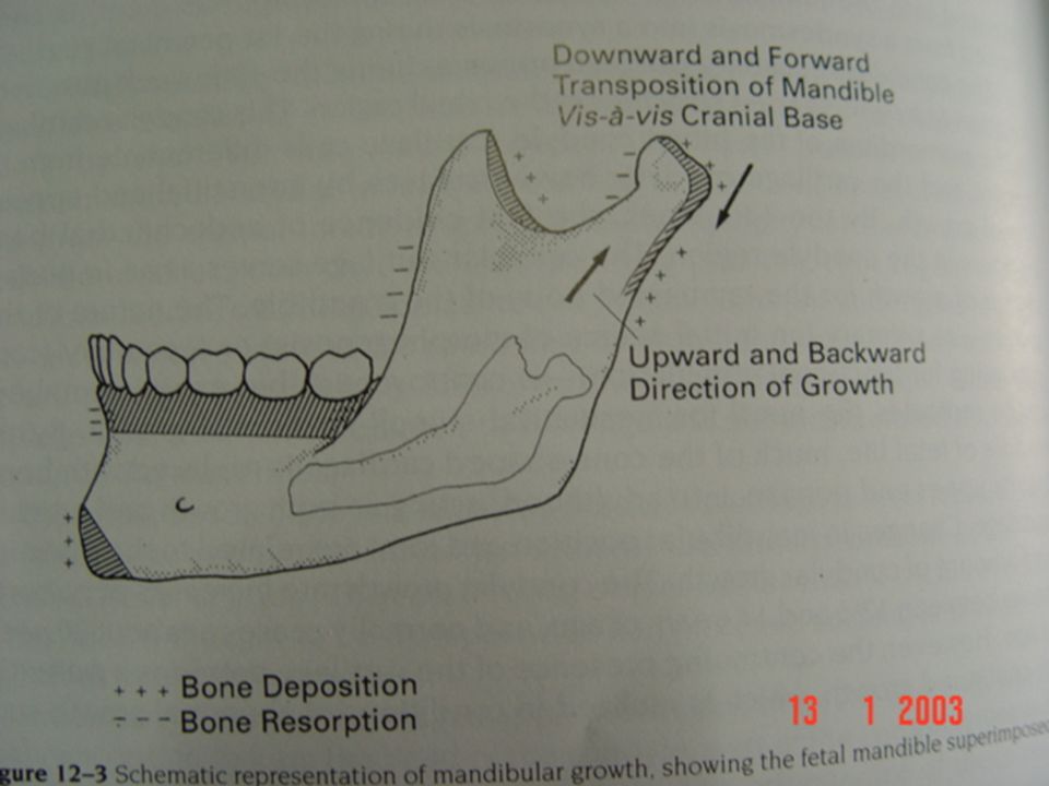
146
B. mandible growth: forward ,downward Height > depth > Width

148
the rate of Craniofacial growth

149
5. Maxillary Growth and Development
A. Passive displacement The cranial base promote the growth of the maxillary, more important for child .

150
Passive displacement

151
depth: maxillary tuberosity Alveolar bone growth
B. active growth: depth: maxillary tuberosity Alveolar bone growth Horizontal part of palatine bone growth

153
The suture between maxilla and cranium
the direction of maxillary movement

154
颧额缝 额颌缝 鼻颌缝 颧颌缝 颞颧缝

155
The site of maxillary growth and absorption
palatal vault moves downward

156
width:median palatine suture growth
B. active growth: width:median palatine suture growth Buccal surface of maxillary bone hyperplasia Alveolar bone growth

157
height:frontozygomatic and zygomaticomaxillary suture growth
Orbital floor reconstruction Basis nasi moves downward Alveolar bone growth

158
The site of maxillary absorption
One side absorption the other side proliferation

159
C. clinical application
high vault Restrict maxillary development maxillary protraction Maxillary arch RPE

163
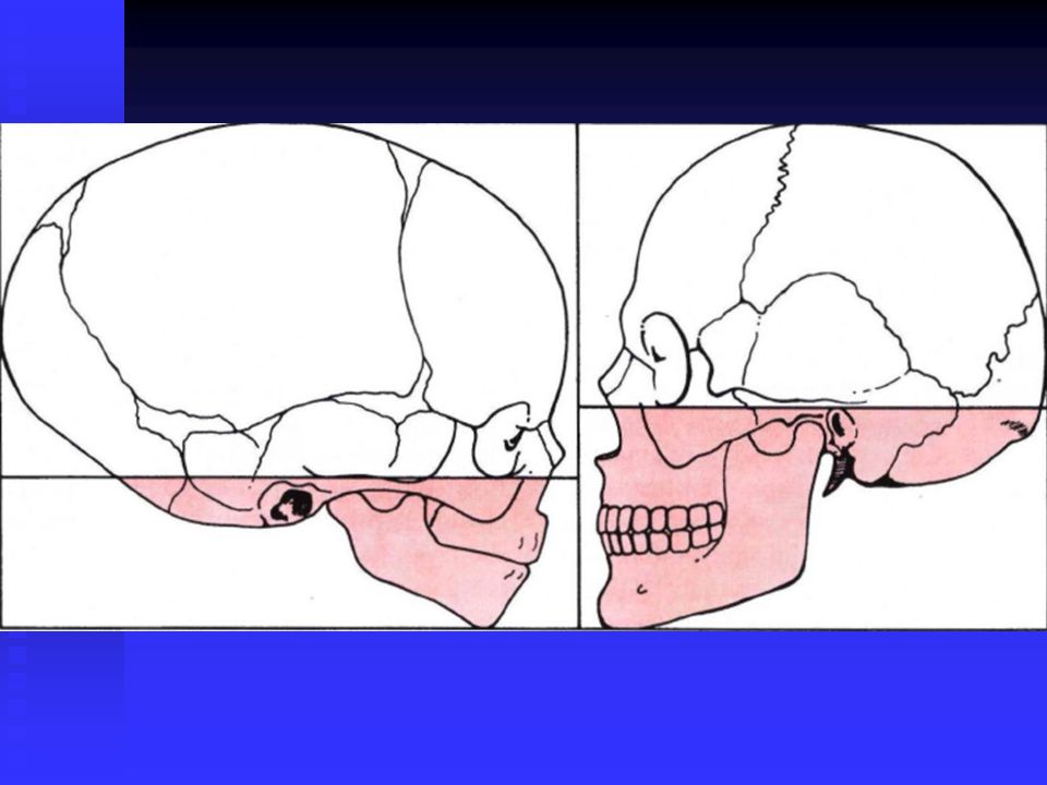
6. Mandibular Growth and Development A. function:
The only movable bone of Craniofacial region、 relevant to mastication 、language 、airway maintenance、countenance。

164
B. growth and development:
Partition:body of mandible alveolar process Mandibular ramus

167
functional protuberance :
Attachment of muscles and teeth condylar process、coronoid process 、 angle of mandible 、alveolar process

168
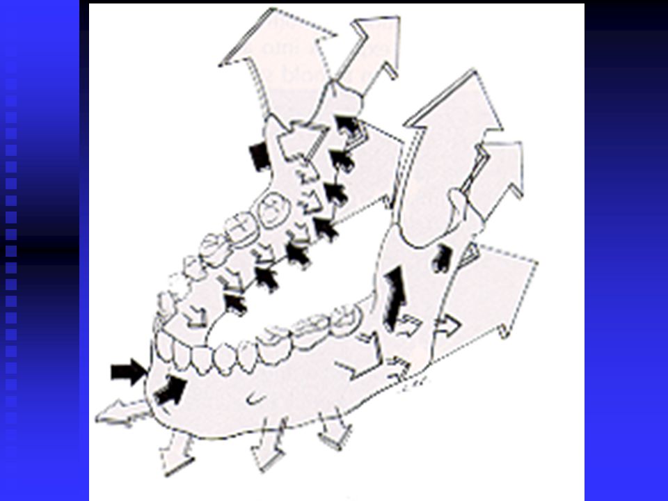
C. The site of growth and mechanisms:
condylar process:fibrocartilage ,growth site 。 body of mandible:outside surface of hyperplasia , inside the absorption

169
C. The site of growth and mechanisms:
Mandibular ramus :posterior margin bone apposition ,anterior margin bone resorption alveolar bone:impact the height of mandible

173
C. The site of growth and mechanisms:
height:condylar process、alveolar bone growth length: posterior margin bone apposition ,anterior margin bone resorption width:condylar process growth,Lateral mandibular hyperplasia

174
D. characteristic: angle of mandible :it will be different with age,growth and masticatory function 。 newborn : 140 – 160 degree Adults : 125 degree the elderly : obtuser

177
D. characteristic: the height of mandibular ramus :the length of mandibular body newborn: 35 :100 adults: 65 :100 mental region: protrusion vary due to the differences of race

178
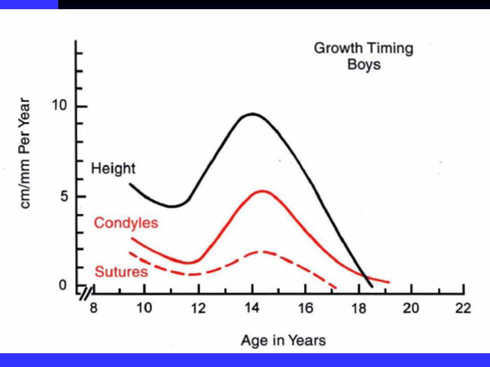
E. Growth time : the growth peak of mandibular height and length is basically the same with physical growth ,or a little ealier. the peak time of adolescent period is the most important in growing period.The time for girls which is 1.5 years earlier than boys,come before menarche .

179
F. clinical application:
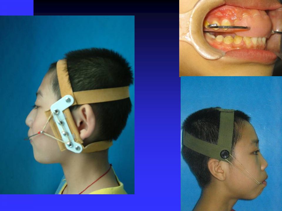
change mandibular growth and developmen functional appliance occlusal pad

181
“V”shaped osteogenesis phenomenon
Enlow, Proposed the "V"-shaped Principle: Many facial bone and cranium have a "V" shaped structure . There are bone apposition in the medial "V"-shape and bone absorption , lateral. So "V" shape move from one location to another , while all have increased in diameter.

182
The way of “V”shaped bone growth

185
When things go wrong Congenital craniofacial malformations: cleft lip/palate, syndromes (Apert, Crouzon, etc..), craniosynostosis Non-syndromic craniosynostosis Trauma Ankylosis Juvenile rheumatoid arthritis

186
When things go wrong Blow to one side of the mandible may fracture the condylar process on the opposite side pull of the lateral pterygoid muscle distracts the condylar fragment including all the cartilage = resorption occurs Trauma

187
五、Theories of growth and development
It is a truism that growth is strongly influnced by genetic factor.In order to understand the etiologic processes of malocclusion and dentofacial deformity,it is necessary to learn how facial growth is influncend and controlled.Exactly what determines the growth of the jaws,however,remains unclear and continus to be the subject of intensive research.

188
Bone theory It implies that genetic control is expressed directly at the level of the bone,and therefore its locus should be the periosteum。

189
Cartilage theory Genetic control is expressed in the cartilage,while bone responds passively to being displaced.This indirect genetic control is called epigenetic.

190
Soft tissue matrix theroy
Genetic control is mediated to a large extent outside the skeletal system and that growth of both bone and cartilage is controlled epigenetically,occurring only in response to s signal from other tissues. In contemporary thought, the truth is to be found in some synthesis of the second and third theories,while the first ,though it was the dominant view until 1960s,has largerly been discarded.

191
六、Dentition , occlusal growth and development
(一). Eruption of the primary teeth 1 eruption begins when the root has been formed. 2 the time of eruption are not different in gender ,are related to race and little relation with nutrition. 3 pairs of the same name erupt in the same time.
. Eruption of the primary teeth. 1 eruption begins when the root has been formed. 2 the time of eruption are not different in gender ,are related to race and little relation with nutrition. 3 pairs of the same name erupt in the same time.")
192
六、Dentition , occlusal growth and development
(一). Eruption of the primary teeth the timing and sequece of eruption the mandibular central incisors will erupt first— 6 – 8months the maxillary second molars erupt at last— 2 -3years maxillary teeth erupt late than Mandibular teeth. sequence : I II IV III V
. Eruption of the primary teeth 4 the timing and sequece of eruption. the mandibular central incisors will erupt first— 6 – 8months. the maxillary second molars erupt at last— 2 -3years. maxillary teeth erupt late than Mandibular teeth. sequence : I II IV III V.")
193
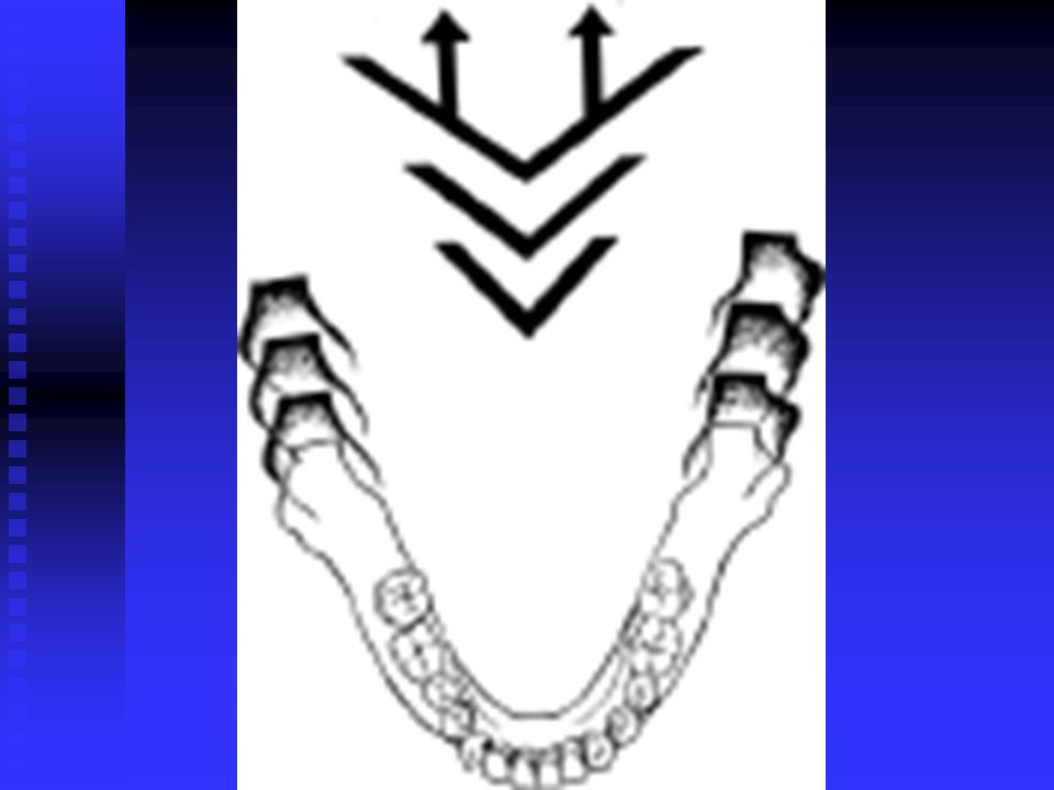
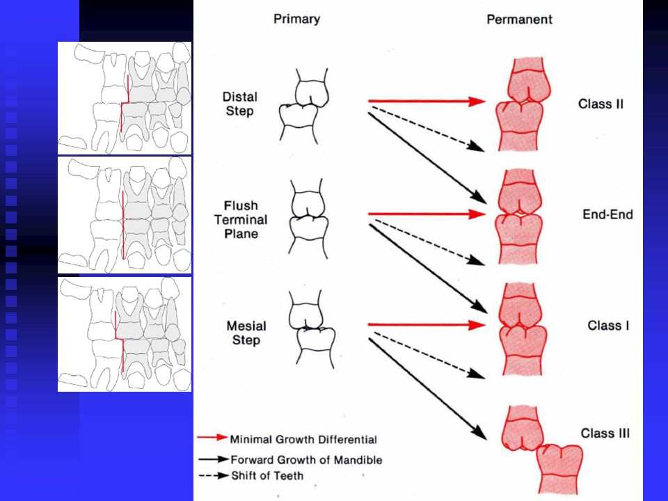
六、Dentition , occlusal growth and development
(二). Characteristics of primary dentition 5 flush terminal plane: Look at the distal aspect of the 2nd primary molar Mesial step : % Mesio step Disto step
. Characteristics of primary dentition. 5 flush terminal plane: Look at the distal aspect of the 2nd primary molar. Mesial step : % Mesio step. Disto step.")
194
Positioning of Primary Teeth

195
Classification of Occlusion of the Primary Second Molar
Look at the distal aspect of the 2nd molar Flush terminal plane Mesial step Mesio step Disto step

196
Flush Terminal Plane Distal Mesial

197
Mesial Step Distal Mesial

198
Mesio Step Distal Mesial

199
Disto Step Distal Mesial

200
THE THREE TYPES OF TERMINAL PLANES
FLUSH PLANE MESIAL STEP DISTAL STEP TYPE TYPE TYPE

202
六、Dentition , occlusal growth and development
(二). Characteristics of primary dentition 1 anterior teeth space 2 Primate space 3 shallow overjet ,overbite 4 ML side of maxillary primary canine contacts the DB side of madibular primary canine.
. Characteristics of primary dentition. 1 anterior teeth space. 2 Primate space. 3 shallow overjet ,overbite. 4 ML side of maxillary primary canine contacts the DB side of madibular primary canine.")
204
No Primary Spacing

206
(三). mixed dentition period 1 The eruption of permanent teeth:
tooth germ moves in the alveolar bone, and finally comes out of bone 。 the deciduous root absorpted and root of permanent teeth continue to grow during eruption with the height of alveolar bone increasing. Eruption conditions: crown fully formed ,roots start to form.

208
The eruption of the first permanent molar 6years
the eruption of the maxillary lateral incisor 8years

209
The complete eruption of the lateral incisor 9years
The eruption of first premolars, mandibular canines ,11years

210
Deciduous teeth have all been replaced 12years
Permanent roots are fully formed 15years

211
1 .The eruption of permanent teeth:
Degree in the formation of the root is different First permanent molars: % Canine: 70%; first premolar: 50% Second premolar: 50% Second Molar: %

212
2 Eruption time and sequence
time: 6 — 12years sequence:U L

213
3. gap relationships in the process of tooth replacement :
The whole maxillary deciduous dentition: 68.2 The whole maxillary permanent dentition: 74.0 The whole mandibular deciduous dentition : 61.8 The whole mandibular permanent dentition : 64.4

214
3 space relations in replacement of teeth:
When the replacement of anterior teeth: Gap between deciduous anterior teeth Permanent incisor when erupting tip forward Deciduous canine displace Arch width increase

215
Replacement of the posterior teeth :
Premolar erupt more buccally than deciduous teeth (Milk canine + the first and second deciduous molars )Width> Replacement permanent teeth
Width> Replacement permanent teeth.")
216
Leeway space Length change

217
Leeway Space: upper: mm each side lower: mm each side

218
4 occlusal adjustment in the course of tooth relapment
The early replacement: apex to apex relationship between molars reason:a the mesial movement L > U b growth to the forward L > U a neutral relationship.

219
5 temporary malocclusion in the mixed dentition years:
Gap between Maxillary central incisor Maxillary lateral incisor tilt distally when erupting Permanent anterior teeth (especially mandibular) crowding mildly Mild distal molar relationship (early mixed dentition) Temporary deep overbite (early mixed dentition)
 crowding mildly. Mild distal molar relationship (early mixed dentition) Temporary deep overbite (early mixed dentition)")
220
7 years old 9 years old 14 years old
Changes in the axial inclination due to the eruption of the maxillary anterior teeth (Broadbent, 1957).
.")
225
The factors that affect occlusal bulding
Power balance : Muscle Periodontal tissue Craniomaxillary Growth Genetic Nutrition Chronic diseases Bad habits Function of factors

226
Summary Growth way of craniofacial bones
cellular level: Hypertrophy Hyperplasia Increased production of extracellular matrix Growth of the Cranial Vault and Base Growth of Maxilla (Nasomaxillary Complex) Resorption 、Apposition Growth of Mandible (Length、Width、Height) Theories of Growth Control Bone Cartilage The soft tissue matrix in which the skeletal elements are embedded - 60’s “Functional Matrix Theory” by Moss Growth of Occlusion
 Resorption 、Apposition. Growth of Mandible (Length、Width、Height) Theories of Growth Control. Bone. Cartilage. The soft tissue matrix in which the skeletal elements are embedded - 60’s Functional Matrix Theory by Moss. Growth of Occlusion.")
227
Questions the methods of Craniofacial Growth and Development?
The development of maxilla and mandible , how to complete in three dimensions? What is leeway space and what is its clinical significane ? The manifestation of temporary malocclusion ,they can be adjusted at the process of growth and development ,why ?

Similar presentations