Download presentation
Presentation is loading. Please wait.

1
Dermatology “Doc, I Have This Rash…” Joseph S. Baler M.D. September 7, 2013

3
Guttate Psoriasis Small pink papules with scale. Scalp, face, trunk, and ext.

4
Guttate Psoriasis 2-3 wks post group A strep. Personal or family h/o psoriasis. May be initial psoriatic event. Post sunburn. Viral URI.

5
Guttate Psoriasis Look for Strep. Pen VK 500mg BID. Topical mid potency steroids such as TAC 0.1% cream. NBUVB, Sunlight. Do not use Prednisone!

6
Photodermatitis

7
Phototoxicity Photoallergy Sunburn reaction, erythema, edema. Direct tissue injury Occurs after first exposure. Onset minutes to hours. Large dose of agent needed for eruption. Pruritic, eczematous lesions. Type IV delayed hypersensitivity. Does not occur after first exposure. Onset 24-48 hours. Small dose of agent.

8
Phototoxicity Photoallergic

9
Phototoxic agents Systemic: Tetracyclines, Phenothiazines, Thiazides, Furosemide, Sulfonylureas. Topical: Furocoumarins: lime, lemon, celery, tar.

10
Photosensitivity/Doxycycline

11
Photoallergic agents Systemic: Quinolones,NSAIDs, sulfonamides. Topical: Fragrances.

12
Photocontact/Allergic Contact Dermatitis

13
?

14
At beach. Having a refreshing drink with a twist of lime.

15
Phytophotodermatitis Limes have psoralens containing compounds that are phototoxic. Oil of Bergamot

16
Phytophotodermatitis Initial onset erythema or blisters after contact and sun exposure. May be absent. 48-72 hrs later hyperpigmentation at sites of contact. May persist up to 4-6 weeks

17
Phytophotodermatitis Biting into lemon Squeezing limes

19
?

20
Allergic Contact Dermatitis-Mangos Mango skins have urushiol which is same as poison ivy Oil from skin of mango drips onto skin creating contact dermatitis

21
Poison Ivy Leaves and vine can cause rashes Any season Sensitivity varies from person to person Three leaflets

23
Poison Ivy Look for linear blisters or erythema Very pruritic Blister fluid is not contagious

24
Poison Ivy New areas may develop over time due to small areas of chemical contact taking longer to react.

27
Resin from plant called urushiol can oxidize and turn black on the skin which is called a “black lacquer spot”

28
Treatment Ultra potent topical steroids if a localized area such as clobetasol propionate 0.05% Systemic steroids often needed Medrol dose pack too little, and too short Prednisone 40-60mg with a slow taper over 12-18 days, maybe longer if reactivation

29
Ivy Block etc. may help prevent the oils from the plant getting to the skin by acting as a barrier which you apply as a lotion prior to potential contact Wash all clothes, tools, shoes, and gloves after contact since resin may last for years even when air dried.

32
Swimmer’s Itch (clam digger’s itch) Sea Bather’s Eruption
 Sea Bather’s Eruption")
33
Swimmer’ Itch Sea Bather’s Erupt. Water: fresh or salt Body part: uncovered Locale: North US and Canada Cause: cercarial forms of nonhuman schistosomes(snails) Water: salt Body part: covered Locale: Florida and Cuba Cause: larval forms of marine coelenterates (sea anemone, jellyfish)
 Water: salt Body part: covered Locale: Florida and Cuba Cause: larval forms of marine coelenterates (sea anemone, jellyfish).")
34
Treatment/Prevention Swimmer’s itch: symptomatic Rx for itch. Vigorous towel drying may prevent penetration of the cercariae Sea Bather’s eruption: symptomatic Rx for itch. Remove swimwear before shower since fresh water may cause discharge of nematocysts. Heat dry swimwear

35
Swimmer’s Itch (clam digger’s itch) Sea Bather’s Eruption
 Sea Bather’s Eruption")
36
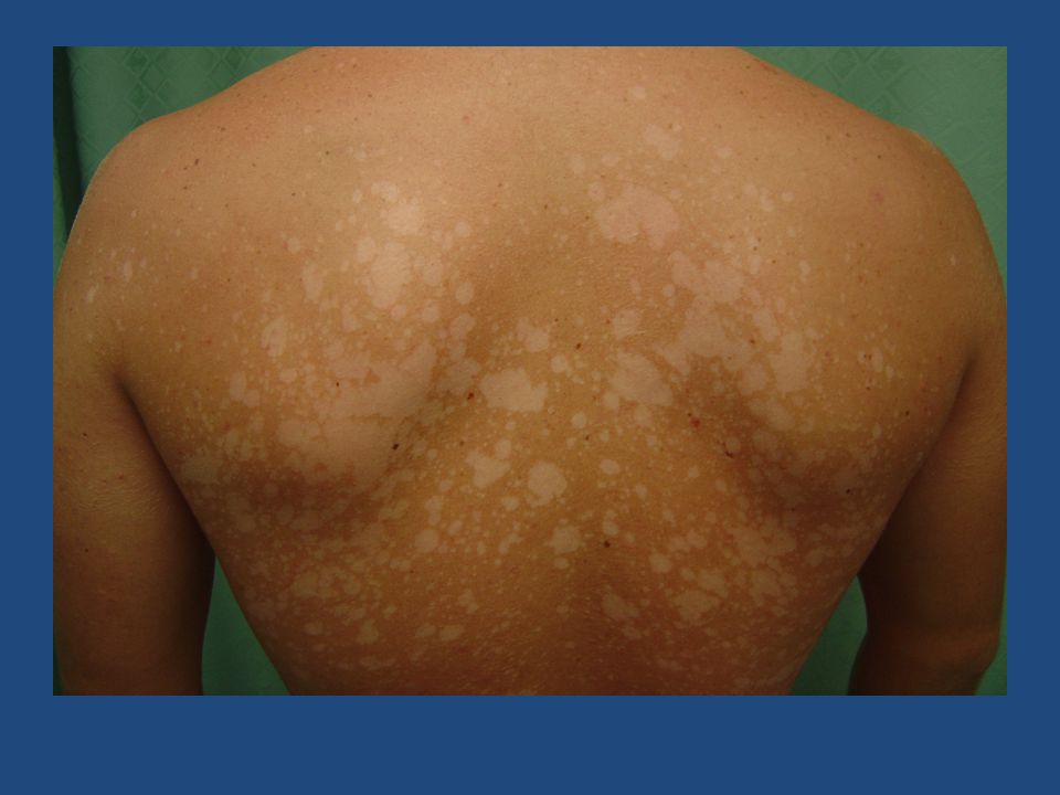
Tinea Versicolor

37
Malassezia furfur Normal cutaneous flora 2-8% US population Warm, humid enviroment Very common in tropical regions of world Immunosupression, Cushings disease

38
Hyper and hypopigmented macules with fine scale Hypopigmentation caused by tyrosinase inhibition Hyperpigmentation caused by enlarged melanosomes

40
KOH: short hyphae and spores “spaghetti and meatballs”

41
Tinea Versicolor - Treatment Topical anti-fungals Selenium sulfide 2.5% lotion Oral ketoconazole 400mg single dose, repeat in 1 week Fluconazole 200-400mg weekly 2-4 weeks Itraconazole 200mg QD x 7 days

42
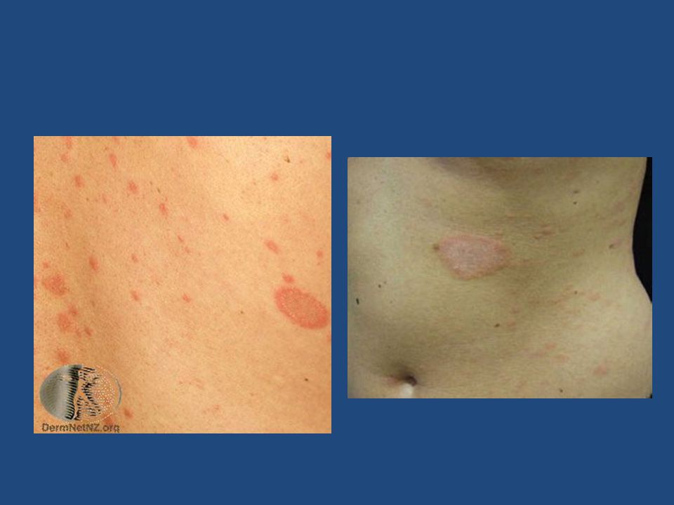
Pityriasis Rosea

43
Pruritic, oval, salmon- colored macules with collarette scale Herald patch on neck or trunk, then 1-2 wks later smaller lesions Lasts approx. 12 weeks

44
Pityriasis Rosea Follows skin creases Can have atypical cases which are more papular, vesicular, or widespread

47
Pityriasis Rosea Probable viral etiology, but no definitive data Clusters during spring common Not contagious

48
PR- Treatment Symptomatic for itch: antihistamines, topical steroids UVB, sunlight helpful

49
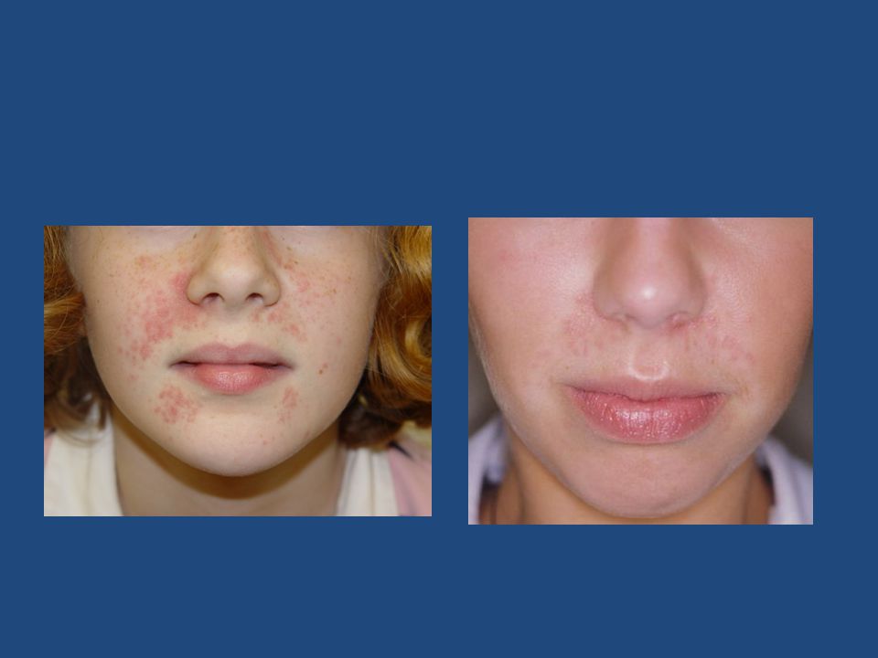
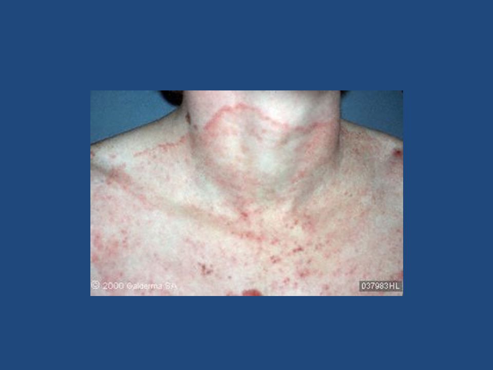
Perioral Dermatitis Acneiform lesions. Erythema and scale. Common in women. Etiology unclear, but topical steroid use often the cause.

50
Perioral Dermatitis Perioral and perinasal most common. Occasional periocular.

52
Treatment Perioral Dermatitis Avoid high potency topical steroids. If topical steroids have been used for longer than 1 month prior to diagnosis, may need to use a mild (1%) hydrocortisone cream to prevent rebound flare.
 hydrocortisone cream to prevent rebound flare..")
53
Treatment Perioral Dermatitis Oral antibiotics: Doxycycline, and minocycline good choices Topicals: Clindamycin lotion, metronidazole gel, lotion Elidel and Protopic have shown some promise

54
Perioral Dermatitis Remember : topical steroids are most often the cause, not the cure.

55
What is it ?

56
Annular Raised scaly boarder Central clearing Pruritic

57
Tinea Corporis

59
Differential Diagnosis Tinea Nummular eczema

60
Annular No central clearing Pruritic

61
What to do? KOH Look for other signs of eczema: dry skin, atopy, h/o eczema If in doubt treat with topical antifungal first If you use topical steroid first, it will flare a fungal infection

63
Be careful with betamethasone/clotrimazole combination Never more than 2 weeks The betamethasone component too potent for most fungal infections, and high risk of steroid atrophy Worsening tinea Striae

65
Treatment Topicals: Econazole, Ketoconazole etc…. Keep dry Be patient

66
Tinea Capitis Most common under age 15. It is rare in adults. Sebaceous gland maturity is protective against Tinea in scalp

67
Scale, loss of hair, other siblings with same If healthy adult with similar clinical picture, think psoriasis or seborrhea

68
Tinea Capitis US: Trichophyton tonsurans most common Europe: Microsporum canis and audouinii most common

69
Woods lamp not always helpful since Trichophyton do not fluoresce. (microsporum do) KOH and culture to diagnose
 KOH and culture to diagnose.")
70
Treatment of Tinea Capitis Systemic rx needed Griseofulvin ultra microsized 250mg bid 8-16wks until clear Griseofulvin Suspension 20-25 mg/kg/day for younger children Lamisil 10-20kg, 62.5mg/d 20-40kg, 125mg/d >40kg, 250mg/d

71
KERATOSIS PILARIS

72
Keratosis Pilaris Hyperkeratotic erythematous follicular papules Cheeks, arms, thighs, occ. trunk Genetic: Autosomal Dominant

73
Keratosis Pilaris Cheeks improve at puberty. Other sites persist Improves in summer, worse in winter Treatment – Ammonium lactate 12%, salicylic acid 6%, urea 40-50%. – Topical retinoids occasionally

74
Pityriasis Alba Don’t confuse Pityriasis alba with Tinea Versicolor Pityriasis alba more often associated with atopic dermatitis Hypopigmented, erythematous dry patches. Face and arms.

75
Pityriasis Alba Treatment Emollients and keratolytics: Ammonium lactate 12%, Salicylic acid 6%. Low potency topical corticosteroids: 1% HC, Desonide 0.05%.

78
Lichen Planus Purple, pruritic papules Wrists, legs, trunk, genitals, and scalp May hyperpigment as it resolves

79
White streaks over surface – Wickham’s striae Adults > children

80
Oral involvement with whitish lacy patches May ulcerate Risk oral SCC

81
Nail LP may cause chronic changes Scalp LP called Lichen Planopilaris. May cause scarring alopecia

82
Immune mediated. May be associated with Hep C

83
Treatment LP Topical corticosteroids Oral steroids if severe UVB, PUVA Oral Retinoids Protopic ointment 0.1% for oral disease Hydroxychloroquine, Mycophenolate Mofetil, and recently Pioglitazone for Scalp LPP

84
SCABIES (Sarcoptes Scabiei var Hominis)
")
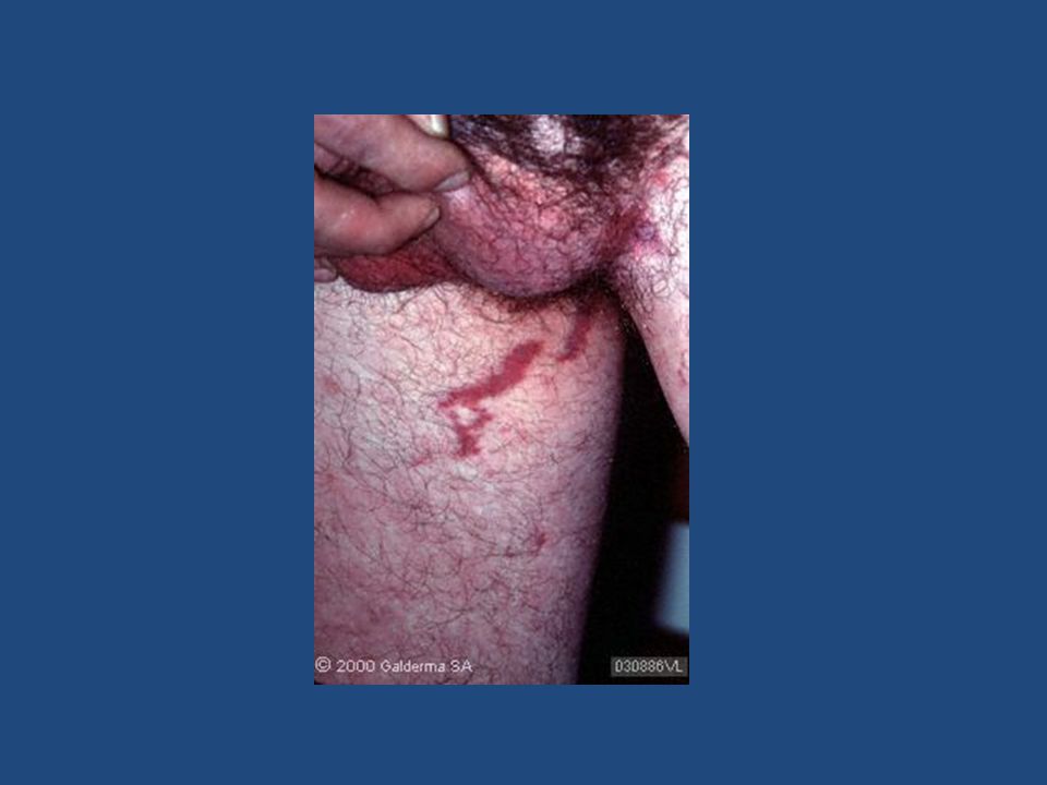
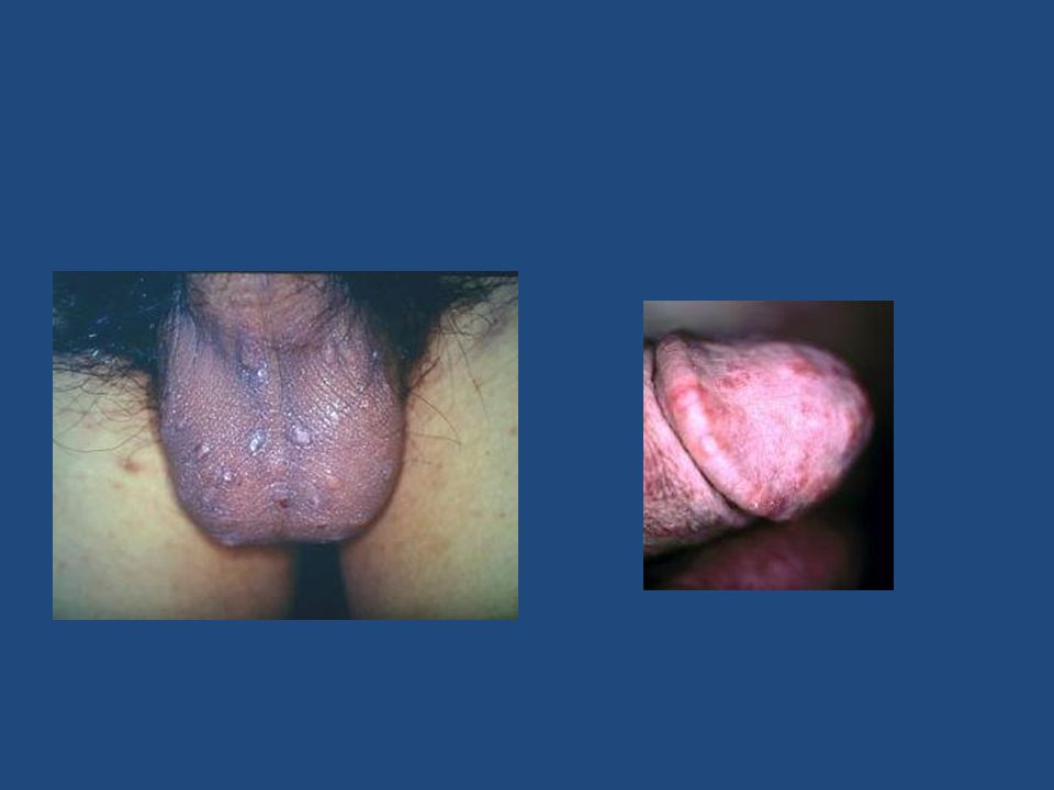
86
SCABIES Very pruritic Burrows and erythematous papules Nipples, areola Glans penis, scrotum Finger webs, axilla

91
Female mite causes symptoms. Male dies after fertilization 5-15 mites per patient Ova or feces may be found

92
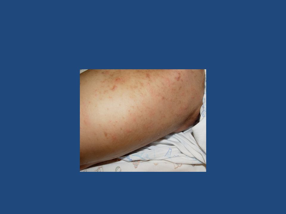
Crusted (Norwegian) scabies has hundreds to millions of mites
 scabies has hundreds to millions of mites")
94
Live up to 48hrs off host Nursing homes, group homes etc. Scraping may be negative

95
If your patient has: Chronic itch Worse at night Others with itch in household No other obvious cause Be Suspicious of Scabies

96
Scabies Treatment 5% Permethrin cream (Elimite) neck to toes (occasionally face and scalp ) overnight. Repeat in 1 week Treat others in house No need for lindane Change bedding after each treatment Ivermectin 150-200 mcg/kg single dose. May repeat in 1 week

97
Dermatology “Doc, I Have This Rash…” Joseph S. Baler M.D. September 7, 2013

Similar presentations


, malaise, arthralgias, GI issues,>")
.>")


