Download presentation
Presentation is loading. Please wait.

1
Glycogen Metabolism Purpose: Glycogen is a branched polymer of glucose; it is the stored form of G. Purpose: Glycogen is a branched polymer of glucose; it is the stored form of G. The many branches each have a C#4 end at which GP and GS can act for rapid response. The many branches each have a C#4 end at which GP and GS can act for rapid response. Glycogen is stored after a meal for release: Glycogen is stored after a meal for release: From liver when blood [G] is low to supply brain; OR From liver when blood [G] is low to supply brain; OR In muscle for rapid activity. In muscle for rapid activity.

3
Main Enzymes of Glycogen Metabolism 1. Glycogen Phosphorylase (GP): releases G as G1P: 1. Glycogen Phosphorylase (GP): releases G as G1P: G n + Pi G n-1 + G1P (no ATP cost) G n + Pi G n-1 + G1P (no ATP cost) GP removes G only from C#4 ends of chains that are at least five G’s from a branch GP removes G only from C#4 ends of chains that are at least five G’s from a branch G1P equilibrates with G6P; this is not regulated G1P equilibrates with G6P; this is not regulated G1P G6P G1P G6P 2. Glycogen Synthase (GS): adds G (as UDP – G) only to C#4 ends of chains. 2. Glycogen Synthase (GS): adds G (as UDP – G) only to C#4 ends of chains. a) Preliminary: G G6P G1P ; then: G1P + UTP PPi + UDP – G a) Preliminary: G G6P G1P ; then: G1P + UTP PPi + UDP – G b)GS rxn: G n + UDP – G UDP + G n +1 b)GS rxn: G n + UDP – G UDP + G n +1
: releases G as G1P: G n + Pi G n-1 + G1P (no ATP cost) G n + Pi G n-1 + G1P (no ATP cost) GP removes G only from C#4 ends of chains that are at least five G’s from a branch GP removes G only from C#4 ends of chains that are at least five G’s from a branch G1P equilibrates with G6P; this is not regulated G1P equilibrates with G6P; this is not regulated G1P G6P G1P G6P 2. Glycogen Synthase (GS): adds G (as UDP – G) only to C#4 ends of chains. 2. Glycogen Synthase (GS): adds G (as UDP – G) only to C#4 ends of chains. a) Preliminary: G G6P G1P ; then: G1P + UTP PPi + UDP – G a) Preliminary: G G6P G1P ; then: G1P + UTP PPi + UDP – G b)GS rxn: G n + UDP – G UDP + G n +1 b)GS rxn: G n + UDP – G UDP + G n +1.")
4
Other Enzymes of Glycogen Metabolism 1. Debranching enzyme: after GP has removed all but the last 4 G residues from a branch, this enzyme: 1. Debranching enzyme: after GP has removed all but the last 4 G residues from a branch, this enzyme: 1) catalyses transfer of 3 G residues to the C#4 end of a nearby branch and 1) catalyses transfer of 3 G residues to the C#4 end of a nearby branch and 2) catalyses hydrolysis of the 1 6 linkage, producing G 2) catalyses hydrolysis of the 1 6 linkage, producing G
 catalyses transfer of 3 G residues to the C#4 end of a nearby branch and 1) catalyses transfer of 3 G residues to the C#4 end of a nearby branch and 2) catalyses hydrolysis of the 1 6 linkage, producing G 2) catalyses hydrolysis of the 1 6 linkage, producing G.")
5
Other Enzymes of Glycogen Metabolism 2. Branching Enzyme: transfers C#1 of a 7G residue segment (from a branch at least 11 G long) to the C#6 of a residue at least 4 G away. 2. Branching Enzyme: transfers C#1 of a 7G residue segment (from a branch at least 11 G long) to the C#6 of a residue at least 4 G away.
 to the C#6 of a residue at least 4 G away. 2. Branching Enzyme: transfers C#1 of a 7G residue segment (from a branch at least 11 G long) to the C#6 of a residue at least 4 G away..")
6
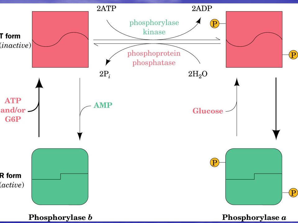
Regulation of GP, GS 1. GP is designated by 2 systems a/b and m/o, which we will not use. Instead, we will refer to the enzymes as: phosporylated (P) or dephosphorylated (DP) ( GS is also P, DP). 1. GP is designated by 2 systems a/b and m/o, which we will not use. Instead, we will refer to the enzymes as: phosporylated (P) or dephosphorylated (DP) ( GS is also P, DP). 2. GP and GS are phosphorylated in response to glucagon (in the liver) (low blood [G]) and adrenalin (muscle) (fight/flight), activating GP for release of G and inactivating GS. 2. GP and GS are phosphorylated in response to glucagon (in the liver) (low blood [G]) and adrenalin (muscle) (fight/flight), activating GP for release of G and inactivating GS. 3. GP kinase (GPK): GP + ATP ADP + GP – P. 3. GP kinase (GPK): GP + ATP ADP + GP – P. 4. They are dephosphorylated in response to insulin inactivating GP, activating GS to store G. 4. They are dephosphorylated in response to insulin inactivating GP, activating GS to store G.
 or dephosphorylated (DP) ( GS is also P, DP). 1. GP is designated by 2 systems a/b and m/o, which we will not use. Instead, we will refer to the enzymes as: phosporylated (P) or dephosphorylated (DP) ( GS is also P, DP). 2. GP and GS are phosphorylated in response to glucagon (in the liver) (low blood [G]) and adrenalin (muscle) (fight/flight), activating GP for release of G and inactivating GS. 2. GP and GS are phosphorylated in response to glucagon (in the liver) (low blood [G]) and adrenalin (muscle) (fight/flight), activating GP for release of G and inactivating GS. 3. GP kinase (GPK): GP + ATP ADP + GP – P. 3. GP kinase (GPK): GP + ATP ADP + GP – P. 4. They are dephosphorylated in response to insulin inactivating GP, activating GS to store G. 4. They are dephosphorylated in response to insulin inactivating GP, activating GS to store G..")
8
Regulatory effectors of GP, GPK GP-DP is: GP-DP is: 1) activated by AMP. MR: GP provides GlP G6P for ATP production in glycolysis, and in OP via PDH, TCA, ET, OP. ML: [AMP] is high when ATP use is rapid and ATP production is needed. 2) inhibited by ATP. MR in 1). ML: when [ATP] is high, GP doesn’t need to release G to produce more 3) inhibited by G6P. MR: G6P is an indirect product of GP. ML: when [G6P] is high, GP doesn’t need to make more. 1) activated by AMP. MR: GP provides GlP G6P for ATP production in glycolysis, and in OP via PDH, TCA, ET, OP. ML: [AMP] is high when ATP use is rapid and ATP production is needed. 2) inhibited by ATP. MR in 1). ML: when [ATP] is high, GP doesn’t need to release G to produce more 3) inhibited by G6P. MR: G6P is an indirect product of GP. ML: when [G6P] is high, GP doesn’t need to make more. GP-P is inhibited by glucose. MR: G is indirect product: GlP G6P G. ML: no need for more fuel when plenty is available GP-P is inhibited by glucose. MR: G is indirect product: GlP G6P G. ML: no need for more fuel when plenty is available GPK is activated by Ca 2+. MR: GPK activates GP, which provides fuel for ATP production. ML: Ca 2+ triggers muscle contraction, ATP production is needed. GPK is activated by Ca 2+. MR: GPK activates GP, which provides fuel for ATP production. ML: Ca 2+ triggers muscle contraction, ATP production is needed.
 inhibited by ATP. MR in 1). ML: when [ATP] is high, GP doesn’t need to release G to produce more 3) inhibited by G6P. MR: G6P is an indirect product of GP. ML: when [G6P] is high, GP doesn’t need to make more. 1) activated by AMP. MR: GP provides GlP G6P for ATP production in glycolysis, and in OP via PDH, TCA, ET, OP. ML: [AMP] is high when ATP use is rapid and ATP production is needed. 2) inhibited by ATP. MR in 1). ML: when [ATP] is high, GP doesn’t need to release G to produce more 3) inhibited by G6P. MR: G6P is an indirect product of GP. ML: when [G6P] is high, GP doesn’t need to make more. GP-P is inhibited by glucose. MR: G is indirect product: GlP G6P G. ML: no need for more fuel when plenty is available GP-P is inhibited by glucose. MR: G is indirect product: GlP G6P G. ML: no need for more fuel when plenty is available GPK is activated by Ca 2+. MR: GPK activates GP, which provides fuel for ATP production. ML: Ca 2+ triggers muscle contraction, ATP production is needed. GPK is activated by Ca 2+. MR: GPK activates GP, which provides fuel for ATP production. ML: Ca 2+ triggers muscle contraction, ATP production is needed..")
9
Regulatory effectors of GS GS-DP is activated by G6P: MR: G6P is indirect substrate: G6P GlP UPD-G (feed forward) ML: when G6P is plentiful, it’s time to store G. GS-DP is activated by G6P: MR: G6P is indirect substrate: G6P GlP UPD-G (feed forward) ML: when G6P is plentiful, it’s time to store G.
 ML: when G6P is plentiful, it’s time to store G..")
12
Hormonal Regulation of Glycogen Metabolism 1. The “hunger hormone”, glucagon is a signal to release G to blood from the liver via glycogenolysis and gluconeogenesis. Liver GP is activated, GS inhibited. 1. The “hunger hormone”, glucagon is a signal to release G to blood from the liver via glycogenolysis and gluconeogenesis. Liver GP is activated, GS inhibited. 2. Adrenalin (epinephrine) is a signal to “break down” muscle glycogen to produce G6P for ATP production (in G’lys and in OP via PDH, TCA, ET and OP) for fight- or-flight. Muscle GP is activated, GS inhibited. 2. Adrenalin (epinephrine) is a signal to “break down” muscle glycogen to produce G6P for ATP production (in G’lys and in OP via PDH, TCA, ET and OP) for fight- or-flight. Muscle GP is activated, GS inhibited. When either one binds its cell-membrane receptor: When either one binds its cell-membrane receptor: a. the hormone-receptor complex binds to adenylate cyclase and activates it to catalyze: ATP PPi + cAMP a. the hormone-receptor complex binds to adenylate cyclase and activates it to catalyze: ATP PPi + cAMP b. cAMP is the internal or “2nd” messenger. It binds to protein kinase (PrK) regulatory (r) subunits, dissociating them from the catalytic (c) subunits, which are then active. b. cAMP is the internal or “2nd” messenger. It binds to protein kinase (PrK) regulatory (r) subunits, dissociating them from the catalytic (c) subunits, which are then active.
 is a signal to break down muscle glycogen to produce G6P for ATP production (in G’lys and in OP via PDH, TCA, ET and OP) for fight- or-flight. Muscle GP is activated, GS inhibited. 2. Adrenalin (epinephrine) is a signal to break down muscle glycogen to produce G6P for ATP production (in G’lys and in OP via PDH, TCA, ET and OP) for fight- or-flight. Muscle GP is activated, GS inhibited. When either one binds its cell-membrane receptor: When either one binds its cell-membrane receptor: a. the hormone-receptor complex binds to adenylate cyclase and activates it to catalyze: ATP PPi + cAMP a. the hormone-receptor complex binds to adenylate cyclase and activates it to catalyze: ATP PPi + cAMP b. cAMP is the internal or 2nd messenger. It binds to protein kinase (PrK) regulatory (r) subunits, dissociating them from the catalytic (c) subunits, which are then active. b. cAMP is the internal or 2nd messenger. It binds to protein kinase (PrK) regulatory (r) subunits, dissociating them from the catalytic (c) subunits, which are then active..")
13
Hormonal Regulation of Glycogen Metabolism c. PrK catalyses the phosphorylation of a variety of proteins, including: c. PrK catalyses the phosphorylation of a variety of proteins, including: the tandem E (G’lys, G’neo) the tandem E (G’lys, G’neo) pyruvate kinase (G’lys) pyruvate kinase (G’lys) ACoA carboxylase (FA synthesis) ACoA carboxylase (FA synthesis) Glycogen synthase, inactivating it Glycogen synthase, inactivating it Glycogen phophorylase kinase (GPK), activating it. Glycogen phophorylase kinase (GPK), activating it. d. GPK-P catalyses phosphorylation of glycogen phosphorylase (GP), activating it. d. GPK-P catalyses phosphorylation of glycogen phosphorylase (GP), activating it. e. Net hormonal effect: GP activated: Gn G; and GS inactive, preventing opposition to GP. e. Net hormonal effect: GP activated: Gn G; and GS inactive, preventing opposition to GP. PrK also phosphorylates phosphoprotein phosphatase inhibitor 1 (PPI-1) causing it to bind to and inactivate PP1, the enzyme that dephosphorylates GP, GS, etc. PrK also phosphorylates phosphoprotein phosphatase inhibitor 1 (PPI-1) causing it to bind to and inactivate PP1, the enzyme that dephosphorylates GP, GS, etc.
 the tandem E (G’lys, G’neo) pyruvate kinase (G’lys) pyruvate kinase (G’lys) ACoA carboxylase (FA synthesis) ACoA carboxylase (FA synthesis) Glycogen synthase, inactivating it Glycogen synthase, inactivating it Glycogen phophorylase kinase (GPK), activating it. Glycogen phophorylase kinase (GPK), activating it. d. GPK-P catalyses phosphorylation of glycogen phosphorylase (GP), activating it. d. GPK-P catalyses phosphorylation of glycogen phosphorylase (GP), activating it. e. Net hormonal effect: GP activated: Gn G; and GS inactive, preventing opposition to GP. e. Net hormonal effect: GP activated: Gn G; and GS inactive, preventing opposition to GP. PrK also phosphorylates phosphoprotein phosphatase inhibitor 1 (PPI-1) causing it to bind to and inactivate PP1, the enzyme that dephosphorylates GP, GS, etc. PrK also phosphorylates phosphoprotein phosphatase inhibitor 1 (PPI-1) causing it to bind to and inactivate PP1, the enzyme that dephosphorylates GP, GS, etc..")
15
Dephosphorylation of (GPK, GP, GS) Dephosphorylation of these enzymes (GPK, GP, GS) is catalyzed by phosphoprotein phosphatase 1 (PP1). Dephosphorylation of these enzymes (GPK, GP, GS) is catalyzed by phosphoprotein phosphatase 1 (PP1). In muscle, phosphorylation of a regulatory glycogen binding protein, G M in response to insulin (which causes dephosphorylation of other Es) at site 1 activates PP1. This results in the opposite activities to the above (GS active to store the plentiful G, GP not, to prevent opposing GS). In muscle, phosphorylation of a regulatory glycogen binding protein, G M in response to insulin (which causes dephosphorylation of other Es) at site 1 activates PP1. This results in the opposite activities to the above (GS active to store the plentiful G, GP not, to prevent opposing GS). Phosphorylation of G M at site 2 ( alone or in addition to site 1) by PrK inactivates PP1, preventing it from opposing PrK. Phosphorylation of G M at site 2 ( alone or in addition to site 1) by PrK inactivates PP1, preventing it from opposing PrK.
 is catalyzed by phosphoprotein phosphatase 1 (PP1). In muscle, phosphorylation of a regulatory glycogen binding protein, G M in response to insulin (which causes dephosphorylation of other Es) at site 1 activates PP1. This results in the opposite activities to the above (GS active to store the plentiful G, GP not, to prevent opposing GS). In muscle, phosphorylation of a regulatory glycogen binding protein, G M in response to insulin (which causes dephosphorylation of other Es) at site 1 activates PP1. This results in the opposite activities to the above (GS active to store the plentiful G, GP not, to prevent opposing GS). Phosphorylation of G M at site 2 ( alone or in addition to site 1) by PrK inactivates PP1, preventing it from opposing PrK. Phosphorylation of G M at site 2 ( alone or in addition to site 1) by PrK inactivates PP1, preventing it from opposing PrK..")
16
In liver, the switch from the phospho- to the dephospho- state of GP, GPK, and GS cannot occur without the accumulation of glucose. It is said that “GP is the glucose sensor”: In liver, the switch from the phospho- to the dephospho- state of GP, GPK, and GS cannot occur without the accumulation of glucose. It is said that “GP is the glucose sensor”: a) In the phospho (active) form, the P’s on GP are “buried” where PP1 can’t get at them. a) In the phospho (active) form, the P’s on GP are “buried” where PP1 can’t get at them. b) When G binds to active GP-P, its conformation changes, “exposing” the P’s so PP1 can “clip them” off. b) When G binds to active GP-P, its conformation changes, “exposing” the P’s so PP1 can “clip them” off. c) PP1 binds strongly to GP-P in the R form; and is not active toward other phosphoproteins in this state. Only after GP-P binds G and PP1 dephosphorylates GP is PP1 released and active. c) PP1 binds strongly to GP-P in the R form; and is not active toward other phosphoproteins in this state. Only after GP-P binds G and PP1 dephosphorylates GP is PP1 released and active. d) PP1 has a much higher affinity for GP-P than for GS, GPK, etc, so it must first “work its way through” nearly all the GP-P, dephosphorylating it, before it has much effect on GS. d) PP1 has a much higher affinity for GP-P than for GS, GPK, etc, so it must first “work its way through” nearly all the GP-P, dephosphorylating it, before it has much effect on GS.
 In the phospho (active) form, the P’s on GP are buried where PP1 can’t get at them. a) In the phospho (active) form, the P’s on GP are buried where PP1 can’t get at them. b) When G binds to active GP-P, its conformation changes, exposing the P’s so PP1 can clip them off. b) When G binds to active GP-P, its conformation changes, exposing the P’s so PP1 can clip them off. c) PP1 binds strongly to GP-P in the R form; and is not active toward other phosphoproteins in this state. Only after GP-P binds G and PP1 dephosphorylates GP is PP1 released and active. c) PP1 binds strongly to GP-P in the R form; and is not active toward other phosphoproteins in this state. Only after GP-P binds G and PP1 dephosphorylates GP is PP1 released and active. d) PP1 has a much higher affinity for GP-P than for GS, GPK, etc, so it must first work its way through nearly all the GP-P, dephosphorylating it, before it has much effect on GS. d) PP1 has a much higher affinity for GP-P than for GS, GPK, etc, so it must first work its way through nearly all the GP-P, dephosphorylating it, before it has much effect on GS..")
18
Amplification cascade GP and GS are regulated by effectors like AMP, ATP, G, and G6P. So why has regulation by enzyme phosphorylation evolved? The big advantage is speed and magnitude of response: each enzymatic step amplifies the signal, and subsequent ones multiply previous ones: GP and GS are regulated by effectors like AMP, ATP, G, and G6P. So why has regulation by enzyme phosphorylation evolved? The big advantage is speed and magnitude of response: each enzymatic step amplifies the signal, and subsequent ones multiply previous ones: 1) each hormone-receptor complex activates one adenylate cyclase, which can produce, say, 1000 cAMP/sec. 1) each hormone-receptor complex activates one adenylate cyclase, which can produce, say, 1000 cAMP/sec. 2) 4 cAMP can --> 2 active PrK which can produce, say, 100 GPK-P/sec. 2) 4 cAMP can --> 2 active PrK which can produce, say, 100 GPK-P/sec. 3) Each GPK-P can phosphorylate, say, 100 GP/s. 3) Each GPK-P can phosphorylate, say, 100 GP/s. So, one H-R complex results in: 1000 cAMP x 2PrK/4 cAMP x 100 GPK-P/PrK x 100GP-P/GPK-P=5,000,000 activated GP/sec, rather than 1 Enzyme/1 effector. That's why it’s called the" amplification cascade". So, one H-R complex results in: 1000 cAMP x 2PrK/4 cAMP x 100 GPK-P/PrK x 100GP-P/GPK-P=5,000,000 activated GP/sec, rather than 1 Enzyme/1 effector. That's why it’s called the" amplification cascade".
 each hormone-receptor complex activates one adenylate cyclase, which can produce, say, 1000 cAMP/sec. 1) each hormone-receptor complex activates one adenylate cyclase, which can produce, say, 1000 cAMP/sec. 2) 4 cAMP can --> 2 active PrK which can produce, say, 100 GPK-P/sec. 2) 4 cAMP can --> 2 active PrK which can produce, say, 100 GPK-P/sec. 3) Each GPK-P can phosphorylate, say, 100 GP/s. 3) Each GPK-P can phosphorylate, say, 100 GP/s. So, one H-R complex results in: 1000 cAMP x 2PrK/4 cAMP x 100 GPK-P/PrK x 100GP-P/GPK-P=5,000,000 activated GP/sec, rather than 1 Enzyme/1 effector. That s why it’s called the amplification cascade . So, one H-R complex results in: 1000 cAMP x 2PrK/4 cAMP x 100 GPK-P/PrK x 100GP-P/GPK-P=5,000,000 activated GP/sec, rather than 1 Enzyme/1 effector. That s why it’s called the amplification cascade ..")
19
Glucokinase (GK) GK catalyzes G + ATP G6P + ADP, same as HXK. GK catalyzes G + ATP G6P + ADP, same as HXK. GK is a liver enzyme; muscle doesn’t have it (has HXK). GK is a liver enzyme; muscle doesn’t have it (has HXK). The properties of GK are more suited to maximum glycogen synthesis when [G] is high. HXK can also support rapid glycogen synthesis, but not as well as GK. The properties of GK are more suited to maximum glycogen synthesis when [G] is high. HXK can also support rapid glycogen synthesis, but not as well as GK. HXK is “designed” to keep up with extremely rapid glycolysis (if that’s the pace PFK sets): when PFK consumes F6P rapidly, G6P is also consumed rapidly, (G6P F6P) so that HXK is not inhibited by G6P. HXK is “designed” to keep up with extremely rapid glycolysis (if that’s the pace PFK sets): when PFK consumes F6P rapidly, G6P is also consumed rapidly, (G6P F6P) so that HXK is not inhibited by G6P. HXK has a very low Km (high affinity) for G, so it can go at almost Vmax rate even if [G] is low, but high [G] doesn’t increase rate. HXK has a very low Km (high affinity) for G, so it can go at almost Vmax rate even if [G] is low, but high [G] doesn’t increase rate.
. GK is a liver enzyme; muscle doesn’t have it (has HXK). The properties of GK are more suited to maximum glycogen synthesis when [G] is high. HXK can also support rapid glycogen synthesis, but not as well as GK. The properties of GK are more suited to maximum glycogen synthesis when [G] is high. HXK can also support rapid glycogen synthesis, but not as well as GK. HXK is designed to keep up with extremely rapid glycolysis (if that’s the pace PFK sets): when PFK consumes F6P rapidly, G6P is also consumed rapidly, (G6P F6P) so that HXK is not inhibited by G6P. HXK is designed to keep up with extremely rapid glycolysis (if that’s the pace PFK sets): when PFK consumes F6P rapidly, G6P is also consumed rapidly, (G6P F6P) so that HXK is not inhibited by G6P. HXK has a very low Km (high affinity) for G, so it can go at almost Vmax rate even if [G] is low, but high [G] doesn’t increase rate. HXK has a very low Km (high affinity) for G, so it can go at almost Vmax rate even if [G] is low, but high [G] doesn’t increase rate..")
20
GK, HXK But GK is not inhibited by G6P, so when [G] is high it can produce a much higher [G6P] which kinetically pushes glycogen synthase: via G6P GlP UDPG But GK is not inhibited by G6P, so when [G] is high it can produce a much higher [G6P] which kinetically pushes glycogen synthase: via G6P GlP UDPG GK has a much higher Km (lower affinity) for G so its rate is nearly proportional to [G] across the physiological [G] range. GK has a much higher Km (lower affinity) for G so its rate is nearly proportional to [G] across the physiological [G] range.
![GK, HXK But GK is not inhibited by G6P, so when [G] is high it can produce a much higher [G6P] which kinetically pushes glycogen synthase: via G6P GlP UDPG But GK is not inhibited by G6P, so when [G] is high it can produce a much higher [G6P] which kinetically pushes glycogen synthase: via G6P GlP UDPG GK has a much higher Km (lower affinity) for G so its rate is nearly proportional to [G] across the physiological [G] range.](http://images.slideplayer.com/13/4046919/slides/slide_20.jpg "GK has a much higher Km (lower affinity) for G so its rate is nearly proportional to [G] across the physiological [G] range..")
21
FAs, Fatty Acids FAs are a much more efficient form of stored fuel: 9kcal/g (9 Cal/g) vs. (4 Cal/gG); also glycogen binds two times its weight of H 2 O. A typical man would have to store ~ 90 kg of glycogen (~200lbs) if he was to have the same energy as in the ~15 kg fat he stores. FAs are a much more efficient form of stored fuel: 9kcal/g (9 Cal/g) vs. (4 Cal/gG); also glycogen binds two times its weight of H 2 O. A typical man would have to store ~ 90 kg of glycogen (~200lbs) if he was to have the same energy as in the ~15 kg fat he stores. Although glycolysis is a major fuel consuming pathway, FAs are the main fuel (except in brain, RBCs, rapid muscle activity). Although glycolysis is a major fuel consuming pathway, FAs are the main fuel (except in brain, RBCs, rapid muscle activity). Because of the above, glycogen storage is limited and “xs G” is converted to fat via glycolysis, PDH, CS and FA synthesis. Because of the above, glycogen storage is limited and “xs G” is converted to fat via glycolysis, PDH, CS and FA synthesis.
; also glycogen binds two times its weight of H 2 O. A typical man would have to store ~ 90 kg of glycogen (~200lbs) if he was to have the same energy as in the ~15 kg fat he stores. FAs are a much more efficient form of stored fuel: 9kcal/g (9 Cal/g) vs. (4 Cal/gG); also glycogen binds two times its weight of H 2 O. A typical man would have to store ~ 90 kg of glycogen (~200lbs) if he was to have the same energy as in the ~15 kg fat he stores. Although glycolysis is a major fuel consuming pathway, FAs are the main fuel (except in brain, RBCs, rapid muscle activity). Although glycolysis is a major fuel consuming pathway, FAs are the main fuel (except in brain, RBCs, rapid muscle activity). Because of the above, glycogen storage is limited and xs G is converted to fat via glycolysis, PDH, CS and FA synthesis. Because of the above, glycogen storage is limited and xs G is converted to fat via glycolysis, PDH, CS and FA synthesis..")
22
FA Use as Fuel FAs are released from storage (as triacylglycerol) by the hydrolytic action of hormone-sensitive lipase, which is activated by phosphorylation by PrK in response to adrenalin or glucagon, deactivated by dephosphorylation by PP1 in response to insulin. FAs are released from storage (as triacylglycerol) by the hydrolytic action of hormone-sensitive lipase, which is activated by phosphorylation by PrK in response to adrenalin or glucagon, deactivated by dephosphorylation by PP1 in response to insulin. FA’s then travel from adipose cells (“cytosol” is mainly a fat globule) via blood to cells that use them. FA’s then travel from adipose cells (“cytosol” is mainly a fat globule) via blood to cells that use them. FA’s are prepared in the cytosol (cytoplasm) for transport to the mitochondrial matrix, where they are converted to ACoA in oxidation. FA’s are prepared in the cytosol (cytoplasm) for transport to the mitochondrial matrix, where they are converted to ACoA in oxidation. FA activation: (costs 2 ATP) FA activation: (costs 2 ATP) a) FA + ATP PPi + FA – AMP; a) FA + ATP PPi + FA – AMP; b) FA – AMP + CoASH AMP + FA – SCoA b) FA – AMP + CoASH AMP + FA – SCoA
 by the hydrolytic action of hormone-sensitive lipase, which is activated by phosphorylation by PrK in response to adrenalin or glucagon, deactivated by dephosphorylation by PP1 in response to insulin. FA’s then travel from adipose cells ( cytosol is mainly a fat globule) via blood to cells that use them. FA’s then travel from adipose cells ( cytosol is mainly a fat globule) via blood to cells that use them. FA’s are prepared in the cytosol (cytoplasm) for transport to the mitochondrial matrix, where they are converted to ACoA in oxidation. FA’s are prepared in the cytosol (cytoplasm) for transport to the mitochondrial matrix, where they are converted to ACoA in oxidation. FA activation: (costs 2 ATP) FA activation: (costs 2 ATP) a) FA + ATP PPi + FA – AMP; a) FA + ATP PPi + FA – AMP; b) FA – AMP + CoASH AMP + FA – SCoA b) FA – AMP + CoASH AMP + FA – SCoA.")
23
CoA from cytosol doesn’t enter matrix (or vice-versa). CoA from cytosol doesn’t enter matrix (or vice-versa). Instead, on the outer surface of the inner membrane, FA is transferred to carnitine (releasing CoASH to the cytosol) in a reaction catalyzed by carnitine acyltransferase I (CATI) (aka Carnitine Palmitoyl TransferaseI, or CPTI). Instead, on the outer surface of the inner membrane, FA is transferred to carnitine (releasing CoASH to the cytosol) in a reaction catalyzed by carnitine acyltransferase I (CATI) (aka Carnitine Palmitoyl TransferaseI, or CPTI). A transport protein in the inner membrane brings fatty-acyl carnitine into the matrix (in exchange for carnitine delivered outside). A transport protein in the inner membrane brings fatty-acyl carnitine into the matrix (in exchange for carnitine delivered outside). CATII (CPTII) on inner surface transfers FAcyl group from carnitine to CoASH. (Palmitate is the 16C saturated FA) CATII (CPTII) on inner surface transfers FAcyl group from carnitine to CoASH. (Palmitate is the 16C saturated FA)
. Instead, on the outer surface of the inner membrane, FA is transferred to carnitine (releasing CoASH to the cytosol) in a reaction catalyzed by carnitine acyltransferase I (CATI) (aka Carnitine Palmitoyl TransferaseI, or CPTI). Instead, on the outer surface of the inner membrane, FA is transferred to carnitine (releasing CoASH to the cytosol) in a reaction catalyzed by carnitine acyltransferase I (CATI) (aka Carnitine Palmitoyl TransferaseI, or CPTI). A transport protein in the inner membrane brings fatty-acyl carnitine into the matrix (in exchange for carnitine delivered outside). A transport protein in the inner membrane brings fatty-acyl carnitine into the matrix (in exchange for carnitine delivered outside). CATII (CPTII) on inner surface transfers FAcyl group from carnitine to CoASH. (Palmitate is the 16C saturated FA) CATII (CPTII) on inner surface transfers FAcyl group from carnitine to CoASH. (Palmitate is the 16C saturated FA).")
24
oxidation + TCA + ET + OP ATP oxidation converts the fatty acyl group to ACoA. Net reaction for complete oxidation of palmitate: oxidation converts the fatty acyl group to ACoA. Net reaction for complete oxidation of palmitate: C 15 H 31 COSCoA + 7CoASH + 7FAD + 7NAD + 8ACoA + 7FADH 2 + 7NADH C 15 H 31 COSCoA + 7CoASH + 7FAD + 7NAD + 8ACoA + 7FADH 2 + 7NADH ATP production from palmitate ATP production from palmitate 8XTCA: +8GTP + 24NADH + 8FADH2 8XTCA: +8GTP + 24NADH + 8FADH2 ET: -31NADH – 15FADH2 ET: -31NADH – 15FADH2 OP: [+3(31) + 2(15)] ATP = + 123 ATP OP: [+3(31) + 2(15)] ATP = + 123 ATP 123ATP + 8GTP – 2 “ATP” (ATP AMP in FA acivation ) = 129ATP 123ATP + 8GTP – 2 “ATP” (ATP AMP in FA acivation ) = 129ATP
 + 2(15)] ATP = ATP OP: [+3(31) + 2(15)] ATP = ATP 123ATP + 8GTP – 2 ATP (ATP AMP in FA acivation ) = 129ATP 123ATP + 8GTP – 2 ATP (ATP AMP in FA acivation ) = 129ATP.")
25
Ketone Body Production The moderate rate of production of acetoacetate, hydroxybutyrate and acetone that occurs normally in the liver mitochondrial matrix delivers “water soluble FA fragments” to cells via blood for use as fuel. The moderate rate of production of acetoacetate, hydroxybutyrate and acetone that occurs normally in the liver mitochondrial matrix delivers “water soluble FA fragments” to cells via blood for use as fuel. Since this process involves unregulated enzymes, the buildup of ACoA in diabetes overproduces these compounds to toxic levels. Since this process involves unregulated enzymes, the buildup of ACoA in diabetes overproduces these compounds to toxic levels.

26
FA Synthesis FA synthesis is a liver pathway FA synthesis is a liver pathway The net effect is to build up the CH 2 chain by joining ACoAs’ acetyl gps. and reducing (and hydrogenating) the C=O of ACoA. The net effect is to build up the CH 2 chain by joining ACoAs’ acetyl gps. and reducing (and hydrogenating) the C=O of ACoA. The ACoAs for FA synth don’t come from oxidation. Rather it’s the “xs G” that enters liver cells after a meal and goes through insulin stimulated glycolysis and PDH. The ACoAs for FA synth don’t come from oxidation. Rather it’s the “xs G” that enters liver cells after a meal and goes through insulin stimulated glycolysis and PDH.
 the C=O of ACoA. The net effect is to build up the CH 2 chain by joining ACoAs’ acetyl gps. and reducing (and hydrogenating) the C=O of ACoA. The ACoAs for FA synth don’t come from oxidation. Rather it’s the xs G that enters liver cells after a meal and goes through insulin stimulated glycolysis and PDH. The ACoAs for FA synth don’t come from oxidation. Rather it’s the xs G that enters liver cells after a meal and goes through insulin stimulated glycolysis and PDH..")
27
But PDH is in matrix, FA sythase is in cytosol: (ACoA doesn’t cross inner membrane) But PDH is in matrix, FA sythase is in cytosol: (ACoA doesn’t cross inner membrane) 1. high ACoA from PDH stimulates PC high oxac. 1. high ACoA from PDH stimulates PC high oxac. 2. (ACoA + oxac citrate) in matrix; then transport citrate to cytosol. 2. (ACoA + oxac citrate) in matrix; then transport citrate to cytosol. 3. in cyto: citrate + ATP ACoA + oxac + ADP + Pi (catalyzed by citrate lyase) 3. in cyto: citrate + ATP ACoA + oxac + ADP + Pi (catalyzed by citrate lyase) 4. oxac + NADH malate + NAD+ then, malate can enter matrix, OR 4. oxac + NADH malate + NAD+ then, malate can enter matrix, OR 5. in cyto: mal + NADP+ NADPH + pyr + CO 2 ; (pyr goes to matrix). This rxn is catalyzed by the malic enzyme. 5. in cyto: mal + NADP+ NADPH + pyr + CO 2 ; (pyr goes to matrix). This rxn is catalyzed by the malic enzyme. 6. The NADPH is needed for FA synthesis (below) 6. The NADPH is needed for FA synthesis (below)
 in matrix; then transport citrate to cytosol. 2. (ACoA + oxac citrate) in matrix; then transport citrate to cytosol. 3. in cyto: citrate + ATP ACoA + oxac + ADP + Pi (catalyzed by citrate lyase) 3. in cyto: citrate + ATP ACoA + oxac + ADP + Pi (catalyzed by citrate lyase) 4. oxac + NADH malate + NAD+ then, malate can enter matrix, OR 4. oxac + NADH malate + NAD+ then, malate can enter matrix, OR 5. in cyto: mal + NADP+ NADPH + pyr + CO 2 ; (pyr goes to matrix). This rxn is catalyzed by the malic enzyme. 5. in cyto: mal + NADP+ NADPH + pyr + CO 2 ; (pyr goes to matrix). This rxn is catalyzed by the malic enzyme. 6. The NADPH is needed for FA synthesis (below) 6. The NADPH is needed for FA synthesis (below).")
28
7. The cyto ACoA is activated for joining by conversion to malonyl CoA (carboxylated) by ACoA carboxylase (ACoAC): ACoA + CO 2 +ATP ---> ADP + Pi + malCoA 7. The cyto ACoA is activated for joining by conversion to malonyl CoA (carboxylated) by ACoA carboxylase (ACoAC): ACoA + CO 2 +ATP ---> ADP + Pi + malCoA
 by ACoA carboxylase (ACoAC): ACoA + CO 2 +ATP ---> ADP + Pi + malCoA 7. The cyto ACoA is activated for joining by conversion to malonyl CoA (carboxylated) by ACoA carboxylase (ACoAC): ACoA + CO 2 +ATP ---> ADP + Pi + malCoA.")
29
FA Synthase In E Coli, this consists of a number of separate enzymes, but in animals 2 identical subunits each contain the enzymatic activities for all the rxns ( oxidation has a different enzyme for each step). In E Coli, this consists of a number of separate enzymes, but in animals 2 identical subunits each contain the enzymatic activities for all the rxns ( oxidation has a different enzyme for each step). The substrate remains bound to the long phosphopantethein prosthetic group (Fig. 25-29, p931), which “carries” it to each of the various active sites. This is on ACP (acyl-carrier protein) The substrate remains bound to the long phosphopantethein prosthetic group (Fig. 25-29, p931), which “carries” it to each of the various active sites. This is on ACP (acyl-carrier protein)
. The substrate remains bound to the long phosphopantethein prosthetic group (Fig , p931), which carries it to each of the various active sites. This is on ACP (acyl-carrier protein) The substrate remains bound to the long phosphopantethein prosthetic group (Fig , p931), which carries it to each of the various active sites. This is on ACP (acyl-carrier protein).")
30
Phases of FA Synthase Reaction “Cycle” Loading: the acetyl group of ACoA is transferred to a cys-S (viaACP) and the malonyl group of mal-CoA to ACP-S. Loading: the acetyl group of ACoA is transferred to a cys-S (viaACP) and the malonyl group of mal-CoA to ACP-S. Condensation, Reduction: C 2 chains (of malCoA from ACoA) are linked, releasing CO 2, then reduced to –CH 2 -CH 2. Condensation, Reduction: C 2 chains (of malCoA from ACoA) are linked, releasing CO 2, then reduced to –CH 2 -CH 2. Reloading: existing chain transferred to cys-S; next malonyl group to ACP-S (each mal of mal CoA goes onto ACP, only acetyl group of ACoA (and existing chain) go onto cys-S Reloading: existing chain transferred to cys-S; next malonyl group to ACP-S (each mal of mal CoA goes onto ACP, only acetyl group of ACoA (and existing chain) go onto cys-S Release: FA hydrolyzed from ACP Release: FA hydrolyzed from ACP
 and the malonyl group of mal-CoA to ACP-S. Condensation, Reduction: C 2 chains (of malCoA from ACoA) are linked, releasing CO 2, then reduced to –CH 2 -CH 2. Condensation, Reduction: C 2 chains (of malCoA from ACoA) are linked, releasing CO 2, then reduced to –CH 2 -CH 2. Reloading: existing chain transferred to cys-S; next malonyl group to ACP-S (each mal of mal CoA goes onto ACP, only acetyl group of ACoA (and existing chain) go onto cys-S Reloading: existing chain transferred to cys-S; next malonyl group to ACP-S (each mal of mal CoA goes onto ACP, only acetyl group of ACoA (and existing chain) go onto cys-S Release: FA hydrolyzed from ACP Release: FA hydrolyzed from ACP.")
31
Regulation of FA Metabolism ACoA Carboxylase (ACoA mal CoA FA synthesis) ACoA Carboxylase (ACoA mal CoA FA synthesis) 1.Inhibited by palmitoyl CoA. MR: indirect product (mal CoA palmitate pal CoA). 1.Inhibited by palmitoyl CoA. MR: indirect product (mal CoA palmitate pal CoA). ML: If [pal CoA] is high, it is being produced faster than it can be used, production can slow) ML: If [pal CoA] is high, it is being produced faster than it can be used, production can slow) 2.Activated by citrate. MR: indirect substrate; citrate oxac + AcoA, the substrate 2.Activated by citrate. MR: indirect substrate; citrate oxac + AcoA, the substrate ML: when[citrate] cyto is high, [citrate] mito is very high, fuel is plentiful, time to store it ML: when[citrate] cyto is high, [citrate] mito is very high, fuel is plentiful, time to store it 3. Inhibited by phosphorylation in response to glucagon or adrenalin. These hormones promote fuel mobilization to make fuel available, so they inhibit storage. 3. Inhibited by phosphorylation in response to glucagon or adrenalin. These hormones promote fuel mobilization to make fuel available, so they inhibit storage. 4. Activated by dephosphorylation in response to insulin. Insulin “signals fed state”, when [G] is high it’s time to store C as glycogen and FAs. 4. Activated by dephosphorylation in response to insulin. Insulin “signals fed state”, when [G] is high it’s time to store C as glycogen and FAs.
. 1.Inhibited by palmitoyl CoA. MR: indirect product (mal CoA palmitate pal CoA). ML: If [pal CoA] is high, it is being produced faster than it can be used, production can slow) ML: If [pal CoA] is high, it is being produced faster than it can be used, production can slow) 2.Activated by citrate. MR: indirect substrate; citrate oxac + AcoA, the substrate 2.Activated by citrate. MR: indirect substrate; citrate oxac + AcoA, the substrate ML: when[citrate] cyto is high, [citrate] mito is very high, fuel is plentiful, time to store it ML: when[citrate] cyto is high, [citrate] mito is very high, fuel is plentiful, time to store it 3. Inhibited by phosphorylation in response to glucagon or adrenalin. These hormones promote fuel mobilization to make fuel available, so they inhibit storage. 3. Inhibited by phosphorylation in response to glucagon or adrenalin. These hormones promote fuel mobilization to make fuel available, so they inhibit storage. 4. Activated by dephosphorylation in response to insulin. Insulin signals fed state , when [G] is high it’s time to store C as glycogen and FAs. 4. Activated by dephosphorylation in response to insulin. Insulin signals fed state , when [G] is high it’s time to store C as glycogen and FAs..")
32
Regulation of FAActive ACoAC Metabolism Phosphorylation shifts ACoAC from active polymer form to inactive monomer form. Phosphorylation shifts ACoAC from active polymer form to inactive monomer form. Carnitine Acyl Transferase I (CATI) (transport of FAs into matrix for oxid’n). Carnitine Acyl Transferase I (CATI) (transport of FAs into matrix for oxid’n). Inhibited by mal CoA. MR: mal CoA is the product of the committed step in the opposing pathway, FA synthesis. Inhibited by mal CoA. MR: mal CoA is the product of the committed step in the opposing pathway, FA synthesis. ML: When [mal CoA] is high, FA synthesis is rapid (in liver), with the purpose of export of these FA’s for storage. Inhibition of CATI prevents consumption from working against synthesis. ML: When [mal CoA] is high, FA synthesis is rapid (in liver), with the purpose of export of these FA’s for storage. Inhibition of CATI prevents consumption from working against synthesis.
 (transport of FAs into matrix for oxid’n). Carnitine Acyl Transferase I (CATI) (transport of FAs into matrix for oxid’n). Inhibited by mal CoA. MR: mal CoA is the product of the committed step in the opposing pathway, FA synthesis. Inhibited by mal CoA. MR: mal CoA is the product of the committed step in the opposing pathway, FA synthesis. ML: When [mal CoA] is high, FA synthesis is rapid (in liver), with the purpose of export of these FA’s for storage. Inhibition of CATI prevents consumption from working against synthesis. ML: When [mal CoA] is high, FA synthesis is rapid (in liver), with the purpose of export of these FA’s for storage. Inhibition of CATI prevents consumption from working against synthesis..")
36
Figure 25-41All of cholesterol’s carbon atoms are derived from acetate. Page 942

37
Amino Acid (AA) oxidation Introduction Introduction 1. Part of the C’s of some of the AAs are convertible to ACoA, either directly or via acetoacetate or pyr. (and less directly, so are the others via TCA int oxac PEP pyr ACoA.) (These AAs are “ketogenic) 1. Part of the C’s of some of the AAs are convertible to ACoA, either directly or via acetoacetate or pyr. (and less directly, so are the others via TCA int oxac PEP pyr ACoA.) (These AAs are “ketogenic) So, these C’s of xs AA intake (in relation to need for protein synth) are used as fuel, just like dietary CH 2 O’s, fats. So, these C’s of xs AA intake (in relation to need for protein synth) are used as fuel, just like dietary CH 2 O’s, fats. 2. Part (or all) of the C’s of 18 of the AAs can be converted to TCA intermediates, which can be converted to G (TCA int oxac PEP G). These are referred to as the “glucogenic” AAs. 2. Part (or all) of the C’s of 18 of the AAs can be converted to TCA intermediates, which can be converted to G (TCA int oxac PEP G). These are referred to as the “glucogenic” AAs. AAs from digestion of muscle protein are the main source of C for gluconeogenesis in CH 2 O starvation AAs from digestion of muscle protein are the main source of C for gluconeogenesis in CH 2 O starvation
 (These AAs are ketogenic) 1. Part of the C’s of some of the AAs are convertible to ACoA, either directly or via acetoacetate or pyr. (and less directly, so are the others via TCA int oxac PEP pyr ACoA.) (These AAs are ketogenic) So, these C’s of xs AA intake (in relation to need for protein synth) are used as fuel, just like dietary CH 2 O’s, fats. So, these C’s of xs AA intake (in relation to need for protein synth) are used as fuel, just like dietary CH 2 O’s, fats. 2. Part (or all) of the C’s of 18 of the AAs can be converted to TCA intermediates, which can be converted to G (TCA int oxac PEP G). These are referred to as the glucogenic AAs. 2. Part (or all) of the C’s of 18 of the AAs can be converted to TCA intermediates, which can be converted to G (TCA int oxac PEP G). These are referred to as the glucogenic AAs. AAs from digestion of muscle protein are the main source of C for gluconeogenesis in CH 2 O starvation AAs from digestion of muscle protein are the main source of C for gluconeogenesis in CH 2 O starvation.")
38
Transaminations (trnsams) Each AA can be converted to the corresponding keto acid by at least one transaminase. This AA is oxidized in this rxn, but kg is reduced to glutamate at the same time so there’s not a net AA oxidation Each AA can be converted to the corresponding keto acid by at least one transaminase. This AA is oxidized in this rxn, but kg is reduced to glutamate at the same time so there’s not a net AA oxidation The amino group transferred to kg (---> glu) is toxic when released as NH 3, this ammonia is detoxified by conversion to urea in the urea cycle (NH 3 can be excreted). Net oxidation occurs by coupling of trnsam with glutamate dehydrogenase (GDH). The amino group transferred to kg (---> glu) is toxic when released as NH 3, this ammonia is detoxified by conversion to urea in the urea cycle (NH 3 can be excreted). Net oxidation occurs by coupling of trnsam with glutamate dehydrogenase (GDH). GDH: glu + NAD + kg + NADH + NH 3 GDH: glu + NAD + kg + NADH + NH 3 This rxn running in reverse when [NH 3 ] is very high depletes TCA ints, interferes with TCA +ET + OP in brain cells and causes the delirium/dementia in liver damaged patients. This rxn running in reverse when [NH 3 ] is very high depletes TCA ints, interferes with TCA +ET + OP in brain cells and causes the delirium/dementia in liver damaged patients.
 is toxic when released as NH 3, this ammonia is detoxified by conversion to urea in the urea cycle (NH 3 can be excreted). Net oxidation occurs by coupling of trnsam with glutamate dehydrogenase (GDH). The amino group transferred to kg (---> glu) is toxic when released as NH 3, this ammonia is detoxified by conversion to urea in the urea cycle (NH 3 can be excreted). Net oxidation occurs by coupling of trnsam with glutamate dehydrogenase (GDH). GDH: glu + NAD + kg + NADH + NH 3 GDH: glu + NAD + kg + NADH + NH 3 This rxn running in reverse when [NH 3 ] is very high depletes TCA ints, interferes with TCA +ET + OP in brain cells and causes the delirium/dementia in liver damaged patients. This rxn running in reverse when [NH 3 ] is very high depletes TCA ints, interferes with TCA +ET + OP in brain cells and causes the delirium/dementia in liver damaged patients..")
39
GDH Regulation Inhibited by ATP and GTP; Activated by ADP and GDP Inhibited by ATP and GTP; Activated by ADP and GDP MR: GDH + trnsam TCA ints TCA ET OP: ATP production MR: GDH + trnsam TCA ints TCA ET OP: ATP production ML: If [ATP] or [GTP] is high, more is not needed; if [ADP] or [GDP] is high, ATP synthesis is needed; TCA ATP ML: If [ATP] or [GTP] is high, more is not needed; if [ADP] or [GDP] is high, ATP synthesis is needed; TCA ATP
![GDH Regulation Inhibited by ATP and GTP; Activated by ADP and GDP Inhibited by ATP and GTP; Activated by ADP and GDP MR: GDH + trnsam TCA ints TCA ET OP: ATP production MR: GDH + trnsam TCA ints TCA ET OP: ATP production ML: If [ATP] or [GTP] is high, more is not needed; if [ADP] or [GDP] is high, ATP synthesis is needed; TCA ATP ML: If [ATP] or [GTP] is high, more is not needed; if [ADP] or [GDP] is high, ATP synthesis is needed; TCA ATP](http://images.slideplayer.com/13/4046919/slides/slide_39.jpg "GDH Regulation Inhibited by ATP and GTP; Activated by ADP and GDP Inhibited by ATP and GTP; Activated by ADP and GDP MR: GDH + trnsam TCA ints TCA ET OP: ATP production MR: GDH + trnsam TCA ints TCA ET OP: ATP production ML: If [ATP] or [GTP] is high, more is not needed; if [ADP] or [GDP] is high, ATP synthesis is needed; TCA ATP ML: If [ATP] or [GTP] is high, more is not needed; if [ADP] or [GDP] is high, ATP synthesis is needed; TCA ATP")
40
Carbamoyl Phosphate Synthetase I Regulation CPSI is activated by N-acetylglutamate NAG CPSI is activated by N-acetylglutamate NAG MR: NAG is produced from ACoA and glu: MR: NAG is produced from ACoA and glu: ACoA + glu NAG. ACoA + glu NAG. A high [ACoA] and/or a high [glu] increases the rate of NAG production and the [NAG], so the [NAG] indicates the levels of AcoA and glu. A high [ACoA] and/or a high [glu] increases the rate of NAG production and the [NAG], so the [NAG] indicates the levels of AcoA and glu. MR, ML for ACoA: when [ACoA] is high there is a need for oxac to react with ACoA in the CS rxn. GDH + trnsam can produce TCA ints from AAs at a high rate only if CPSI consumes the ammonia product of GDH. MR, ML for ACoA: when [ACoA] is high there is a need for oxac to react with ACoA in the CS rxn. GDH + trnsam can produce TCA ints from AAs at a high rate only if CPSI consumes the ammonia product of GDH. MR, ML for glu: when glu is high it has been produced by a high rate of trnsam and there is a need to convert it to kg in GDH to maintain [ kg] for TCA and trnsam. CPSI must consume the ammonia product of GDH. MR, ML for glu: when glu is high it has been produced by a high rate of trnsam and there is a need to convert it to kg in GDH to maintain [ kg] for TCA and trnsam. CPSI must consume the ammonia product of GDH.

Similar presentations


 ~15%>")
![Fatty Acid Metabolism. Introduction of Clinical Case n 10 m.o. girl –Overnight fast, morning seizures & coma –[glu] = 20mg/dl –iv glucose, improves rapidly.](/13/4088684/big_thumb.jpg "Fatty Acid Metabolism. Introduction of Clinical Case n 10 m.o. girl –Overnight fast, morning seizures & coma –[glu] = 20mg/dl –iv glucose, improves rapidly.>")
Biosynthesis of glycerol. 2)Biosynthesis of fatty acids. 3)Biosynthesis of the triacylglycerol. It occurs in.>")


>")

>")

