Download presentation
Presentation is loading. Please wait.

1
Histology + Integument Lab Microscope –Microscope “rules” Introduction to Tissues –Selected Epithelial Tissues –Areolar C.T.

2
Occular (w/ lens) Nosepiece Objective lens Course focus Fine focus Stage Stage clip adjustment knobs Iris control Light source condenser Stage clip Arm base
 Nosepiece Objective lens Course focus Fine focus Stage Stage clip adjustment knobs Iris control Light source condenser Stage clip Arm base")
3
Rules for microscope use 1.ALWAYS carry with two hands; one on base, one on arm 2.ALWAYS begin and end with the low power lens in place 3.ALWAYS begin and end with the stage completely lowered 4.NEVER use course focus with the high power lens

4
Plasma Membrane nucleus Cytoplasm 3 MAIN PARTS OF THE CELL VISIBLE WITH THE MICROSCOPE

5
Histology: the study of tissues Tissue = a group of cells performing related functions and the material around/between them Matrix Fibers Ground substance

6
Epithelial Tissues Characteristics (partial list) Very cellular (E.T. has very little matrix) Covers/lines surfaces Has a distinct top and bottom (polarity) –Top surface = apical surface –Bottom surface = basal surface Basal surface is held in place/fused to another material by a basement membrane
 Covers/lines surfaces Has a distinct top and bottom (polarity) –Top surface = apical surface –Bottom surface = basal surface Basal surface is held in place/fused to another material by a basement membrane.")
7
3 common shapes Amount of cytoplasm related to cell’s ability to be active and perform work squamous cuboidalcolumnar

8
Simple Squamous: Simple = 1 layer Squamous = flattened 1 layer of flattened cells Thin “quick and easy to pass through” Involved with diffusion and filtration Example locations: –lungs, kidneys, capillaries, serous membranes Key visual characteristic is a flattened nucleus pressed/compressed against the adjacent tissue. Plasma membrane unlikely to be seen

9
Simple squamous in kidney (w/ glomerulus)
")
10
Simple squamous in Kidney

13
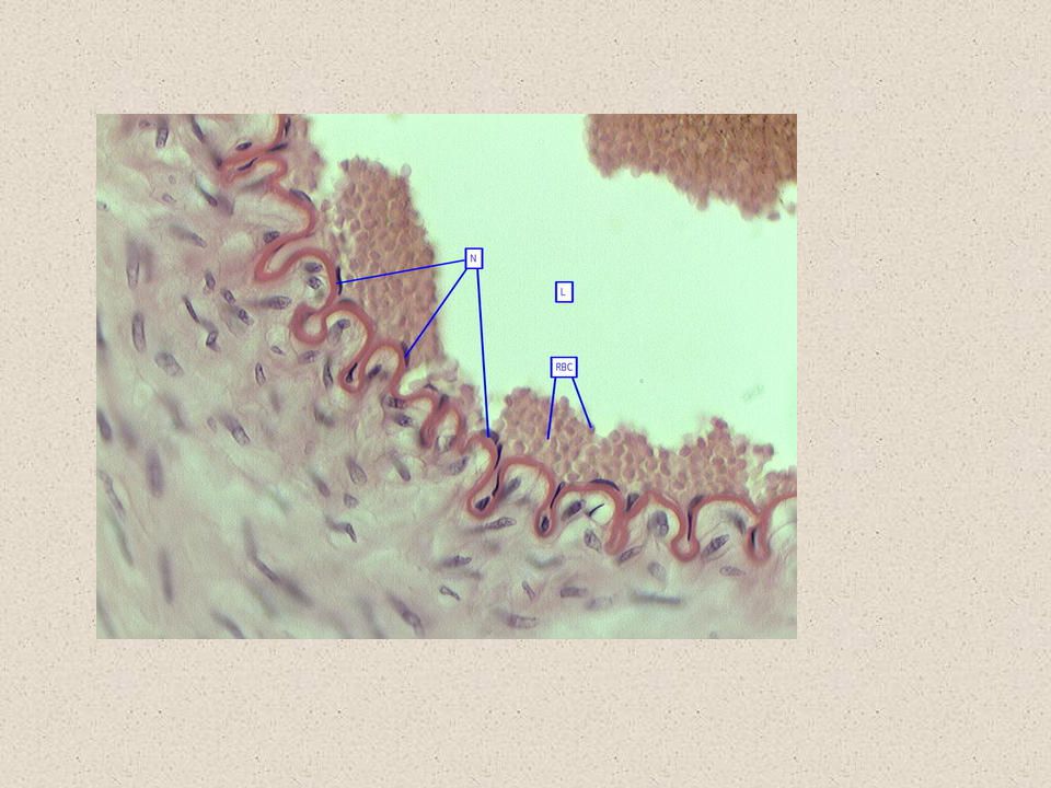
FINDING SIMPLE SQUAMOUS IN BLOOD VESSELS

14
LOOK HEAR FOR SIMPLE SQUAMOUS

16
Simple Squamous

19
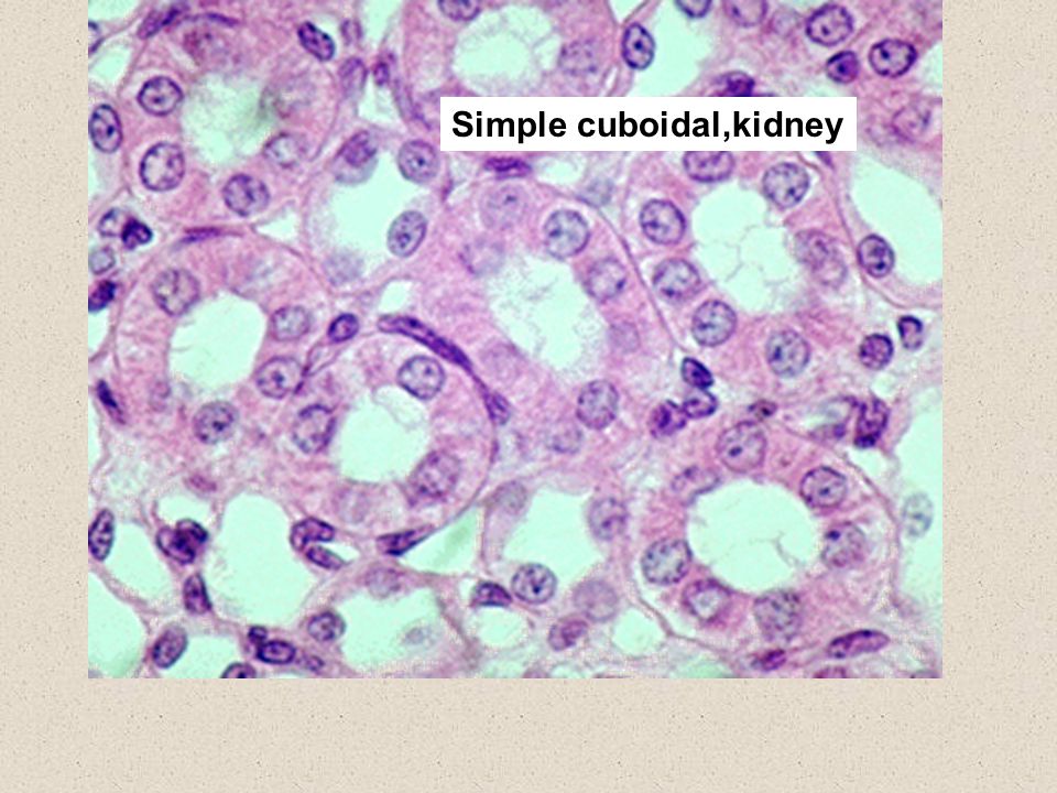
Simple Cuboidal cuboidal = “cube” shaped 1 layer of ‘square’ cells Absorption and secretion Layer thing enough to readily pass through E.g., Kidneys, glands Key visual characteristic is a round nucleus, usually located near the center of cell Cell about as wide as it is tall. Apical surface sometimes appears rounded

20
Simple Cuboidal in Kidney

22
Simple cuboidal,kidney

26
Simple columnar columnar = tall and narrow 1 layer of tall, thin cells Absorption and secretion Layer thing enough to readily pass through Goblet cells = produce mucus (mucin) E.g., stomach and intestines Key visual characteristic is an elongated nucleus located in the basal half of the cell.
 E.g., stomach and intestines Key visual characteristic is an elongated nucleus located in the basal half of the cell.")
27
Finding Simple Columnar in Small Intestine the red line represents the location of the E.T.

28
Simple Columnar

33
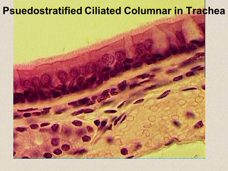
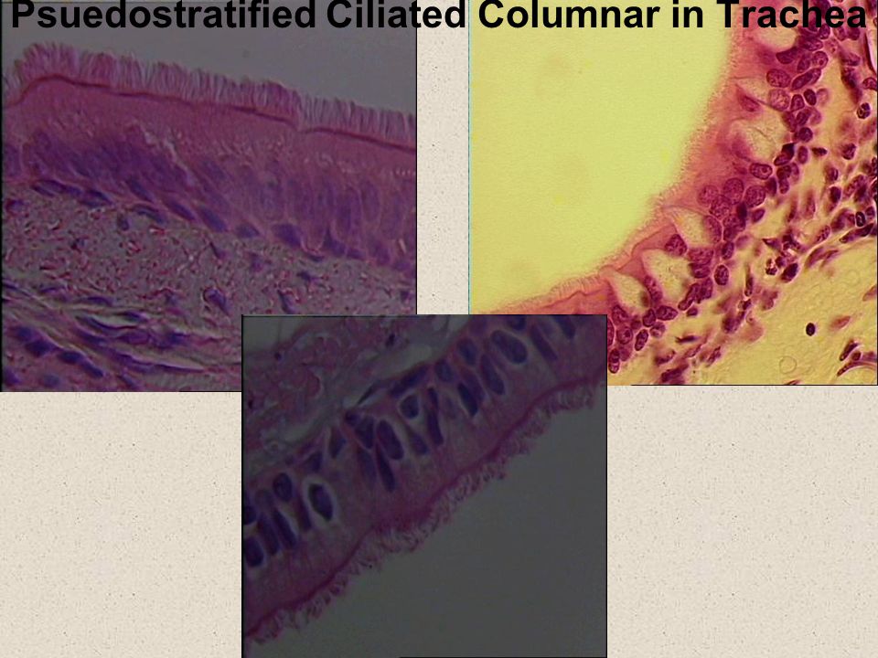
Psuedostratified cilliated columnar 1 layer of tall, irregular cells Goblet cells mucus Cilia = ‘hairs’ that move to sweep mucus away E.g. trachea, bronchi, nasal cavity Tissue cleans air as it passes through respiratory tract Key visual characteristic is cilia –It is the only tissue we look at that has cilia

34
Psuedostratified Ciliated Columnar in Trachea: E.T. is located at red line

35
Psuedostratified Ciliated Columnar in Trachea

41
Stratified squamous stratified = more than one layer of cells Many layers of cells, upper cells flattened Thick layer Protective, relatively difficult to pass through Skin, oral cavity, vagina, pharynx, tongue Visual feature is a thick tissue with thin apical layers. Many layers of nuclei between apical surface and basal surface

42
Stratified Squamous E.T. location in dark red

44
Stratified Squamous (non-keratinized):
:")
45
Connective Tissues Most connective tissue has widely spaced cells with lots of matrix in between –Matrix = fibers + ground substance

46
Areolar Connective Tissue STRUCTURE Cells = fibroblasts Produce proteins fibers Fibers –Collagen: strong, but inelastic –Elastic: weak, but elastic –Both fibers widely spaced and oriented in many directions Function: provides a supportive layer that loosely binds structures together

47
Areolar CT

48
Areolar

Similar presentations


 – 2.Connective (CT) – 3.Muscle – 4.Nervous.>")


 types of tissue: – 1. Epithelial – 2. Connective – 3. Muscle – 4. Nervous.>")


 Connective tissue Muscle.>")


