Download presentation
Presentation is loading. Please wait.

1
Introduction to Microbiology
Introduction to Clinical Laboratory Sciences Introduction to Microbiology CLS 245

2
Introduction to Clinical Laboratory Sciences
What is Microorganism? Microorganisms ( or Microbes) are vey small organism that can’t be seen by naked eyes. However, these Microbes can be seen with the aid of microscope. These microorganisms can be divided into 4 subgroups: Bacteria,, Fungi, Viruses and Parasites. Microbiology is a broad field that include the study of all types of microorganisms.
 are vey small organism that can’t be seen by naked eyes. However, these Microbes can be seen with the aid of microscope. These microorganisms can be divided into 4 subgroups: Bacteria,, Fungi, Viruses and Parasites. Microbiology is a broad field that include the study of all types of microorganisms.")
4
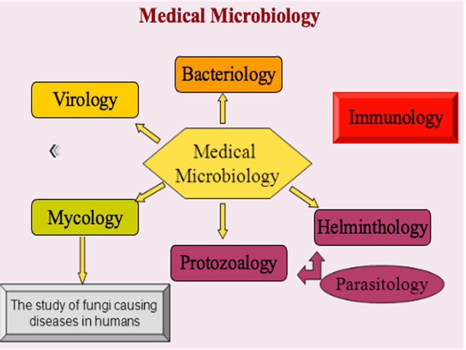
Cont.. Medical Microbiology is the study of microorganisms: Bacteria
Fungus Parasites Viruses Most can only be seen with the microscope!

5
Cont.. Medical Microbiology studies are usually performed on human blood and body fluids. Microorganisms can cause disease in humans. Microbiologists determine the type of microorganism causing the disease and find a drug, usually an antibiotic, to inhibit the microorganism. Microbiologists continue to study the microorganisms through research to determine new antibiotics

6
Cont.. Microorganisms are studied in clinical hospital laboratories, reference laboratories, and research facilities.

7
Prokaryotes vs Eukaryotes
Introduction to Clinical Laboratory Sciences Prokaryotes vs Eukaryotes Before getting into Microbiology, it is important to understand the differences between Prokaryotes and Eukaryotes. Adapted from:

8
Prokaryotes vs Eukaryotes
Introduction to Clinical Laboratory Sciences Prokaryotes vs Eukaryotes -ER , Golgi , Mitochondria and Lysosome are in Eukaryotes but not in pro -human are eukaryotes Yeast also are eukaryotes, so when you get fungal infection it is hard to treat with antibiotics.

9
Introduction to Clinical Laboratory Sciences
Viruses -Virology is study of viruses. -Viruses are very small microorganisms (smaller than bacteria) and they can’t grow or reproduce apart from living cells. -They are too small to be seen by microscope. -Viruses are not alive, why? Viruses are not a live, why? They can’t reproduce or grow apart from living cell, they also can’t produce energy.
 and they can’t grow or reproduce apart from living cells. -They are too small to be seen by microscope. -Viruses are not alive, why Viruses are not a live, why They can’t reproduce or grow apart from living cell, they also can’t produce energy.")
10
Viruses Viruses have wide variety of structure
but they all share 2 important element: Genetic material (DNA or RNA) Protein coat or capsid
 Protein coat or capsid.")
11
Introduction to Clinical Laboratory Sciences
Fungi The study of fungi is called Mycology. The fungi are a group of eukaryotic microorganisms, some of which are capable of causing superficial, cutaneous, subcutaneous, or systemic disease. However, some fungi are also directly important as a food for human (making a bread) or as a treatment ( producing penicillin).
 or as a treatment ( producing penicillin).")
12
Introduction to Clinical Laboratory Sciences
Fungi cont Introduction to Clinical Laboratory Sciences Mykes (Greek word) means Mushroom. Fungi are eukaryotes , differ from bacteria and other prokaryotes. Fungi digest dead organic matter. Molds and mushroom are multicellular while Yeast are unicellular Adapted from DR T.V Rao MD Yeast: unicellular fungi Molds: Multicellular fungi
 means Mushroom. Fungi are eukaryotes , differ from bacteria and other. prokaryotes. Fungi digest dead organic matter. Molds and mushroom are multicellular while Yeast are. unicellular. Adapted from DR T.V Rao MD. Yeast: unicellular fungi. Molds: Multicellular fungi.")
13
Introduction to Clinical Laboratory Sciences

14
Introduction to Clinical Laboratory Sciences
Parasites Introduction to Clinical Laboratory Sciences Medical parasitology: “the study and medical implications of parasites that infect humans” A parasite: “a living organism that acquires some of its basic nutritional requirements through its intimate contact with another living organism”. Parasites may be simple unicellular protozoa or complex multicellular metazoa Eukaryote: a cell with a well-defined chromosome in a membrane-bound nucleus. All parasitic organisms are eukaryotes Protozoa: unicellular organisms, e.g. Plasmodium (malaria) Metazoa: multicellular organisms, e.g. helminths (worms) and arthropods (ticks, lice) Adapted from Angela Allen, Research Assistant, The School of Medicine, Swansea University, Swansea, UK Dr. Stephen Allen, Senior Lecturer in Paediatrics and Honorary Consultant Paediatrician, The School of Medicine, Swansea University, Swansea, UK
 Metazoa: multicellular organisms, e.g. helminths (worms) and arthropods (ticks, lice) Adapted from. Angela Allen, Research Assistant, The School of Medicine, Swansea University, Swansea, UK. Dr. Stephen Allen, Senior Lecturer in Paediatrics and Honorary Consultant Paediatrician, The School of Medicine, Swansea University, Swansea, UK.")
15
Introduction to Clinical Laboratory Sciences
Cont. Parasite Host: “the organism in, or on, which the parasite lives and causes harm” Intermediate host: “the organism in which the parasite lives during a period of its development only” Vector: “a living carrier (e.g.an arthropod) that transports a pathogenic organism from an infected to a non-infected host”. A typical example is the female Anopheles mosquito that transmits malaria Adapted from Angela Allen, Research Assistant, The School of Medicine, Swansea University, Swansea, UK Dr. Stephen Allen, Senior Lecturer in Paediatrics and Honorary Consultant Paediatrician, The School of Medicine, Swansea University, Swansea, UK
 that transports a pathogenic organism from an infected to a non-infected host . A typical example is the female Anopheles mosquito that transmits malaria. Adapted from. Angela Allen, Research Assistant, The School of Medicine, Swansea University, Swansea, UK. Dr. Stephen Allen, Senior Lecturer in Paediatrics and Honorary Consultant Paediatrician, The School of Medicine, Swansea University, Swansea, UK.")
16
Introduction to Clinical Laboratory Sciences
E. Coli O157:H7 can make you very sick. What are bacteria? Single celled organisms Very small Need a microscope to see Can be found on most materials and surfaces Billions on and in your body right now Streptococcus can cause strep throat. USDA NIFSI Food Safety in the Classroom© University of Tennessee, Knoxville 2006 This E. coli helps you digest food.

17
Introduction to Clinical Laboratory Sciences
What do they look like? Three basic shapes Rod shaped called bacilli (buh-sill-eye) Round shaped called cocci (cox-eye) Spiral shaped Some exist as single cells, others cluster together Bacilli Cocci Spiral Cluster of cocci
 Round shaped called cocci (cox-eye) Spiral shaped. Some exist as single cells, others. cluster together. Bacilli. Cocci. Spiral. Cluster of cocci.")
18
Introduction to Clinical Laboratory Sciences
Bacteria are ALIVE! What does it mean to be alive? They reproduce (make more of themselves) They need to eat
 They need to eat.")
19
Introduction to Clinical Laboratory Sciences
How do bacteria eat? Photosynthetic bacteria Some make their own food from sunlight—like plants Some are scavengers Share the environment around them Example: The bacteria in your stomach are now eating what you ate for breakfast Some are warriors (pathogens) They attack other living things Example: The bacteria on your face can attack skin causing infection and acne Harmless bacteria on the stomach lining E. Coli O157:H7 is a pathogen
 They attack other living things. Example: The bacteria on your face can attack skin causing infection and acne. Harmless bacteria on the stomach lining. E. Coli O157:H7. is a pathogen.")
20
Introduction to Clinical Laboratory Sciences
What is a pathogen? Bacteria that make you sick Why do they make you sick? To get food they need to survive and reproduce How do they make you sick? They produce poisons (toxins) that result in fever, headache, vomiting, and diarrhea and destroy body tissue
 that result in fever, headache, vomiting, and diarrhea and destroy body tissue.")
21
Where do you get a pathogen?
Introduction to Clinical Laboratory Sciences Where do you get a pathogen? Indirect contact Contact with people who are sick Direct or indirect Food, Water, or other Surfaces that are contaminated Foods that could be contaminated Direct contact

22
Are all bacteria pathogens?
Introduction to Clinical Laboratory Sciences Are all bacteria pathogens? No, most are harmless Some are even helpful Examples of helpful bacteria: Lactobacillus: makes cheese, yogurt, & buttermilk and produces vitamins in your intestine Leuconostoc: makes pickles & sauerkraut Pediococcus: makes pepperoni, salami, & summer sausage USDA NIFSI Food Safety in the Classroom© University of Tennessee, Knoxville 2006

23
Tools and instruments in Microbiology lab
Safety hoods are used to avoid splashing and inhaling possible pathogens. Agar plate for bacterial growth Slide used to make Bacterial smear And stain them Microscope to visualize the bacteria

24
Introduction to Clinical Laboratory Sciences
Types of staining techniques Simple staining (use of a single stain) Differential staining (use of two contrasting stains separated by a decolorizing agent) For visualization of morphological shape & arrangement. Identification Visualization of structure Gram stain Acid fast stain Spore stain Capsule stain
 Differential staining. (use of two contrasting stains. separated by a decolorizing agent) For visualization of morphological. shape & arrangement. Identification. Visualization. of structure. Gram. stain. Acid fast. stain. Spore. stain. Capsule. stain.")
25
Introduction to Clinical Laboratory Sciences
Gram Stain: It is the most important differential stain used in bacteriology because it classified bacteria into two major groups: b) Gram negative: Appears red after Gram’s stain Gram positive: Appears violet after Gram’s stain
 Gram negative: Appears red after Gram’s stain. Gram positive: Appears violet after Gram’s stain.")
26
Introduction to Clinical Laboratory Sciences
Crystal violet ↓ Iodine Alcohol Safranin

27
Introduction to Clinical Laboratory Sciences
Gram –ve E.coli Gram +ve S.aureus Step 1: Crystal Violet Step 2: Gram’s Iodine Step 3: Decolorization (Aceton-Alcohol) Step 4: Safranin Red
 Step 4: Safranin Red.")
28
Introduction to Clinical Laboratory Sciences
Gram’s +ve Bacteria Gram’s -ve Bacteria

29
Introduction to Clinical Laboratory Sciences
Gram-positive bacteria Have a thick peptidoglycan layer surrounds the cell. The stain gets trapped into this layer and the bacteria turned purple. Retain the color of the primary stain (crystal violet) after decolorization with alcohol Gram-negative bacteria have a thin peptidoglycan layer that does not retain crystal violet stain. Instead, it has a thick lipid layer which dissolved easily upon decoulorization with Aceton-Alcohol. Therefore, cells will be counterstained with safranin and turned red.
 after decolorization with alcohol. Gram-negative bacteria. have a thin peptidoglycan layer that does not retain crystal violet stain. Instead, it has a thick lipid layer which dissolved easily upon decoulorization with Aceton-Alcohol. Therefore, cells will be counterstained with safranin and turned red.")
30
Types of culture media Based on their consistency a) solid medium
b) liquid medium (ex: Blood culture) c) semi solid medium (for motility) -Some media are used for bacterial growth, some for Bacterial differentiation and others for bacterial selection. Macconkey agar which is selective for gram negative bacteria and differential them for lactose Fermentation.
 liquid medium (ex: Blood culture) c) semi solid medium (for motility) -Some media are used for bacterial growth, some for Bacterial differentiation and others for bacterial selection. Macconkey agar which is selective for gram negative bacteria and differential them for lactose. Fermentation.")
31
Introduction to Clinical Laboratory Sciences
Blood Culture Blood is sterile in normal condition Blood cultures are incubated and monitored electronically for bacterial and fungus growth.

Similar presentations


, parasites (parasitology), viruses (virology)>")
 –algae.>")


 Very small - need a microscope to see Can be found on most materials and surfaces.>")