Download presentation
Presentation is loading. Please wait.

1
Introduction to tromatodes
Phylum Platyhelminthes Class Trematoda Order Digenea

2
Morphology Adult worm Flattened (flatworm) and leaf like
Sucker: oral & ventral (fluke) Body wall: musculo-tegumental sac Parenchyma (structure between body wall and internal organs): connective tissue fibers, cells and space between them
 Body wall: musculo-tegumental sac. Parenchyma (structure between body wall and internal organs): connective tissue fibers, cells and space between them.")
4
Digestive tract: not intact i.e. no anal opening, caecum
Reproductive system: hermaphrodite (monoecious) exception of schistosome Muscular system Nervous system Excretory system
 exception of schistosome. Muscular system. Nervous system. Excretory system.")
5
Egg Size divergent Ovoid Operculum (exception of that of schistosome)
Content: ovum , vitelline cells, or miracidium

6
Egg of Clonorchis sinensis
Clonorchis sinensis egg. These are small operculated eggs. Size 27 to 35 µm by 11 to 20 µm. The operculum, at the smaller end of the egg, is convex and rests on a visible "shoulder". At the opposite (larger, abopercular) end, a small knob or hooklike protrusion is often visible (as is the case here). The miracidium is visible inside the egg. Egg of Clonorchis sinensis
 end, a small knob or hooklike protrusion is often visible (as is the case here). The miracidium is visible inside the egg. Egg of Clonorchis sinensis.")
7
Egg of Paragonimus westermani
And a third example of a Paragonimus egg. (Original image from a Japanese language site tentatively titled Internet Atlas of Human Parasitology.) 来源 Egg of Paragonimus westermani
 来源 Egg of Paragonimus westermani.")
8
Egg of Fasciolopsis buski
F.buski eggs are released in feces unembryonated. The operculated eggs are oval, brown and measure by um Egg of Fasciolopsis buski

9
S. japonicum S. Mansoni S. haematobium
Schistosome egg

10
Features Reflecting Adaptation to Parasitism
Organs of attachment highly developed Retardation of digestive system Highly developed reproductive system

11
Life Cycle Complex Alteration of generation sexual generation and asexual generation alter in the life cycle of parasite Asexual multiplication in larval stage in snail host Multiple hosts transfer and having reservoir hosts in majority Water environment is essential

12
Important Species Liver fluke: Clonorchis sinensis
Intestinal fluke: Fasciolopsis buski Lung fluke: Paragonimous westermani P. skrjabini Blood fluke: Schistosoma spp.

13
The Liver Fluke 肝吸虫 Clonorchis sinensis 中华支睾吸虫

14
Introduction Parasite of biliary passage Cause “clonorchiasis”
A common trematode in Far East First report oversea Chinese in India

15
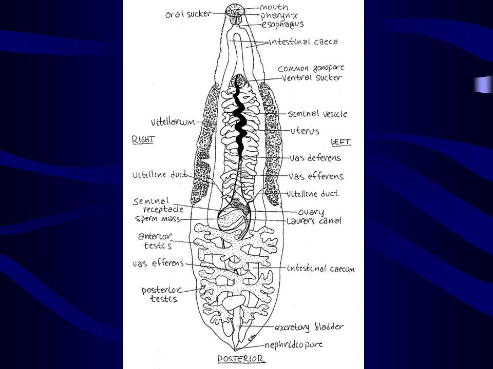
Morphology Adult worm Size & Shape like the seed of sunflower
Sucker: oral = ventral 2 dendritic testes lie in tandem to each other in the posterior region (clonorchis)
")
16
Adults of Clonorchis sinensis
Clonorchis sinensis adult Adults of Clonorchis sinensis

17
Clonorchis sinensis, adult, stained whole mount; approximate size = 15 mm. Click here to view a labeled image of this parasite, or here to view a labeled line drawing of this parasite. 来源

19
Cross section of Clonorchis sinensis adult in the hepatic bile duct
Clonorchis sinensis, liver biopsy: most infections are asymptomatic. Cross section of Clonorchis sinensis adult in the hepatic bile duct

20
Egg Size: smallest Shape: just like sesame Color: yellowish brown
Operculum distinct: shoulder, knob Content: miracidium

21
Clonorchis sinensis egg. These are small operculated eggs
Clonorchis sinensis egg. These are small operculated eggs. Size 27 to 35 µm by 11 to 20 µm. The operculum, at the smaller end of the egg, is convex and rests on a visible "shoulder". At the opposite (larger, abopercular) end, a small knob or hooklike protrusion is often visible (as is the case here). The miracidium is visible inside the egg. Clonorchis sinensis egg. These are small operculated eggs. Size 27 to 35 µm by 11 to 20 µm. The operculum, at the smaller end of the egg, is convex and rests on a visible "shoulder". At the opposite (larger, abopercular) end, a small knob or hooklike protrusion is often visible (as is the case here). The miracidium is visible inside the egg.
 end, a small knob or hooklike protrusion is often visible (as is the case here). The miracidium is visible inside the egg. Clonorchis sinensis egg. These are small operculated eggs. Size 27 to 35 µm by 11 to 20 µm. The operculum, at the smaller end of the egg, is convex and rests on a visible shoulder . At the opposite (larger, abopercular) end, a small knob or hooklike protrusion is often visible (as is the case here). The miracidium is visible inside the egg.")
22
Egg of Clonorchis sinensis
Clonorchis sinensis/ O.viverrini egg: eggs of the two species are similar. They measure by um are operculated at one end and have a small knob on the other end. The colour is yellow Egg of Clonorchis sinensis

23
Life cycle of Clonorchis sinensis
Embryonated eggs are discharged in the biliary ducts and in the stool . Eggs are ingested by a suitable snail intermediate host ; there are more than 100 species of snails that can serve as intermediate hosts. Each egg releases a miracidia , which go through several developmental stages (sporocysts , rediae , and cercariae ). The cercariae are released from the snail and after a short period of free-swimming time in water, they come in contact and penetrate the flesh of freshwater fish, where they encyst as metacercariae . Infection of humans occurs by ingestion of undercooked, salted, pickled, or smoked freshwater fish . After ingestion, the metacercariae excyst in the duodenum and ascend the biliary tract through the ampulla of Vater . Maturation takes approximately 1 month. The adult flukes (measuring 10 to 25 mm by 3 to 5 mm) reside in small and medium sized biliary ducts. In addition to humans, carnivorous animals can serve as reservoir hosts. Life cycle of Clonorchis sinensis
. The cercariae are released from the snail and after a short period of free-swimming time in water, they come in contact and penetrate the flesh of freshwater fish, where they encyst as metacercariae . Infection of humans occurs by ingestion of undercooked, salted, pickled, or smoked freshwater fish . After ingestion, the metacercariae excyst in the duodenum and ascend the biliary tract through the ampulla of Vater . Maturation takes approximately 1 month. The adult flukes (measuring 10 to 25 mm by 3 to 5 mm) reside in small and medium sized biliary ducts. In addition to humans, carnivorous animals can serve as reservoir hosts. Life cycle of Clonorchis sinensis.")
24
Life Cycle A model pattern of trematode Main points
Definitive host: human being Reservoir host: dog, cat, etc. Residing: hepatic bile duct Discharge of eggs with feces

25
Hatching in the host small intestine
2 intermediate host I: snails, such as Bithynia,Parafossarulus II: freshwater fishes, such as Cyprinus 2 generation of asexual proliferation Infective stage: metacercaria in fish Infective route: oral consumption

26
Pathogenesis Due to adult worm Mechanism Mechanical: sucker
Chemical: excretions, secretions, metabolite Biological: nutrition deprivation

27
Pathological process Inflammation Proliferation ThickeningOcclusion
Extensive involvementFibrosis of the liver

28
Clinical Manifestations
Acute stage: allergic reaction Chronic stage: functional impairment of liver (Cholangitis, Cholecystitis, Bile stone, Jaundice, etc) Advanced stage: portal cirrhosis & malignancy
 Advanced stage: portal cirrhosis & malignancy.")
29
Laboratory Diagnosis Etiological Immunological
Examination of egg in feces by sedimentation method Duodenal aspiration Immunological ELISA to detect antiboby or antigen

30
Epidemiology Distribution Far East (China, South Korea, Japan, etc.)
24 provinces in China (Guangdon: 5 million infected etc.)
")
31
Clonorchis sinensis/Opisthorchis viverrini: geographic distribution.

32
Endemic Factors Source of infection: mainly wild carnivores
I,II intermediate host in the same water-field Mode of fish breeding Dinning habit & Customs

33
Principle of Control Cure patients & carrier
praziquantel:25mg/kg, tid, 2 days Control reservoir host Carry out scientific fish-breeding Hygienic education not eating raw or undercooked fishes

34
Paragonimus westermani 卫氏并殖吸虫 Paragonimus skrjabini (Paragonimus szechuanensis) 斯氏狸殖吸虫
 斯氏狸殖吸虫")
35
The Lung Fluke Genus paragonimus Zoonotic parasite (cause zoonosis)
Animal infection> human infection 2 major species in China

36
Introduction Pathogen of lung disease Endemic hemoptysis
Favorite lodging site: lung Ectopic site: brain, abdomen, muscle, etc.

37
Morphology Adult worm Body thick (a half piece of a bean grain)
Tegument: spinous Sucker: oral = ventral Parallel arrangement of reproductive organ lobular testes (posterior) lobular ovary & uterus (anterior)
 lobular ovary & uterus (anterior)")
38
Paragonimus westermani, adult, stained whole mount; approximate size = 9 mm in length. Click here to view an image in which some of the internal organs are labeled, or click here to view a labeled line drawing 来源

39
Cross section of lung containing adult Paragonimus westermani.

40
Egg Median size, ovoid (water pot) Golden yellow
Distinctive & wide operculum Contain 1 germ cell & several yolk cells

41
Egg of Paragonimus westermani.
And a third example of a Paragonimus egg. (Original image from a Japanese language site tentatively titled Internet Atlas of Human Parasitology.) 来源 Egg of Paragonimus westermani.
 来源 Egg of Paragonimus westermani.")
42
Life cycle of Paragonimus westermani.
Crab or crayfish The eggs are excreted unembryonated in the sputum, or alternately they are swallowed and passed with stool . In the external environment, the eggs become embryonated , and miracidia hatch and seek the first intermediate host, a snail, and penetrate its soft tissues . Miracidia go through several developmental stages inside the snail : sporocysts , rediae , with the latter giving rise to many cercariae , which emerge from the snail. The cercariae invade the second intermediate host, a crustacean such as a crab or crayfish, where they encyst and become metacercariae. This is the infective stage for the mammalian host . Human infection with P. westermani occurs by eating inadequately cooked or pickled crab or crayfish that harbor metacercariae of the parasite . The metacercariae excyst in the duodenum , penetrate through the intestinal wall into the peritoneal cavity, then through the abdominal wall and diaphragm into the lungs, where they become encapsulated and develop into adults (7.5 to 12 mm by 4 to 6 mm). The worms can also reach other organs and tissues, such as the brain and striated muscles, respectively. However, when this takes place completion of the life cycles is not achieved, because the eggs laid cannot exit these sites. Time from infection to oviposition is 65 to 90 days. Infections may persist for 20 years in humans. Animals such as pigs, dogs, and a variety of feline species can also harbor P. westermani. Life cycle of Paragonimus westermani.
. The worms can also reach other organs and tissues, such as the brain and striated muscles, respectively. However, when this takes place completion of the life cycles is not achieved, because the eggs laid cannot exit these sites. Time from infection to oviposition is 65 to 90 days. Infections may persist for 20 years in humans. Animals such as pigs, dogs, and a variety of feline species can also harbor P. westermani. Life cycle of Paragonimus westermani.")
43
Life cycle Definitive host: human being
Reservoir host: carnivorous animals Habitation: lung & ectopic site Intermediate host: I: Melania snails II: stream crabs, crayfish

44
Infective stage: metacercaria
Infective mode: oral route, may via paratenic host (swine) Migration & Preadult wondering Ectopic parasitism: cerebral, abdominal,etc. Eggs discharged with sputum & feces 3 generation of asexual multiplication
 Migration & Preadult wondering. Ectopic parasitism: cerebral, abdominal,etc. Eggs discharged with sputum & feces. 3 generation of asexual multiplication.")
45
Metacercariae of Paragonimus westermani.
来源

46
Pathogenesis Stage take responsibility: adult & preadult
Pathological processes Abscess stage(脓肿期) Cystic stage(囊肿期) Scar formation stage(纤维疤痕期)
 Cystic stage(囊肿期) Scar formation stage(纤维疤痕期)")
47
4 clinical types Thoracic (pulmonary type):chest pain, coughing, blood-tinged sputum(hemoptysis) Abdominal (hepatic type):hepatomegaly Cranial type: dizzy, headache, epilepsy Musculocutaneous type: migratable subskin nodule

48
Laboratory diagnosis Disease history + physical examination
Etiological diagnosis eggs in sputum or feces by sedimentation Immunological diagnosis for ectopic infections

49
Epidemiology Global main continent except Europe China 23 provinces

50
Paragonimus westermani infection occurs in Asia (especially in China, Corea, India, Japan, Laos, Philippines, Sri Lanka, Taiwan, Thailand, Viet-Nam), Central-West Africa, South America (Ecuador, Per? Venezuela). Paragonimus westermani infection occurs in Asia (especially in China (Taiwan), Corea, India, Japan, Laos, Philippines, Sri Lanka, Thailand, Viet-Nam), Central-West Africa, South America (Ecuador, Peru Venezuela).
, Corea, India, Japan, Laos, Philippines, Sri Lanka, Thailand, Viet-Nam), Central-West Africa, South America (Ecuador, Peru Venezuela).")
51
Principle of control Treat patient: praziquantel Hygienic education
Social construction, economic refinement

52
The Ginger Fluke 姜片虫 Fasciolopsis buski 布氏姜片吸虫 Intestinal fluke 肠道吸虫

53
Morphology Adult worm Like a ginger piece Big muscular trematode
Have strong suckers,ventral >> oral

54
Adult fluke of Fasciolopsis buski
Adult fluke of Fasciolopsis buski. The adult flukes range in size: 20 to 75 mm by 8 to 20 mm. Image contributed by Georgia Division of Public Health. Adult fluke of Fasciolopsis buski The adult flukes range in size: 20 to 75 mm by 8 to 20 mm

55
Fasciolopsis buskii adult worm

56
Egg Biggest Ovoid Minute operculum Yellowish Germ cell inclusions

57
F. buski eggs are released in feces unembryonated
F.buski eggs are released in feces unembryonated. The operculated eggs are oval, brown and measure by um F.buski eggs are released in feces unembryonated. The operculated eggs are oval, brown and measure by um

58
Life cycle of Fascilopsis buski
Immature eggs are discharged into the intestine and stool . Eggs become embryonated in water , eggs release miracidia , which invade a suitable snail intermediate host . In the snail the parasites undergo several developmental stages (sporocysts , rediae , and cercariae ). The cercariae are released from the snail and encyst as metacercariae on aquatic plants . The mammalian hosts become infected by ingesting metacercariae on the aquatic plants. After ingestion, the metacercariae excyst in the duodenum and attach to the intestinal wall. There they develop into adult flukes (20 to 75 mm by 8 to 20 mm) in approximately 3 months, attached to the intestinal wall of the mammalian hosts (humans and pigs) . The adults have a life span of about one year. Life cycle of Fascilopsis buski
. The cercariae are released from the snail and encyst as metacercariae on aquatic plants . The mammalian hosts become infected by ingesting metacercariae on the aquatic plants. After ingestion, the metacercariae excyst in the duodenum and attach to the intestinal wall. There they develop into adult flukes (20 to 75 mm by 8 to 20 mm) in approximately 3 months, attached to the intestinal wall of the mammalian hosts (humans and pigs) . The adults have a life span of about one year. Life cycle of Fascilopsis buski.")
59
The adult flukes (20 to 75 mm by 8 to 20 mm) reside in the duodenum and jejunum of mammalian hosts (humans and pigs), where they are attached to the intestinal wall. Immature eggs are discharged into the intestine and stool. After development in water, each egg releases a miracidium which invades a suitable snail intermediate host. In the snail the parasites undergo several developmental stages (sporocysts, rediae, and cercariae). The cercariae are released from the snail and encyst as metacercariae on aquatic plants. The mammalian hosts become infected by ingesting metacercariae on the aquatic plants. After ingestion, the metacercariae excyst in the duodenum and attach to the intestinal wall. They develop in approximately 3 months into adults, which have a life span of one year. Fasciolopsis buski infects humans and pigs.

60
Life Cycle Definitive host: human being Reservoir host: swine, etc.
Intermediate host: Planorbis snails Aquatic plant vectors: caltrops, water chestnut, etc. Habitation: small intestine

61
Infective stage: metacercaria
Infective route: oral Developmental stages: as Paragonimus 3 generation of asexual proliferation

62
Pathogenesis Factors Traumatic (suckers) Obstructive (due large size)
Toxic (excretion, secretion, metabolite)
")
63
Clinical Manifestation
Abdominal pain Acute intestinal obstruction Anemia Generalized edema

64
Laboratory Diagnosis Examination of egg in feces by sedimentation method

65
Epidemiology Aquatic plant raising districts

66
Fasciolopsis buski: is endemic in China, Taiwan, South-East Asia, Malaysia and India.

67
Principle of Control Drug for treatment: praziquantel
Water & nightsoil control; Scientific swine raising Hygienic education

68
Introduction 6 species of human schistosomes Schistosoma japonicum
S. mansoni S. haematobium S. intercalatum S. mekongi S. malayi

69
Schistosoma japonicum
日本血吸虫

70
Distribution and Epidemic Situation
Worldwide 200 million of population infected in 74 countries (S.m. 55; S.h. 55; S.j. 4; S.i. 10; S.me. 2; S.ma. 1) China 11 million in 12 provinces 0.7 million in 8 provinces 50 years
 China. 11 million in 12 provinces 0.7 million in 8 provinces. 50 years.")
71
Regional distribution of S. japonicum infection in China
Before control After control (1996) Regional distribution of S. japonicum infection in China
 Regional distribution of S. japonicum infection in China.")
72
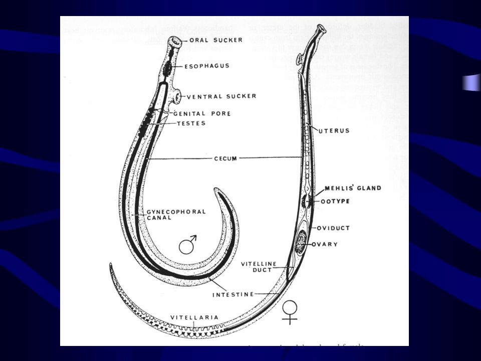
Morphology Difference from other trematodes Dioecious adults
Non-opeculate egg Bifurcated (forked) cercaria invades the final host by skin Adults parasitize blood vessels
 cercaria invades the final host by skin. Adults parasitize blood vessels.")
73
Adult Male (15 mm length) < female (22 mm)
Oral sucker < ventral sucker 2 paralleled guts form a blind caecum in the posterior ends 7 testes in male and single ovary with a tubule uterus in female Gynecophoric canal (male) in which female repose
 in which female repose.")
75
adult schistosomes live in pairs in the portal system and in mesenteric venules; adults of S.japonicum are bigger than adults of S.mansoni. males are mm in lenght and 0,5 wide, and have a ventral infolding from the ventral sucker to the posterior end forming the gynecophoric canal. Adult male with female in the copulatory groove

76
A scanning electron micrograph of schistosomes in copula

77
Egg Miracidium Cercaria Ovoid and non-opeculate 74~106 m × 55~80 m
Contains one miracidium Bear a minute lateral knob. Miracidium Cercaria

78
S. japonicum S. Mansoni S. haematobium
Schistosome egg

79
Schistosome miracidium

80
cercarae are the infective forms. They measure about 500 micron
cercarae are the infective forms. They measure about 500 micron. After encountering the skin, the cercariae penetrate and lose the tail transforming into schistosomulae Schistosome cercaria

81
Life Cycle Eggs discharged fresh water (hatch) miracidia penetrate oncomelania(I.H.)mother sporocysts (multiplication) daughter sporocysts (multiplication) cercariae (infective form) skin penetration of D.H. schistosomulum right heart lungs left heart systemic circulation portal system pairing and sexual maturation mesenteric veins lay eggs eggs develop and live in tissue for 21 days( 23% in liver tissue, 60% in intestinal tissue, 17% discharged)
 miracidia penetrate oncomelania(I.H.)mother sporocysts (multiplication) daughter sporocysts (multiplication) cercariae (infective form) skin penetration of D.H. schistosomulum right heart lungs left heart systemic circulation portal system pairing and sexual maturation mesenteric veins lay eggs eggs develop and live in tissue for 21 days( 23% in liver tissue, 60% in intestinal tissue, 17% discharged)")
83
Cercarial dermatisis due to avian schistosome
Typically, hosts of avian schistosomes are migratory water birds, including shorebirds, ducks, and geese. Adult worms are found in the blood vessels and produce eggs that are swallowed and passed in the feces. On exposure to water, the eggs hatch and liberate a ciliated miracidium that infects a suitable molluscan intermediate host. The parasite develops the in this intermediate host, usually a certain species of snail, to produce free-swimming cercariae that are released under appropriate conditions and penetrate the skin of the birds to complete the cycle. Humans are inadvertent and inappropriate hosts; cercariae may penetrate the skin but do not develop further. A number of species of dermatitis-producing cercariae have been described from both freshwater and saltwater environments, and exposure to either type of cercariae will sensitize persons to both. Cercarial dermatisis due to avian schistosome

84
Main Points of Life Cycle
Residing site: mesenteric vein I.H.(Only one): Oncomelania hupensis No metacercaria and redia stage Two generation of sporocyst Infective stage: cercaria Route of infection: skin penetration
: Oncomelania hupensis. No metacercaria and redia stage. Two generation of sporocyst. Infective stage: cercaria. Route of infection: skin penetration.")
85
Adult of Schistosome in mesenteric veins of hamster
S.mansoni : Females are slender (1 mm in diameter) and longer (9-17 mm in lenght), and are held in the ginecophoric canal during copulation. Each female lays about 300 eggs per day. Adult male with female in the copulatory groove. Adult of S.mansoni in mesenteric veins of hamster. Adult of Schistosome in mesenteric veins of hamster
 and longer (9-17 mm in lenght), and are held in the ginecophoric canal during copulation. Each female lays about 300 eggs per day. Adult male with female in the copulatory groove. Adult of S.mansoni in mesenteric veins of hamster. Adult of Schistosome in mesenteric veins of hamster.")
86
Onchomelania hupensis
intermediate host of S.japonicum are snails of the genus Onchomelania, hupensis spp. Onchomelania hupensis

87
Skin penetration of cercaria first appearance of eggs: 30~35 days
Life spans in human: 4~5 years, longest: 35 years

88
Significance of tissue egg
The egg which can develop and live in tissue Significance of tissue egg Major pathogenic stage, inflammation and granuloma around the egg Diagnosis and evaluation of therapeutic efficacy

89
Immunity Concomitant immunity
Host carrying an initial infection of adult schistosomes shows the protection to a cercarial challenge infection (攻击性感染)and this protective immunity will disappear with eradication of schistosomes in the host
and this protective immunity will disappear with eradication of schistosomes in the host.")
90
Immuno-evasion: It’s an ability by which the schistosome adult can evade the host immune response. The possible mechanism of evasion. Acquire host antigen on it surface Host-like antigen produced by parasite Changing of tegument very quickly Parasite may inactivate or down-regulate immune effectors

91
Pathogenesis Schistosomiasis at each stage of the life cycle in human body Cercaria (skin-penetration) Dermatitis Schistosomula (migration) Larva migrans Adult: (immunocomplex) Immuno-nephropathy
 Larva migrans. Adult: (immunocomplex) Immuno-nephropathy.")
92
Tissue egg: principal pathogenic stage
Miracidium within eggSEASensitization of T Cell Th1IL-2, INF-, TNF activate macrophage, induce cell-mediated immunity Th2IL-4, IL-5 stimulate IgE production or eosinophilia inflammation and granuloma fibrosis portal hypertension intestinal polyp

93
Clinical Form Acute schistosomiasis Chronic schistosomiasis
Fever, diarrhea, abdominal pain, enlargement of liver or spleen Chronic schistosomiasis asymptomatic or diarrhea, abdominal pain, enlargement of liver or spleen Advanced schistosomiasis Ascites; splenomegaly;collateral circulation; dwarfism; Ectopic lesion: encephalitis; focal epilepsy

94
Advanced schistosomiasis patient with portal hypertension and ascites
Brazilian with portal hypertension and ascites due to S.mansoni. Advanced schistosomiasis patient with portal hypertension and ascites

95
A patient with S. j has marked ascites, splenomegaly, umbilical hernia and distended superficial abdominal veins.

96
Diagnosis Parasitological diagnosis (etiological, definitive diagnosis) Demonstrating eggs by stool examination Stool examination after concentration (sedimentation) *Miracidium-hatching from eggs Rectal biopsy — eggs-demonstration
 *Miracidium-hatching from eggs. Rectal biopsy — eggs-demonstration.")
97
Miracidium-hatching from eggs
Necessity: tissue ova; low egg burden due to mass treatment Possibility: Hatch quickly (T:25; limpid water; free of Cl2;light; pH= ) Three tropism:limpidity, phototrophic, ascendancy Swimming in a zigzag way
 Three tropism:limpidity, phototrophic, ascendancy. Swimming in a zigzag way.")
98
Immunodiagnosis (indirect diagnosis)
Detection of antibodies in serum, urine, saliva by ELISA, but is impossible to distinguish current infection from past infection. Detection of antigens (circulation anodic and cathodic antigen)
")
99
Combined diagnosis: including epidemiological antecedent, symptoms and signs, parasitological and immunological examination.

100
Treatment Praziquantel 60 mg/kg divided in 3 doses.

101
Factors of Transmission and Prevention
Source of infection: patients and reservoir host Intermediate host: Oncomelania Contact with cercaria-infected water

102
Preventive measures Detection and treatment of patients and reservoir host Elimination or control of oncomelania Protection of susceptible population and avoidance of contact with cercaria-infected water Prevention of water contamination by human night soil

103
Distinguishing of 3 major schistosomes
(see page 54, Table 3-1)
")
Similar presentations


>")
>")


 Doç.Dr.Hrisi BAHAR.>")

