Download presentation
Presentation is loading. Please wait.

1
Dr.Abdulaziz Al-Abdulwahed
Endodontic Mishaps “Procedural Accidents” Causes, Prevention, Treatment and Prognosis. Dr.Abdulaziz Al-Abdulwahed

2
Introduction Collective mishaps “Procedural Accidents”, are considered as unwanted or unforeseen events that can encounter a practitioner in any complex disciplinary of dentistry. Knowledge of factors, prevention, effect on prognosis must be in mind. Sticking with the basic principles of diagnosis, case selection, treatment planning and operative procedures (from access cavity to obturation and post preparation will ensure the prevention of most accidents.

4
In case of an accident occurs:
Inform the patient about the accident. Explain how it can be corrected. Telling the patient about alternative modalities of treatment. Clarifying the effect of this accident on prognosis. Proper documentation is necessary. Knowing your limitations as a practitioner.

5
Perforations during access cavity
Causes In most cases, the pulp chamber is located in the center of the anatomic crown and the canal extends parallel to the long axis of the tooth. Lack of attention in the inclination of tooth in relation to adjacent tooth or alveolar bone will cause gouging and perforation. Failure to orient the bur parallel to the long axis of the tooth will cause gouging and perforation. That can happen most of the time with indirect approaches. Trans-illumination, magnification, radiographs all aiding means of guiding to a proper access.

8
Causes Failure to recognize when the bur passes the pulp chamber (small or flattened), especially in multi-rooted teeth will cause furcation perforation. Prevention Knowledge of internal and surface anatomy and their relationship. Location and angulation comparing to adjacent tooth or alveolar bone to avoid misaligned preparation. Radiographs: multiple radiographs with different horizontal angulations.
, especially in multi-rooted teeth will cause furcation perforation. Prevention. Knowledge of internal and surface anatomy and their relationship. Location and angulation comparing to adjacent tooth or alveolar bone to avoid misaligned preparation. Radiographs: multiple radiographs with different horizontal angulations.")
11
Operative procedures Sometimes access cavity can be started without rubber dam when problems are anticipated with tilted or misaligned teeth. During this procedure files should be secured with floss. Then after finishing the access cavity, rubber dam is placed. Failure to recognize that the bur has passed the pulp chamber especially in constricted or calcified pulp chambers >>> safe ended access bur, Endo Z bur. Using apex locators and angulated radiographs. Another method of isolation is “split dam”. Radiographing a bur placed in the preparation and secured with cotton pellets, beware of patient aspiring or swallowing it. Using fiberopitc lights. As beam is directed through the access opening (reflected light) and illuminates the pulp chamber floor (transmitted light) which will appear dark spotted. The usage of magnification loops (3x).
 and illuminates the pulp chamber floor (transmitted light) which will appear dark spotted. The usage of magnification loops (3x).")
14
Recognition and treatment
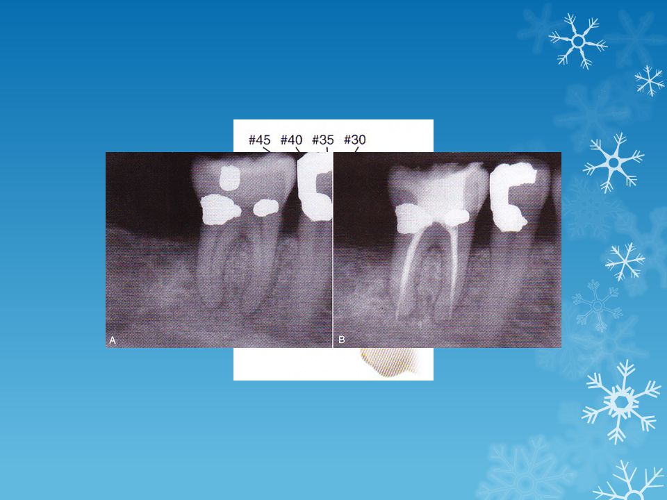
Injury to the PDL and alveolar bone may give continuous bleeding, sometimes paper points tends to ‘refresh’ the bleeding process. Perforations can be recognized by: Sudden pain during working length determination, especially when profound anesthesia was established. Sudden appearance of hemorrhage. Burning pain, or bad taste with irrigation (NaOCl). False reading of the apex locator with the initial file entry. Or malpositioned file appears in radiograph. Evaluation of the prognosis, with factors in mind: size, location, extent, and the ability to be repaired.
. False reading of the apex locator with the initial file entry. Or malpositioned file appears in radiograph. Evaluation of the prognosis, with factors in mind: size, location, extent, and the ability to be repaired.")
15
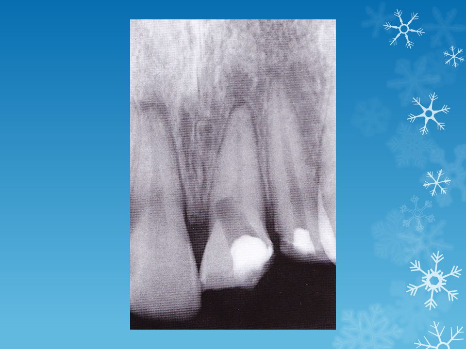
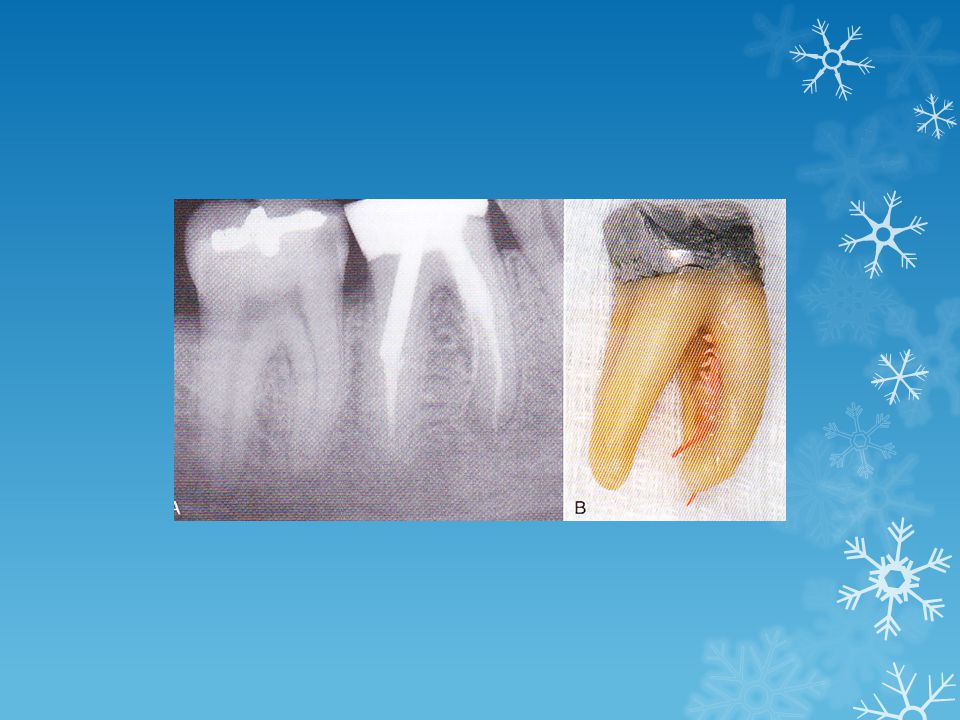
Lateral root perforation
If the location of the defect is at or above the height of crestal bone, repairing with amalgam or GIC, or crowns with subgingival margins. If located below the height of crestal bone, MTA (Mineral Trioxide Aggregate) is a material of choice in sealing perforation site internally. Orthodontic extrusion, crown lengthening are other alternatives. Furcation Perforation Direct: punched out appearance, caused by burs in order to find canal orifices. Easily accessible, and with immediate repair with MTA, or composite, GIC will have favorable prognosis. Stripping: furcation side of the coronal part of the root, due to excessive flaring of the walls, difficult to be accessed, with negative prognosis compared with the direct type, due to the inflammatory process and the development of a perio pocket. MTA is the material of choice in sealing the perforation site.
 is a material of choice in sealing perforation site internally. Orthodontic extrusion, crown lengthening are other alternatives. Furcation Perforation. Direct: punched out appearance, caused by burs in order to find canal orifices. Easily accessible, and with immediate repair with MTA, or composite, GIC will have favorable prognosis. Stripping: furcation side of the coronal part of the root, due to excessive flaring of the walls, difficult to be accessed, with negative prognosis compared with the direct type, due to the inflammatory process and the development of a perio pocket. MTA is the material of choice in sealing the perforation site.")
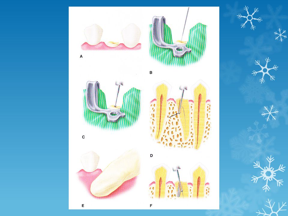
21
Nonsurgical treatment
Especially with furcation perforation is preferred when it’s possible. Amalgam, Cavit, ZOE, Calcium Hydroxide all have been used and evaluated clinically. MTA immediate repair offers one of the best results. Surgical treatment Hemisection, Premolarization (Bicuspidization). Especially with divergent multirooted teeth. Root amputation Intentional replantation. Prognosis Factors are: location of the defect, size, time elapsed from accident to repair, periodontal status, length of the root trunk and skills of the practitioner. Early detection will favor prognosis. Late detection >> periodontal communication, pocket formation.
. Especially with divergent multirooted teeth. Root amputation. Intentional replantation. Prognosis. Factors are: location of the defect, size, time elapsed from accident to repair, periodontal status, length of the root trunk and skills of the practitioner. Early detection will favor prognosis. Late detection >> periodontal communication, pocket formation.")
23
Accidents during cleaning and shaping
Ledge formation Occurs when working length cannot be negotiated and the patency of the canal is lost. Factors: No straight access. Inadequate lubrication or irrigation. Excessive enlargement of curved canals with larger files (especially Stainless Steel). Packing debris in the apical portion of the canal.
. Packing debris in the apical portion of the canal.")
24
How to prevent ledge formation?
Peroperative radiographic evaluation (curves, length, initial size). Curvatures: Apical ledging is more associated with curved canals in the coronal third. After achieving straight line access, coronal flaring is required before the negotiation of the apical area. Length: Longer canals are more prone for ledging than shorter canals. Initial size: Smaller diameter canals are more prone to be ledged than wider diameter canals. The Challenging canal is small in diameter, long in length and curved.
. Curvatures: Apical ledging is more associated with curved canals in the coronal third. After achieving straight line access, coronal flaring is required before the negotiation of the apical area. Length: Longer canals are more prone for ledging than shorter canals. Initial size: Smaller diameter canals are more prone to be ledged than wider diameter canals. The Challenging canal is small in diameter, long in length and curved.")
25
Determination of working length continuously is vital
Determination of working length continuously is vital. Frequent irrigation and recapitulation as well as using lubricants all minimize ledge formation. NaOCl has some lubricating properties, but can be enhanced with e.g. EDTA in water soluble paste (RC prep) or glycol or glycerin. In addition using Nickel Titanium files will reduce ledge formation due to increased flexibility and decreased stress . Anticurvature filing. Balanced force technique. Initial preparation of the apical third can be achieved with passive step back technique with a selection of a small MAF to begin with, as this will reduce stress on files. If using stainless steel files, precurve the file before insertion, and using copious amount of irrigation and lubricants is highly recommended.
 or glycol or glycerin. In addition using Nickel Titanium files will reduce ledge formation due to increased flexibility and decreased stress . Anticurvature filing. Balanced force technique. Initial preparation of the apical third can be achieved with passive step back technique with a selection of a small MAF to begin with, as this will reduce stress on files. If using stainless steel files, precurve the file before insertion, and using copious amount of irrigation and lubricants is highly recommended.")
27
Management of ledge Prognosis Once formed it is difficult to correct.
1st bypass attempt, by placing No. 10 file (with the 2- 3 mm apical end of the is sharply bent) to negotiate the original canal and to reestablish working length. Lubricants are helpful. After engaging the canal (by pecking motion). Quick reaming and filing motion is done with NaOCl flushing. 2nd by pass attempt if the 1st one fails. Progressively flaring the coronal and middle third with larger files. After that a smaller file (smaller than MAF) is inserted in a pecking motion to engage the original canal. If all attempts fail. Obturation to the point of ledge, with recall of the case Prognosis Depends on the amount of debris, and the area of uninstrumented canal. Short cleaned ledges have good pronosis. Recall appointments.
 to negotiate the original canal and to reestablish working length. Lubricants are helpful. After engaging the canal (by pecking motion). Quick reaming and filing motion is done with NaOCl flushing. 2nd by pass attempt if the 1st one fails. Progressively flaring the coronal and middle third with larger files. After that a smaller file (smaller than MAF) is inserted in a pecking motion to engage the original canal. If all attempts fail. Obturation to the point of ledge, with recall of the case. Prognosis. Depends on the amount of debris, and the area of uninstrumented canal. Short cleaned ledges have good pronosis. Recall appointments.")
29
Creating artificial canals
The same factors that leads to the formation of ledge. As an attempt form the operator to tread files apical to gain back working length. It is very difficult to repair. If succeeded in the negotiation of the original canal, cleaning and shaping is continued. In case of obturating the canal, evaluation of perforation must be performed, by the use of apex locators, hemorrhage control by paper points. If perforated an apical stop with a large file is created, then obturation begins. Recall appointments. >>> Surgery. Prognosis Depends on the ability to renegotiate the original canal as well as instrumenting this part and obturating it.

30
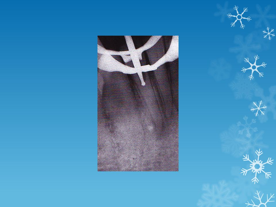
Apical root perforations
Occur through the apical foramen or through the newly created canal. Overinstrumentation beyond the apical foramen. Zipping and apical transportation and caused when files’ (especially stainless steel ) restoring force exceeds the threshold of cutting dentin in a cylindrical shaped curved canal. Apical transportation involves moving the apical foramen from a physiologic to an iatrogenic position. Apical transportation categorized in three types Types I, II and III. To prevent apical perforation proper working length must be established and maintained. Not to use larger files for curved canals to the WL. To correct this situation an apical seat is formed and new WL is established by 1-2 mm. an MTA is placed prior to obturation to serve as a barrier. Prognosis depends upon the size and shape of the defect, as type I has a more favorable prognosis.
 restoring force exceeds the threshold of cutting dentin in a cylindrical shaped curved canal. Apical transportation involves moving the apical foramen from a physiologic to an iatrogenic position. Apical transportation categorized in three types Types I, II and III. To prevent apical perforation proper working length must be established and maintained. Not to use larger files for curved canals to the WL. To correct this situation an apical seat is formed and new WL is established by 1-2 mm. an MTA is placed prior to obturation to serve as a barrier. Prognosis depends upon the size and shape of the defect, as type I has a more favorable prognosis.")
32
Lateral perforations The same reasons that causes apical perforation can be applied on lateral (midroot perforation). Using large and stiffed files on curved canals. In addition, applying misdirected forces and not inserting files in a passive way. Indicators for lateral perforation are the same for apical perforation (fresh hemorrhage on files, pain with cleaning and shaping with a previously non-symptomatic tooth, deviation of files radiographically). To treat this condition: perform bypassing the perforated area with smaller files. If failed, concentrating on cleaning and shaping the coronal portion and using low concentration of NaOCl. Prognosis depends on the size of the uninstrumented part of the canal, with diificulty of creating an apical stop, thus extruding obturating materials. Accessibility plays an important role in prognosis. Thus, facial perforation are more easy to be repaired. Recall appointments with its procedures are essential in following up cases.
. To treat this condition: perform bypassing the perforated area with smaller files. If failed, concentrating on cleaning and shaping the coronal portion and using low concentration of NaOCl. Prognosis depends on the size of the uninstrumented part of the canal, with diificulty of creating an apical stop, thus extruding obturating materials. Accessibility plays an important role in prognosis. Thus, facial perforation are more easy to be repaired. Recall appointments with its procedures are essential in following up cases.")
34
Coronal root perforations
Occur during preparation with files, G.G, and Peeso Reamers. Good illumination and cautious exploration of calcified canals will prevent these kind of situations. Careful (step-back or passive step-back) instrumentation during cleaning and shaping. The accessibility of stripping perforations in coronal part of the root is challenging. Therefore, have the poorest prognosis of all root perforations. An attempt to fix this issue internally is preferred, even though prognosis is guarded. Poorest prognosis.
 instrumentation during cleaning and shaping. The accessibility of stripping perforations in coronal part of the root is challenging. Therefore, have the poorest prognosis of all root perforations. An attempt to fix this issue internally is preferred, even though prognosis is guarded. Poorest prognosis.")
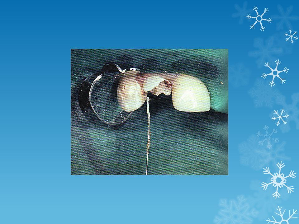
36
Separated Instruments
Limited flexibility (especially Stainless Steel Files). S.S. or Nickel Titanium files, rotary or hand files may get separated. Improper use (excessive force, overuse). Detecting it with blunt end of the withdrawing file. Loss of patency of the canal. Inform the patient. Continuous lubrication, irrigation, recognition of twisted or defected files are necessary precautions. Working the canal with files until it is loose, then use the next larger file.
. S.S. or Nickel Titanium files, rotary or hand files may get separated. Improper use (excessive force, overuse). Detecting it with blunt end of the withdrawing file. Loss of patency of the canal. Inform the patient. Continuous lubrication, irrigation, recognition of twisted or defected files are necessary precautions. Working the canal with files until it is loose, then use the next larger file.")
37
Treating this condition by attempting to bypass the separated part then removing it with Hedstrom files or ultrasonics. Or cleaning and shaping then obturating to the area.

38
Aspiration or ingestion:
Considered a very serious event. Preventing it by obligatory use of rubber dam. Forceful irrigation of NaOCl causes penetration into the PA regions causing inflammatory process and pain or discomfort. Loose irrigation is a must.

39
Accidents during obturation: Underfilling:
Causes may include: improper canal shaping and flaring, ledging, poor adaptation of master cone. To treat this issue: removal of underfilled gutta percha and reatreatment. Overfilling: Caused by overinstrumentation, apex widely open, faulty selection of MAC with no tug-back. To treat this issue: apical surgery to remove excess GP and using retrograde filling (e.g. MTA) Vertical root fracture: Faulty post preparation technique, excessive condensation forces. Usually vertical fracture is associated with focal deep periodontal pocket (in one area) as well as lateral radiolucency. Poorest prognosis. Extraction or root amputation.
 Vertical root fracture: Faulty post preparation technique, excessive condensation forces. Usually vertical fracture is associated with focal deep periodontal pocket (in one area) as well as lateral radiolucency. Poorest prognosis. Extraction or root amputation.")
40
Accidents during post space preparation:
Perforation due to faulty technique in removing GP. Appearing with fresh blood on site. Radiographs should be reviewed. Poor (hopeless prognosis). Surgical management is required if post cannot be removed. If post can be removed a non surgical approach can be done. Follow up these cases for the determination of repair success.
. Surgical management is required if post cannot be removed. If post can be removed a non surgical approach can be done. Follow up these cases for the determination of repair success.")
41
References M.Torabinejad, Walton. Endodontics, Principals and Practice. 4th Ed.

Similar presentations