Download presentation
Presentation is loading. Please wait.

1
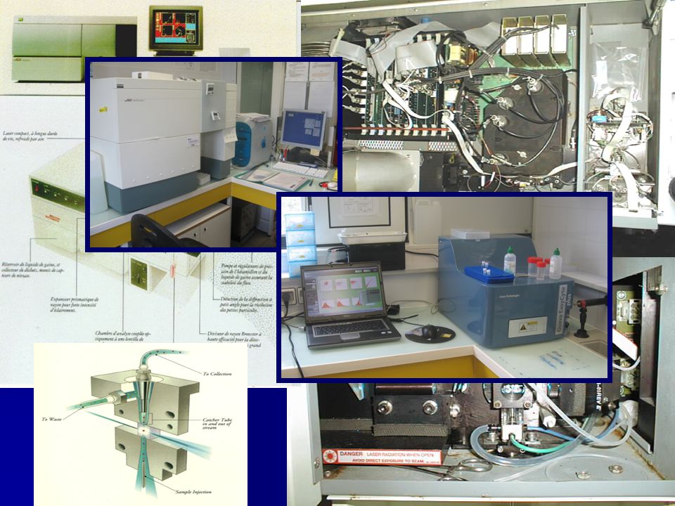
Introduction to Flow cytometry and applications Introduction to Flow cytometry and applications First part : principles First part : principles Second part : main applications Second part : main applications –Focused on LEMAR applications Third part : available equipments in the lab Third part : available equipments in the lab –FACScalibur Becton-Dickinson –GUAVA EasyCyte plus –Utilisation rules Formation à la cytométrie en flux – 9 novembre 2011 C. Lambert, P. Soudant, N. Le Goïc, C. Paillard, H. hégaret, L. donaghy, M. Auffret

2
What is flow cytometry? Measurement of cell characteristics in a stream of fluid A laser beam is focused on the moving cells Scattered light and emitted fluorescence are detected and converted in an electronic pulse

3
Basic Scheme

4
Classical flow cytometer is combining three systems 1- Fluidics 1- Fluidics –introduction of the cells in the analyze chamber 2 - Optics 2 - Optics –Production and collection of scattered light and emitted fluorescence 3 - Electronics 3 - Electronics –Conversion to digital values and storage on a computer for data analysis

5
1-Fluidics-FACSCalibur TM Need to have cells in suspension flow in single file through an illuminated volume

6
Fluidics: hydrodynamic focusing Accomplished by injecting sample into a sheath fluid as it passes through a small (50-300µm) orifice (laminar coaxial flow - Bernoulli Effect) needle Facs : 80 µm Capillary Guava : 100 µm 12 µL/min 60 µL/min
 orifice (laminar coaxial flow - Bernoulli Effect) needle Facs : 80 µm Capillary Guava : 100 µm 12 µL/min 60 µL/min")
7
Laminar Coaxial Flow Direction of flow The Bernoulli Effect Velocity Gradient Viscous drag along walls. Sheath fluid hoy@cf.ac.uk Lower pressure Particles move to low pressure area Fluidics: hydrodynamic focusing

8
New technologies : without fluid Capillary

9
A suspension of single cells or other particles in a suitable buffer, usually PBS (phosphate buffer saline) Typical density : 10 5 - 10 7 cells / ml (FACS) 10 4 - 5.10 5 (Guava) Acquisition speeds :up to 1000 events / sec typical 300/800 (FACS) (max 500 : Guava) Particles size (range) : from ~1 µm to ~40/50 µm hoy@cf.ac.uk Fluidics: Sample requirements for flow cytometry
 Typical density : cells / ml (FACS) (Guava) Acquisition speeds :up to 1000 events / sec typical 300/800 (FACS) (max 500 : Guava) Particles size (range) : from ~1 µm to ~40/50 µm Fluidics: Sample requirements for flow cytometry")
10
Suspensions: straightforward samples Non-adherent cell cultures Circulating cells (blood cells, haemocytes in haemolymph... Water borne micro-organisms (zooplankton, phytoplankton… Suspensions of bacteria, yeasts, viruses. hoy@cf.ac.uk Fluidics : Sample preparation for flow cytometry

11
Sources requiring more preparation Adherent cultures and solid tissues have to be processed to release cells as a suspension Enzymatic digestion : trypsin, collagenase, pronase. Chelating agents - removal of ‘binding’ ions : EDTA, EGTA. Mechanical : teasing, sieving, aspiration (syringing) and sonication. Chromosomes can be released from mitotic cells Nuclei can also be recovered from tissues stored in paraffin blocks hoy@cf.ac.uk Fluidics : Sample preparation for flow cytometry.
 and sonication. Chromosomes can be released from mitotic cells Nuclei can also be recovered from tissues stored in paraffin blocks Fluidics : Sample preparation for flow cytometry..")
12
2- Optics Light source (excitation) is constituted of Light source (excitation) is constituted of –Laser beam (blue laser 488nm) –lens and prisms allow to focus the laser beam Optical reception is constituted of Optical reception is constituted of –a mirror system and optical filters which direct specific wavelengths to the corresponding detector
 is constituted of Light source (excitation) is constituted of –Laser beam (blue laser 488nm) –lens and prisms allow to focus the laser beam Optical reception is constituted of Optical reception is constituted of –a mirror system and optical filters which direct specific wavelengths to the corresponding detector")
13
Dichroic mirrors 1 2 3 Band Pass Filters Photo- multiplier tubes Laser(s) Cell Collection Lenses Scatter Low & High angle hoy@cf.ac.uk 2- Optics : Basic Optics of a Flow Cytometer
 Cell Collection Lenses Scatter Low & High angle 2- Optics : Basic Optics of a Flow Cytometer")
15
FACS Academy 2- Optics: What kind of information are provided by a flow cytometer? Relative size (Forward Scatter- FSC) : diffracted light on the small angle Relative size (Forward Scatter- FSC) : diffracted light on the small angle FSC Laser Nbr of events Invitrogen Facs academy
 : diffracted light on the small angle Relative size (Forward Scatter- FSC) : diffracted light on the small angle FSC Laser Nbr of events Invitrogen Facs academy.")
16
2- Optics: What kind of information are provided by a flow cytometer? Internal or complexity relative ‘ granularity ’ Internal or complexity relative ‘ granularity ’ (Side Scatter-SSC) : diffracted light on the right angle Laser SSC Invitrogen Nbr of events
 : diffracted light on the right angle Laser SSC Invitrogen Nbr of events.")
17
FACS Academy Relative intensity of fluorescence Relative intensity of fluorescence –FL1 (530 / 30 nm, BP – 525/30 nm BP ) –FL2 (585 / 42 nm, BP – 583/ 26 nm BP) –FL3 (> 670nm, LP – 680/30 nm BP ) 2- Optics: What kind of information are provided by a flow cytometer? Site BDSite BD/ Fluorescence Spectrum Viewer

18
2- Optics: What kind of information are provided by a flow cytometer? Possibility to use several fluorochromes with different emission wavelengths. Possibility to use several fluorochromes with different emission wavelengths. Great diversity of fluorescent probes are Great diversity of fluorescent probes are now available

19
Fluorescence is the property of a molecule to absorb light of a particular wavelength and re-emit light of a longer wavelength. Fluorescence is the property of a molecule to absorb light of a particular wavelength and re-emit light of a longer wavelength. Relative intensity of fluorescence (FL1, FL2, FL3)
.")
20
The intensity of the fluorescence is proportional to the number of antibody fixation sites. The intensity of the fluorescence is proportional to the number of antibody fixation sites. Relative intensity of fluorescence (FL1, FL2, FL3) Applications: Applications: Cell cycle and ploïdy analysis using DNA staining Cell cycle and ploïdy analysis using DNA staining Cell viability, cell functions Cell viability, cell functions Enzymatic activities Enzymatic activities Quantification of pigments Quantification of pigments etc.. etc..
 Applications: Applications: Cell cycle and ploïdy analysis using DNA staining Cell cycle and ploïdy analysis using DNA staining Cell viability, cell functions Cell viability, cell functions Enzymatic activities Enzymatic activities Quantification of pigments Quantification of pigments etc.. etc...")
21
3- Electronics Creation of a pulse : conversion of the optical signals into proportional electronic signal (pulse) Creation of a pulse : conversion of the optical signals into proportional electronic signal (pulse)
 Creation of a pulse : conversion of the optical signals into proportional electronic signal (pulse)")
22
Analyze height, width, and area of the pulse Analyze height, width, and area of the pulse Computer interface for data processing Computer interface for data processing Listed and displayed as Flow Cytometry Standard (FCS) Files Listed and displayed as Flow Cytometry Standard (FCS) Files 3- Electronics
 Files Listed and displayed as Flow Cytometry Standard (FCS) Files 3- Electronics")
23
Interpreting Multi-parameter Data Single parameters (univariate) can be displayed as a histogram. Dual parameter (bivariate) data can be displayed in two dimensions using dot or density plots. Three parameters may be displayed as a 3-D projection, these are not always easy to interpret and can often be ambiguous.
 data can be displayed in two dimensions using dot or density plots. Three parameters may be displayed as a 3-D projection, these are not always easy to interpret and can often be ambiguous..")
24
Introduction to Flow cytometry (Invitrogen ~12 min video) http://probes.invitrogen.com/resources/education/t utorials/4Intro_Flow/player.html http://probes.invitrogen.com/resources/education/t utorials/4Intro_Flow/player.html http://probes.invitrogen.com/resources/education/t utorials/4Intro_Flow/player.html http://probes.invitrogen.com/resources/education/t utorials/4Intro_Flow/player.html
 utorials/4Intro_Flow/player.html utorials/4Intro_Flow/player.html utorials/4Intro_Flow/player.html utorials/4Intro_Flow/player.html")
25
Complementary notions Fluorescence compensation Fret

26
FITCPE Compensation is the process by which the fluorescence “spillover” originating from a fluorochrome other than the one specified for a particular PMT detector is subtracted as a percentage of the signal from other PMT’s.

27
FRET Fluorescence Resonance Energy Transfer If two fluorescent molecules with suitable spectral overlap are in close proximity, then the energy from the excited state of one molecule (the DONOR) can be transferred to the second molecule (the ACCEPTOR), absorbed and then re-emitted.
 can be transferred to the second molecule (the ACCEPTOR), absorbed and then re-emitted.")
28
FRET Fluorescence Resonance Energy Transfer Typical donor/acceptor pairs: –Fluorescein / rhodamine –Fluorescein / eosine –Fluorescein / pyrenebutyrate –Anthranilamide / nitrotyrosine –Coumarin / ethidium bromide Utilisation: detect if two labelled protein or nucleic acids come into contact or a doubly labelled single molecules is hydrolysed detect changed in conformation measure concentration by a competitive binding assay

29
Advantages of FCM High speed analysis High speed analysis Multi-parameter data acquisition Multi-parameter data acquisition Cell sorting Cell sorting

30
Introduction to Flow cytometry (Invitrogen ~12 min video) http://probes.invitrogen.com/resources/education/t utorials/4Intro_Flow/player.html http://probes.invitrogen.com/resources/education/t utorials/4Intro_Flow/player.html http://probes.invitrogen.com/resources/education/t utorials/4Intro_Flow/player.html http://probes.invitrogen.com/resources/education/t utorials/4Intro_Flow/player.html
 utorials/4Intro_Flow/player.html utorials/4Intro_Flow/player.html utorials/4Intro_Flow/player.html utorials/4Intro_Flow/player.html")
31
Cellular parameters measurable by flow cytometry. Without probes : size, shape, cytoplasmic granularity, autofluorescence and pigmentation. With probes : DNA content, DNA composition, DNA synthesis, chromatin structure, RNA, protein, sulphydryl groups, antigens (surface, cytoplasmic & nuclear), lectin binding sites, cytoskeletal components, membrane structure (potential, permeability & fluidity), enzyme activity, endocytosis, surface charge,receptors, bound and free calcium, apoptosis, necrosis, pH, drug kinetics, etc... hoy@cf.ac.uk Part II : Applications of Flow Cytometry
, lectin binding sites, cytoskeletal components, membrane structure (potential, permeability & fluidity), enzyme activity, endocytosis, surface charge,receptors, bound and free calcium, apoptosis, necrosis, pH, drug kinetics, etc... Part II : Applications of Flow Cytometry.")
32
Most frequent applications in marine biology and oceanography Discrimination and identification of different cell types in various matrix Discrimination and identification of different cell types in various matrix Assessment of cell viability, cell cycle, physiological status, cell biochemical composition Assessment of cell viability, cell cycle, physiological status, cell biochemical composition Cell sorting coupled to cell culture or other assays (PCR) Cell sorting coupled to cell culture or other assays (PCR)
 Cell sorting coupled to cell culture or other assays (PCR)")
33
Applications in marine biology and oceanography Cell counting (hemocytes, phytoplancton, bacteria, viruses…) Cell counting (hemocytes, phytoplancton, bacteria, viruses…) Cell morphology (size / complexity) Cell morphology (size / complexity) Autofluorescence (pigments: chlorophyll, phycoerythrin… Autofluorescence (pigments: chlorophyll, phycoerythrin… Mortality / viability Mortality / viability Mmembrane Integrity (propidium iodide; SytoxGreen®)Mmembrane Integrity (propidium iodide; SytoxGreen®) Enzymatic Activities (DCFH-DA)Enzymatic Activities (DCFH-DA) DNA content (Sybr-green®, propidium iodide ) DNA content (Sybr-green®, propidium iodide ) Oxidative Activity Oxidative Activity Apoptosis Apoptosis mitochondrial membrane potential mitochondrial membrane potential …
 Cell counting (hemocytes, phytoplancton, bacteria, viruses…) Cell morphology (size / complexity) Cell morphology (size / complexity) Autofluorescence (pigments: chlorophyll, phycoerythrin… Autofluorescence (pigments: chlorophyll, phycoerythrin… Mortality / viability Mortality / viability Mmembrane Integrity (propidium iodide; SytoxGreen®)Mmembrane Integrity (propidium iodide; SytoxGreen®) Enzymatic Activities (DCFH-DA)Enzymatic Activities (DCFH-DA) DNA content (Sybr-green®, propidium iodide ) DNA content (Sybr-green®, propidium iodide ) Oxidative Activity Oxidative Activity Apoptosis Apoptosis mitochondrial membrane potential mitochondrial membrane potential …")
34
Bivalve Hemocytes

35
Detection – counting bivalves hemocytes Distinction of hemocyte subpopulations based on their size (FSC) and complexity (SSC) granulocytes Hyalinocytes small agranulocytes
 and complexity (SSC) granulocytes Hyalinocytes small agranulocytes")
36
R1 = hemocyte aggregated R2 = hemocytes (one cell) R3 = sperm Other = cell debris and unknow stuff Red = aggregates Green = one cell Blue = sperm Not that simple ! It requires DNA staining with SYBRgreen (fluorescein) of oyster hemocytes granulocytes Hyalinocytes small agranulocytes Gate : R2 Gate : R1*R2*R3
 of oyster hemocytes granulocytes Hyalinocytes small agranulocytes Gate : R2 Gate : R1*R2*R3.")
37
Hemocyte mortality rate Prodidium iodide (PI) : Prodidium iodide (PI) : PI is incorporated only in cells which had lost their membrane integrity = dead cells with a orange/red-fluorescence Orange/ red Fluorescence (PI)
 : Prodidium iodide (PI) : PI is incorporated only in cells which had lost their membrane integrity = dead cells with a orange/red-fluorescence Orange/ red Fluorescence (PI)")
38
Hemocyte viability Double staining (SYBR-green + propidium iodide ) Live hemocytes Dead hemocytes Dead + live hemocytes
 Live hemocytes Dead hemocytes Dead + live hemocytes")
39
Intracellular Oxidative Activity DCFH-DA (dichlorofluorescein diacetate) (485 nm, 535 nm) DCFH-DA is membrane permeant, within the cells the diacetate group is enzymatically cleaved off (esterase). Both DCFH-DA and DCFH is non- fluorescent. In presence of oxidative activity, they are oxidized to highly fluorescent dichlorofluorescein. In presence of oxidative activity, they are oxidized to highly fluorescent dichlorofluorescein.

40
Intracellular Oxidative Activity Anion superoxide : O 2 - Anion superoxide : O 2 - Peroxyde d’hydrogène : H 2 O 2 Peroxyde d’hydrogène : H 2 O 2 Peroxynitrite (NO 3 - ) Oxyde nitrique (NO) Enzymes : – –peroxydase – –xanthine oxydase – –lipoxygenase cytochrome c
 Oxyde nitrique (NO) Enzymes : – –peroxydase – –xanthine oxydase – –lipoxygenase cytochrome c")
41
Intracellular Oxidative Activity increasing IOA granulocytes hyalinocytes agranulocytes Fluorochrome = DCFH-DA (green fluorescence - FL1)
")
42
IOA of hemocyte from sensitive ’S’ oysters is higher than the one from resistant ‘R’ oysters during gametogenesis. gametogenesis Genetic studies: Effect of genetic stock on oyster immune system and physiological status; Lab experiment Activité oxydative intracellulaire (fluorescence verte DCF – FL1)
.")
43
Phagocytosis Principle/ Method: hemocytes Principle/ Method: hemocytes –Use of fluorescent latex beads 2 µm: Detectable on the FL1 detector of the flow cytometer. Detectable on the FL1 detector of the flow cytometer. –Contact between beads and cells (some hours) –Estimate the % of active cells (index)
 –Estimate the % of active cells (index).")
44
Phagocytosis FL1 events Histogram

45
Phagocytosis 2/3 hours After addition of fluorescent beads 2/3 hours After addition of fluorescent beads Beads only R1 Gate R1

46
Phagocytosis Cytochalasin BNo cytochalasin

47
Phagocytosis index: % of active cells (more than 3 beads)
")
48
Result : interaction between toxin from algae and clam hemocytes. Ford, Bricelj, Lambert, Paillard, 2008 (Mar Biol 154, 241-253)
.")
49
Fluorescent probe: LysoTracker® red Freely membrane permeant probe that accumulates in intracytoplasmic lysosomal compartments L ysosomes: - intracytoplasmic acidic organelles which contain hydrolases - can fuse with phagocytic vacuoles (phagosomes) - resulting intra-vacuolar acidification and contact of lysosomal enzymes with ingested material favors its degradation. Donaghy et al., In preparation Low Medium High Distinction between at least 3 hemocyte populations displaying low, medium and high amounts of intracytoplasmic lysosomes. Differential involvement of hemocyte subtypes in mollusc immune response ? Example: Turbo cornutus hemocytes lysosomal amount Lysosomes (Lysotracker red)
.")
50
Mitochondrial membrane potential The electron chain transport, responsible of the ATP production, generate a strong electrochemical gradient across the membrane (negative charge inside). The electron chain transport, responsible of the ATP production, generate a strong electrochemical gradient across the membrane (negative charge inside).electrochemical gradientelectrochemical gradient T his gradient is an indicator of the mitochondrion functionality and of its energetic status. T his gradient is an indicator of the mitochondrion functionality and of its energetic status. To estimate this gradient : use of JC-10 probe (~JC-1) To estimate this gradient : use of JC-10 probe (~JC-1)
.electrochemical gradientelectrochemical gradient T his gradient is an indicator of the mitochondrion functionality and of its energetic status. T his gradient is an indicator of the mitochondrion functionality and of its energetic status. To estimate this gradient : use of JC-10 probe (~JC-1) To estimate this gradient : use of JC-10 probe (~JC-1).")
51
Mitochondrial membrane potential (MMP) JC-10 probe: two iso-forms - Monomer = green fluo (low MMP) - Agregates = orange fluo (high MMP) (1) Cytoplasm loading
 JC-10 probe: two iso-forms - Monomer = green fluo (low MMP) - Agregates = orange fluo (high MMP) (1) Cytoplasm loading")
52
Mitochondrial membrane potential (MMP): oyster Green fluo : monomer Orange fluo : agregates + cccp : ratio ~1 CCCP : Carbonyl cyanide 3-chlorophenylhydrazone : decoupling agent Data from Sébastien Artigaud (Master 2-2010) Ratio green/ orange > 4
: oyster Green fluo : monomer Orange fluo : agregates + cccp : ratio ~1 CCCP : Carbonyl cyanide 3-chlorophenylhydrazone : decoupling agent Data from Sébastien Artigaud (Master ) Ratio green/ orange > 4")
53
DNA content and cell cycle DNA content of normal cockle hemocytes

54
Normal Haemocytes Neoplastic Haemocytes Photos: A. Villalba, Spain DNA content analysis of neoplastic cells 4N <2N

55
3N 4N Examples of heavily diseased cockles 5N DNA content analysis of neoplastic cells in haemolymph of the cockle Cerastoderma edule. 8N 7N 6N

56
Phytoplancton

57
Detection – counting of phytoplancton Based on the fluorescence of pigments (chlorophyll) Based on the fluorescence of pigments (chlorophyll) Karenia mikimotoi
 Based on the fluorescence of pigments (chlorophyll) Karenia mikimotoi")
58
1) Detection FSC Karenia debris Size (FSC) Complexity (SSC) Statistics : n = 6
 Detection FSC Karenia debris Size (FSC) Complexity (SSC) Statistics : n = 6")
59
1) Detection FL3 Kareniadebris Size (FSC) FL3 (chlorophyll) Statistics : n = 4
 Detection FL3 Kareniadebris Size (FSC) FL3 (chlorophyll) Statistics : n = 4")
60
Nano-pico plancton / cyanobacteria phycoerythrine autofluorescence (orange) Chlorophyll autofluorescence (red)
 Chlorophyll autofluorescence (red)")
61
Station 4 Station 3Station 2 Station 1 Charente estuary

62
Ex 2 : Detection of mixture of 8 eukaryotic cell types + 2 cyanobacteria D’après Marie et al., 2004

63
Ex 3 : coccolithophore counting, « Size », chlorophyll level. Gephyrocapsa oceanica

64
Ex 3 : coccolithophore counting, « Size », chlorophyll level. SSC value is a better estimation of cocolithophore volume (R²=0,93) than FSC (R²=0,06)
 than FSC (R²=0,06).")
65
Viability / mortality phytoplancton Principle : membrane integrity loss Principle : membrane integrity loss –Chlorophyll : red fluorescence, FL3 detector. –Sytox-green:, 500-530 nm, FL1 detector –Sytox-green: Yellow-green-fluorescence, 500-530 nm, FL1 detector SYTOX is incorporated only in cells which had lost their membrane integrity = dead cells with a Yellow- green-fluorescence

66
Viability / mortality Karenia mikimotoi

67
Viability Karenia mikimotoi Hansy Haberkorn LIVE CELLS DEAD CELLS Chlorophyll (FL3) Fluorescence SYTOX (FL1)
 Fluorescence SYTOX (FL1)")
68
Micro-algae: lipid content Bodipy stainning (493/503) Autofluorescence of chlorophyll pigments, excited by the laser : red fluo (FL3, 670 LP) lipid droplet stained with Bodipy : green fluo (FL1, 530/30 BP) Image: X 100, immersion, epifluorescence HBO, cube I3 (BP450-490/ BS 500 / LP 515) green fluorescence (Bodipy) With copper without copper Tetraselmis sp. 10µm

69
Bacteria, viruses

70
Bacteria D’après G. Gregory, BD workshop, Marseille, Avril 2009 EB: ethidium bromide, CFA carboxy-fluorescein-diacetate, Rh rhodamine, Pi Propidium oidide, CSE : CHEMUNEX Fluorescent Cell Labeling.

71
Double marquage: Sybr® Green I : ADN/ARN (fluo verte/jaune, FL1) Iodure de Propidium: ADN/ARN (fluo orange/rouge, FL2) si perte intégrité membranaire Viabilité : Bactérie marine Vibrio aestuarianus (pathogène de C. gigas) Bactéries vivante 31,49% Bactéries "mortes“ 64,86% Membranes endommagées 3,65% Sélection taille/ complexité
 Bactéries vivante 31,49% Bactéries mortes 64,86% Membranes endommagées 3,65% Sélection taille/ complexité.")
72
Viruses D’après G. Gregory, BD workshop, Marseille, Avril 2009 background

73
Viruses (Anne-Claire Baudoux) 12 november 2009 Sybr-green staining R1 : viruses (in red) Background Bacteria
 12 november 2009 Sybr-green staining R1 : viruses (in red) Background Bacteria")
74
Some other examples of application in the lab Oyster Sperms Grazing experiment ploidy

75
Sperm viability : using double staining (SYBR-green + propidium iodide ) R2 : intact membrane R3 : damaged membrane R4 : loss of membrane integrity
 R2 : intact membrane R3 : damaged membrane R4 : loss of membrane integrity")
76
Application : interaction GRAZING Oxyrrhis sp. dinoflagellate Dunaliella salina Green micro-algae verte : chlorophyceae GRAZING

77
Grazing: Oxyrrhis / Dunaliella Oxyrrhis sp. Dunaliella salina Post doc: Sabine Schultes

78
Grazing: Oxyrrhis / Dunaliella Post doc: Sabine Schultes

79
Ploidy (oysters) : diploid triploid. On gills cell nucleus : Propidium iodide staining(red fluo FL2)
.")
80
Ploidy (oysters) : i deally Standard beads
 : i deally Standard beads")
81
Ploidy (oysters)
")
82
Flow cytometry : main manufacturers ‘FACS’ has become a generic term for ALL flow cytometry FACS : Fluorescence Activated Cell Sorter Is actually a trade name of Becton Dickinson (BD). FACS I, II, III, IV, 400, 420, 440, FACStar, Vantage, DiVa, Aria : Sorters. FACS Analyser, FACScan, FACSCalibur*, LSR, FACSCanto: ‘Benchtop’. Coulter (Beckman Coulter) have also produced a significant range of machines. EPICS (Electronic Programmable Individual Cell Sorter), Profile, Elite, XL, ALTRA : a mixture of sorters and ‘benchtops’. Other Manufacturers : GUAVA, Ortho (Cytofluorograph, Cytoron), Skatron, Partec, Dako (Galaxy), Cytomation (MoFlo), DakoCytomation : Cyan benchtop.
 have also produced a significant range of machines. EPICS (Electronic Programmable Individual Cell Sorter), Profile, Elite, XL, ALTRA : a mixture of sorters and ‘benchtops’. Other Manufacturers : GUAVA, Ortho (Cytofluorograph, Cytoron), Skatron, Partec, Dako (Galaxy), Cytomation (MoFlo), DakoCytomation : Cyan benchtop..")
83
LEMAR : two systems same optics, various fluidics Facscalibur Becton Dickinson GUAVA Easycyte plus®

84
96 wells micro-plate reader Fluidic part is reduced

85
Coming soon (dec. 2011) Three lasers 10 detectors Universal loader …
 Three lasers 10 detectors Universal loader …")
86
Utilisation des cytomètres Personnes « accréditées » = formées Personnes « accréditées » = formées Inscription préalable (calendrier) Inscription préalable (calendrier) Protocole à respecter Protocole à respecter –Mise en route –Sauvegarde des données –Nettoyage, fermeture –Bilan d’utilisation mis par écrit
 Inscription préalable (calendrier) Protocole à respecter Protocole à respecter –Mise en route –Sauvegarde des données –Nettoyage, fermeture –Bilan d’utilisation mis par écrit")
87
Utilisation des cytomètres Compensation financière (coût des appareils : ~80 K€ ) Compensation financière (coût des appareils : ~80 K€ ) –Entretien hebdomadaire Nettoyage approfondi, calibrage (billes) Nettoyage approfondi, calibrage (billes) –révision annuelle Technicien Becton et/ou Guava Technicien Becton et/ou Guava –Remplacement des pièces usagées –Mise à jour des logiciels
 Compensation financière (coût des appareils : ~80 K€ ) –Entretien hebdomadaire Nettoyage approfondi, calibrage (billes) Nettoyage approfondi, calibrage (billes) –révision annuelle Technicien Becton et/ou Guava Technicien Becton et/ou Guava –Remplacement des pièces usagées –Mise à jour des logiciels")
Similar presentations


>")


 Optical Light Scatter and Flow Cytometry.>")


 Dr. Hayder Kh. Q. Ali 1.>")
