Download presentation
Presentation is loading. Please wait.

1
Diffuse Alveolar Hemorrhage (DAH) Internal Medicine Lecture Howard M. Mintz, M.D. October 14, 2004
 Internal Medicine Lecture Howard M. Mintz, M.D. October 14, 2004")
3
Hallmarks of DAH Hemoptysis (may be absent in 1/3 of cases) Diffuse pulmonary infiltrates Anemia Hypoxemic respiratory failure
 Diffuse pulmonary infiltrates Anemia Hypoxemic respiratory failure")
4
Etiologies of DAH Many causes and clinical syndromes

6
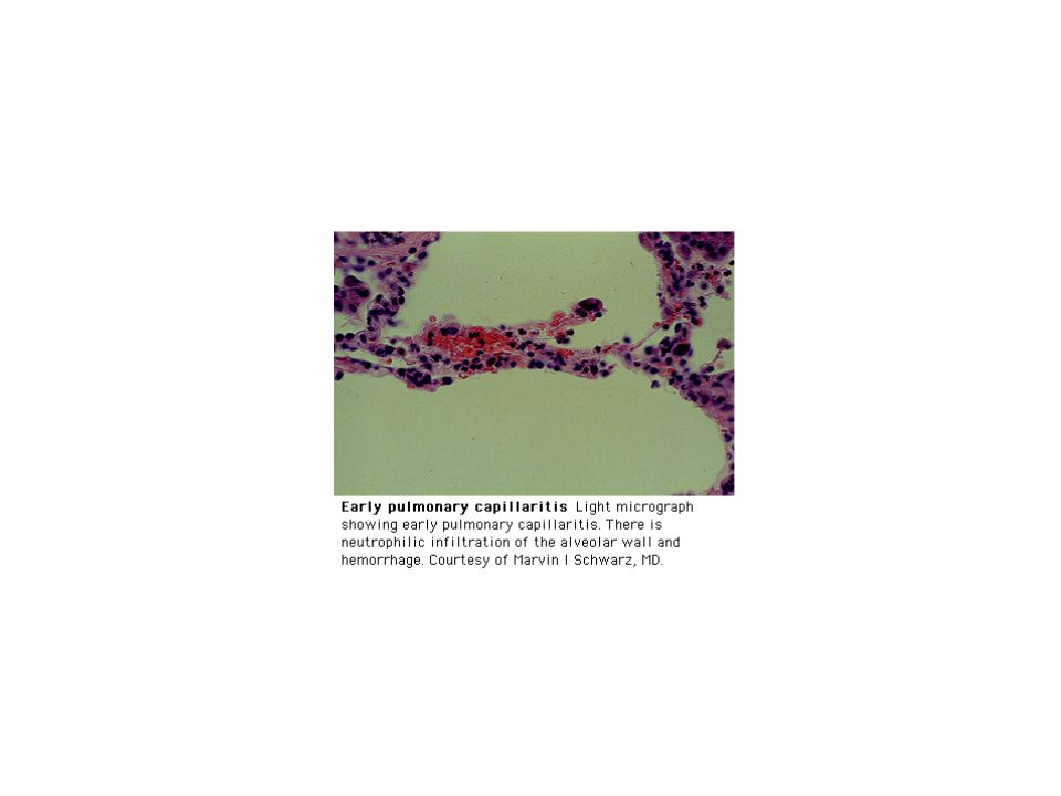
Pathology of DAH 3 Broad histologic patterns identified Pulmonary capillaritis Bland pulmonary hemorrhage Diffuse alveolar damage

7
Pulmonary Capillaritis I Most common of the histologic patterns First described by Spencer in 1957 Neutrophilic interstitial infiltrate with fragmentation of neutrophils (leukocytoclasis) and pyknotic neutrophils with release of cytokines Nuclear dust in interstitium and alveolar spaces
 and pyknotic neutrophils with release of cytokines Nuclear dust in interstitium and alveolar spaces")
8
Pulmonary Capillaritis II Disruption of the interstitium and capillaries with leakage of blood and fibrin into alveolar spaces Edema of basement membrane with subsequent necrosis of interstitium and eventual fibrosis Neutrophils are seen lining the interstitium

11
Diffuse Alveolar Damage Hyaline membrane formation, alveolar and interstitial edema, microthrombi, and capillary congestion are present See slide

13
Bland Alveolar Hemorrhage RBC’s in the alveolar spaces, but alveolar walls appear normal except for type II epithelial cell hyperplasia See slide

16
Clinical Presentation of DAH I Patients often have an underlying known condition Hemoptysis can be acute or subacute, but typically presents within one week of onset Majority of patients are less than forty years old 1/3 do not have hemoptysis, but present with dyspnea and cough

17
Clinical Presentation DAH II In patients without hemoptysis, diagnosis is confirmed by the presence of blood on serial BAL Anemia Pulmonary infiltrates Chest pain, nonspecific Symptoms of underlying disease processes

18
History in DAH Careful drug history Smoking history History of underlying illnesses such as valvular heart disease, cytotoxic agents, drugs Social history, in particular cocaine usage History of any renal, skin, or eye diseases

19
Physical Findings in DAH Nonspecific Fevers, rales, signs of consolidation Synovitis, iridocyclitis, myositis, palpable purpura

21
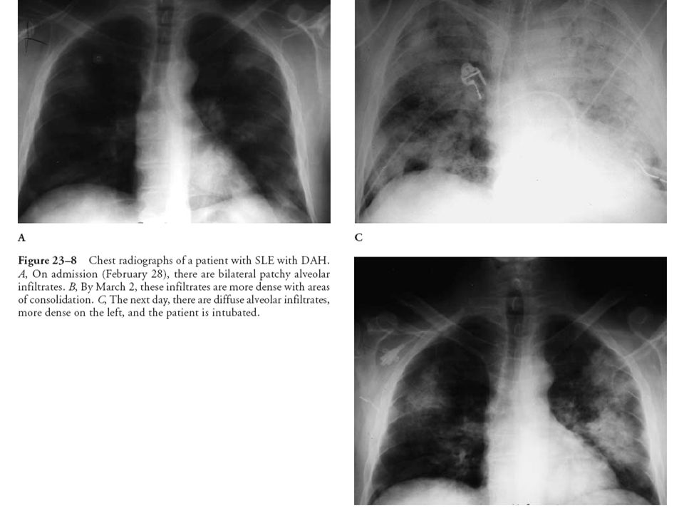
Radiographic Findings in DAH Nonspecific, focal or generalized infiltrates Rapidly progressive bilateral infiltrates Interstitial fibrosis in presence of recurrent disease Kerley’s B line suggestive of valvular etiology, also in conditions associated with myocarditis, venoocclusive disease

25
Laboratory Findings in DAH I Low or falling hematocrit or hemoglobin In the setting of chronic or recurrent episodes, low serum iron Nonspecific elevations of white count Thrombocytopenia Elevation of ESR Proteinuria, microscopic hematuria, casts suggest glomerulonephrits

26
Laboratory Findings in DAH II Hypoxemia Elevation of DLCO Restrictive pattern associated with fibrosis or obstructive patterns with marked emphysematous changes ANCA ABMA, IgG

27
ANCA in DAH Diagnosis Antineutrophilic cytoplasmic antibodies (ANCA)first described in 1982 in association with pauci-immune glomerulonephritis ANCA described in association with Wegener’s granulomatosis in 1985 Subsequently described in microscopic polyangitis (MPA) and limited renal vasculitis
first described in 1982 in association with pauci-immune glomerulonephritis ANCA described in association with Wegener’s granulomatosis in 1985 Subsequently described in microscopic polyangitis (MPA) and limited renal vasculitis")
29
ANCA Testing Indirect immunofluorescence assay (IIA) is more sensitive Enzyme link immunosorbent assay (ELISA) is more specific Best used in conjunction with IIA for screening and ELISA for confirmation Two relative antigens in vasculitic diseases, proteinase 3 (PR3) and myeloperoxidase (MPO) Antigens are found in neutrophils and monocytes PR3-ANCA and MPO-ANCA
 is more sensitive Enzyme link immunosorbent assay (ELISA) is more specific Best used in conjunction with IIA for screening and ELISA for confirmation Two relative antigens in vasculitic diseases, proteinase 3 (PR3) and myeloperoxidase (MPO) Antigens are found in neutrophils and monocytes PR3-ANCA and MPO-ANCA")
30
Immunofluorescence Patterns In Vasculitis Sera from patients with suspected ANCA related vasculitis are incubated in ethanol fixed neutrophils Two distinct patterns of fixation identified, c-ANCA with cytoplasmic pattern and p-ANCA with perinuclear pattern c-ANCA pattern is typically associated with antibodies against PR3 p-ANCA is typically associated with antibodies against MPO See photographics

33
Immunofluorescence Utility and Errors Tests are visually graded and inspected Tests are not specific and false positives and negatives can occur IIA testing should be confirmed with ELISA testing for PR3 and MPO

35
Specific Examples of DAH Capillaritis-Microscopic Polyangiitis Diffuse Alveolar Damage-Crack Cocaine Bland Hemorrhage-Amiodarone

36
Microscopic Polyangiitis (MPA) I Rare disease with prevalence estimated 3 cases per million Etiology is unknown Small vessel involvement including arterioles, venules, and or capillaries Immune complexes are not demonstrated Typical presentation is that of renal failure with glomerulonephritis and hemoptysis with capillaritis Histopathologically segmental distribution, neutrophilic infiltration, and fibrinoid necrosis (See slide)
 I Rare disease with prevalence estimated 3 cases per million Etiology is unknown Small vessel involvement including arterioles, venules, and or capillaries Immune complexes are not demonstrated Typical presentation is that of renal failure with glomerulonephritis and hemoptysis with capillaritis Histopathologically segmental distribution, neutrophilic infiltration, and fibrinoid necrosis (See slide)")
37
MPA II ANCA is positive in about 75% of patients p-ANCA is present with MPO by ELISA in 85% of patients c-ANCA is rare with PR3 by ELISA Systemic disease Skin manifestations including splinter hemorrhages and purpura Musculoskeletal with arthralgias, myalgias, arthritis Gastrointestinal with abdominal pain and GI hemorrhage Neurological with peripheral neuropathy

38
MPA II ANCA is positive in about 75% of patients p-ANCA is present with MPO by ELISA in 85% of patients c-ANCA is rare with PR3 by ELISA Systemic disease Skin manifestations including splinter hemorrhages and purpura Musculoskeletal with arthralgias, myalgias, arthritis Gastrointestinal with abdominal pain and GI hemorrhage Neurological with peripheral neuropathy

39
MPA III Prominent gastrointestinal signs and symptoms and lack of upper airway disease helps distinguish from Wegener’s Classical polyarteritis nodosa rarely involves the lung 45% of patients have circulating immune complexes but tissue localization is rare, pauci-immune disease 33% of patients have antibodies to hepatitis C or B Treatment same as for Wegener’s Survival about 65% with recurrence associated with tapering of therapy Case report in which MPA eventually developed features of WG

40
Maimon, N. et al. Chest 2003;124:2384-2387 The histologic section from a right middle lobe open lung biopsy showed extensive hemorrhaging in the alveolar spaces

41
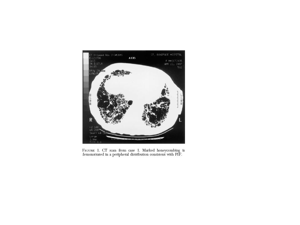
Pulmonary Interstitial Fibrosis & MPA Report of six cases of PIF in which patients were ANCA positive and eventually diagnosed with MPA The diagnosis of PIF may precede that of MPA by many years See CT

43
Maimon, N. et al. Chest 2003;124:2384-2387 Diffuse alveolar consolidation with air bronchogram involving the entire right lung field

45
Cocaine History & Mechanisms of Action First isolated from coca leaves in 1859 Part of the original formulation of Coke, removed in 1906 First reported deaths occurred in 1893 Potent sympathomimetic and CNS stimulant based on its ability to block reuptake of catecholamines & serotonin Cocaine HCL boiled with baking soda and extracted with ether or alcohol, yields heat stable “Crack” or “Rock” Smoked and reaches the CNS within seconds with half life of 60-90 minutes Frequently mixed with marijuana or tobacco

46
Crack Cocaine with Diffuse Alveolar Damage Cocaine is abused in two forms, cocaine HCl and cocaine alkaloidal The alkaloidal form or “crack” cocaine is lipid soluble and resistant to thermal breakdown Rapidly absorbed from the lung via pulmonary capillary network Euphoria is similar to that of intravenous usage Smoking of “crack” is also associated with absorption of impurities, ignition products and thermal breakdown products of cocaine

47
BAL in Crack Lung Users Up to 40% of of “crack” cocaine users have hemosiderin stained alveolar macrophages. <1% of nonsmokers at autopsy have this finding. ~9% of smokers have this finding. Endothelin (ET)-1, an endothelium-derived vasoconstrictor peptide, indicator of cell damage is also found in a higher proportion of “crack” cocaine users. ET-1 is found in a high proportion of BAL samples from “crack” users and is felt to be a marker of alveolar capillary damage BAL in “crack” cocaine users have an absolute increase in hemosiderin stained alveolar macrophages
-1, an endothelium-derived vasoconstrictor peptide, indicator of cell damage is also found in a higher proportion of crack cocaine users. ET-1 is found in a high proportion of BAL samples from crack users and is felt to be a marker of alveolar capillary damage BAL in crack cocaine users have an absolute increase in hemosiderin stained alveolar macrophages.")
48
Janjua, T. M. et al. Chest 2001;119:422-427 Iron content of alveolar macrophages (AM)
")
49
Janjua, T. M. et al. Chest 2001;119:422-427 Ferritin content of alveolar macrophages recovered

50
Baldwin, G. C. et al. Chest 2002;121:1231-1238 Prominent hemosiderin-laden AMs in the BAL fluid of a CS (left, A), which are absent in the BAL fluid of a TS (middle, B) or a NS (right, C)
, which are absent in the BAL fluid of a TS (middle, B) or a NS (right, C).")
51
Baldwin, G. C. et al. Chest 2002;121:1231-1238 Increased percentage of hemosiderin-positive AMs in the BAL specimen of cocaine smokers

52
Acute Lung Injury with Crack Typically develops within 1-48 hours 25% of users with develop respiratory symptoms including fever, cough, nonspecific chest pain, hemoptysis, back pain, hyperpnea, dyspnea, melanoptysis, wheezing Diffuse pulmonary infiltrates, eosinophilic pleural effusions, acute lung injury pattern Eosinophilia

53
Crack Pulmonary Injuries I Barotrauma, ischemia, provocation of inflammatory damage, and direct cellular toxicity Barotrauma is the result of Valsalva maneuver after inhalation and the forceful inhalation of air into partners. Pneumothoraces, pneumomediastinum, and pneumopericardium Ischemia is the result of the vasoconstrictive properties Severe bronchospasm in patients with preexisting asthma

54
Crack Cocaine Pulmonary Injuries II Case of bilateral infiltrates and bilateral hilar adenopathy mimicking sarcoid, probably induced by contaminants in crack The morphologic features of squamous metaplasia and mucus gland hypertrophy similar to that of cigarette smokers, possibly increased risk of lung cancer

55
Microenvironment and Cocaine Cocaine inhibits alveolar macrophages ability to kill most bacteria and tumor cells in vitro Cocaine users are unable to kill bacteria using nitric oxide as an antibacterial effector molecule These changes may predispose to increase pulmonary infections in these users. Marijuana has similar adverse effects. Inhibits phagocytosis Staph aureus Both drugs frequent smoked together

56
Kleerup, E. C. et al. Chest 2002;122:629-638 Distribution of Vc and DMCO in the cross-sectional cohort is shown

57
Chronic Exposure to Crack Pulmonary fibrosis Diffuse alveolar hemorrhage Hemosiderosis Pulmonary infarction Eosinophilic interstitial lung disease Bullous emphysema Medial artery hypertrophy Noncardiogenic pulmonary edema Increased risk of pneumonia, multifactorial problem

58
Treatment of Crack Lung Need to make history of exposure Supportive Role of steroids unproven, helpful in those patients with bronchospasm Screen for HIV and concomitant drugs Drug treatment

59
Amiodarone Lung Disease Drugs characteristics Expanding scope of the problem Clinical presentations Radiographic presentations Diagnosis and therapeutic options

60
Brinker, A. et al. Chest 2004;125:1591-a-1592-a Trends in receipt of clinically serious, domestic spontaneous adverse event reports (1,941) in association with amiodarone (all forms) with further indication of the subset of reports coded for parenchymal lung injury (n = 280)
 in association with amiodarone (all forms) with further indication of the subset of reports coded for parenchymal lung injury (n = 280).")
61
Kerstein, J. et al. Chest 2004;126:716-724 Incidence of atrial fibrillation in the therapy group vs the control group

62
Kerstein, J. et al. Chest 2004;126:716-724 Comparison of length of stay (los) in the amiodarone group and the control group
 in the amiodarone group and the control group.")
63
PHD Amiodarone Usage 1997-2003

64
Characteristics of Amiodarone I Principal metabolite is desethyl-aminodarone or DEAm DEAm and amiodarone are toxic to lung tissue Propensity for accumulation in lung tissue with a ratio of plasma to lung of 1:500 DEAm and amiodarone also have a propensity to accumulate in the liver and skin Amiodarone and DEAm are localized in lysosomes and block the removal of phospholipids

65
Characteristics of Amiodarone II Amiodarone drug levels do not predict the development of pulmonary toxicity DEAm levels are higher in patients who develop amiodarone pneumonitis that controls The foamy macrophage on BAL is characteristic of amiodarone exposure and its absence bespeaks against this diagnosis Clearance of amiodarone is very slow with biopsies demonstrating the drug after one year of cessation of therapy

66
Characteristics of Amiodarone III Clinically the development of amiodarone infiltrates is associated with a prolonged radiographic resolution because of the prolonged deposition of this agent Each molecule of amiodarone and DEAm contain two iodines The presence of the iodines explains the frequent development of thyroid dysfunction in patients receiving amiodarone The iodine also explains the increase in CT density in liver and lung in patients diagnosed with pulmonary toxicity

70
Amiodarone Pulmonary Toxicity First clinical description in 1980, although drug was introduced in 1969 in Europe Rat models have demonstrated the toxicity since 1987 The histological features of amiodarone toxicity are relatively distinctive in nature, foamy macrophages There is a relative dose relationship between total cumulative dose and incidence of toxicity Reintroduction of agent to patient with previous toxicity results in recurrence of syndrome

71
Relationship Between Dose and Incidence of Toxicity to Amiodarone Estimated that 50% of patients receiving 1200 mg per day will develop toxicity Estimated incidence 5% to 15% if dose is greater than or equal to 500 mg per day Estimated incidence 0.1% to 0.5% in dose is less than 200 mg per day Overall incidence is probably in the range of 4% to 6%

72
Onset of Toxicity to Amiodarone Majority of cases will occur within one year of exposure May occur following loading intravenous dose Cases described after 10 years of therapy Can develop following cessation of therapy Toxicity increases in certain patient populations including advanced age, cardiac surgery, pulmonary surgery, ARDS, insertion of ICD Common denominator is exposure to high FIO2 during surgery, similar to situation in bleomycin pulmonary toxicity and radiation

73
Radiographic Presentations in Amiodarone Pulmonary Toxicity Subacute pneumonitis Single of multiple subpleural masses Pulmonary fibrosis Organizing pneumonia ARDS Alveolar hemorrhage

74
Subacute Amiodarone Pneumonitis Patchy or diffuse infiltrates Suggestion of RUL predominance on plain radiographic examination, usually not confirmed by HRCT, bilateral process Alveolar interstitial pattern on HRCT Increased attenuation on HRCT Pleural effusion rare

75
Subpleural Masses in Amiodarone Lung Disease Single or multiple Typically abut the pleura Chest pain and pleural rub DDX includes pulmonary infarction, malignancy, pneumonia, lymphoma

77
Pulmonary Fibrosis with Amiodarone Very uncommon, <0.1% of cases Setting of prior acute pneumonitis from amiodarone or resolving phase of illness or as initial presentation Differs from idiopathic pulmonary fibrosis by the rapidity of disease progression and history of amiodarone usage Reticular infiltrates at the bases with typical signs & symptoms of ILD Irreversible Not felt to be steroids responsive

78
Organizing Pneumonia from Amiodarone Indistinguishable from other forms of organizing pneumonia Migratory infiltrates may occur See radiographs

80
ARDS from Amiodarone Settings cardiac surgery, pulmonary resection, and following defibrillator implantation (J. Intern Medicine Med 2001, Liverani up to 10% incidence) lung biopsy for diagnosis 50% fatality Rapidly progressive pulmonary infiltrates with hypoxemic respiratory failure Poorly responsive to therapy
 lung biopsy for diagnosis 50% fatality Rapidly progressive pulmonary infiltrates with hypoxemic respiratory failure Poorly responsive to therapy.")
81
Subclinical Disease from Amiodarone Setting of chronic therapy Plain radiographs typically normal HRCT ground glass infiltrates Typically responds to cessation of drug Unclear if it is progressive Biopsy foamy cells and inflammatory cells evident Gallium scans positive, but T99 more sensitive

82
Diagnosis and Management I Clinical lab: increase in LDH sensitive, but not specific, increased ESR, and some leukocytosis PFT’s most sensitive with reduction in DLCO as first abnormality. A fall in DLCO does not necessarily indicate pulmonary toxicity, only 1/3 of patients develop overt disease A fall of 15% is cutoff for sensitivity and 30% for specificity according to Mayo study

83
Diagnosis and Management II Stable DLCO is indicative of absence of toxicity, but fall should be confirmed with HRCT/nuclear No clear cut benefit to routine screening, but study for Canada suggests baseline chest x-ray and PFT’s, serial studies in patients with new symptoms BAL is nonspecific, finding of foamy macrophages is not indicative of toxicity, only exposure HRCT and nuclear imaging important in confirmation of diagnosis KL6, high molecular weight, glycoprotein, type II pneumocytes, increased

84
Diagnosis and Management III Cessation of drug is mainstay of therapy Steroids help in certain cases, but not uniformly Steroids must be tapered very slowly and over a prolonged period of time. Early mortality is increased in patients not started on steroids. Radiographic and PFT improvement follow clinical improvement Permanent reduction in DLCO is possible. Recurrent disease with cessation of steroids is frequently resistant to therapy

85
Diagnosis and Management IV Steroids 0.75 mg/kg to 1.0 mg/kg or methylprednisolone or equivalent. Treat at least for six months, preferably for one year No reduction in dosage until objective evidence of improvement Incidence is higher in patients with COPD as is mortality Mortality up to 33% 2/3 of patients will develop recurrent disease if reexposed to the medication, DO NOT RECHALLENGE!!

86
Diagnosis and Management V Clinical clues to suggest condition would include concomitant skin discoloration, thyroid dysfunction, myeloid suppression, photosensitivity, corneal deposits with chronic therapy

87
Potential Mechanism of Toxicity for Amiodarone Hamster alveolar macrophages are sensitive to amiodarone When amiodarone is added to preparation of hamster alveolar macrophages, the membrane potential declines Following the decline in the membrane potential, decline in intracellular ATP Decline in cell viability identified

Similar presentations


 Infections (pneumonia, airways disease)>")


>")


 Dr. Meg-angela Christi Amores.>")


 Dr. Raid Jastania. Vasculitis Inflammation of the walls of the vessels Causes of inflammation: –Infectious, physical, chemical,>")


