Download presentation
Presentation is loading. Please wait.

1
Middle Cerebral Artery Doppler
Nafisa Dajani/M.D Maternal Fetal Medicine Department of Obstetrics and Gynecology University of Arkansas for Medical Sciences

2
Uses Of MCA in Obstetrics
Evaluation of IUGR Evaluation of fetal anemia

3
Location: MCA is the middle branch from the circle of Willis that courses anteriorly and temporally

4
Why the Interest in the MCA?
The MCA bed has attracted investigators for its ability for auto regulation Vasodilatation assures continued supply of glucose and oxygen ( IUGR with brain sparing) The MCA is easy to identify Doppler measurements have satisfactory reproducibility with appropriate training
 The MCA is easy to identify. Doppler measurements have satisfactory reproducibility with appropriate training.")
5
Is this new? Studies in the 90s have shown repeatedly abnormally low PI in 30-54% of IUGR. The mean systolic velocity was also higher in SGA fetuses. And fetal blood sampling indicated a relationship between fetal hypoxemia and MCA PI that was strongest when PI was 2-4 standard deviations below the normal range

6
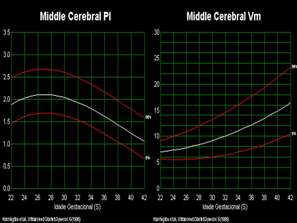
PI, S/D Pulsatility : frequency change
The PI is a reflection of downstream resistance (S-D)/S Normally there is a decrease in diastolic brain flow at weeks (more resistance, incr.PI) correlating with an increase in fetal cerebral cellular multiplication, followed by a progressive decrease in resistance (decr. PI)
/S. Normally there is a decrease in diastolic brain flow at weeks (more resistance, incr.PI) correlating with an increase in fetal cerebral cellular multiplication, followed by a progressive decrease in resistance (decr. PI)")
8
Normal IUGR With hypoxia there is cerebral vasodilatation, so initially the diastolic flow may be in the normal range ,when the vasodilatation ability is exhausted as with fetal acidosis the resistance starts increasing again.

9
The redistribution in hypoxemic fetuses may be transient (PI decreases). With worsening hypoxemia, the PI may increase and diastolic flow may be reversed. This may precede intrauterine death

10
How to Measure MCA dopplers
Enlarge the circle of Willis ( 75% of image) Image the whole vessel if possible Align vessel parallel to the ultrasound beam, angle zero for best measurement Measure close to the exit point from the internal carotid Avoid measuring during fetal breathing, hiccups, or movement
 Image the whole vessel if possible. Align vessel parallel to the ultrasound beam, angle zero for best measurement. Measure close to the exit point from the internal carotid. Avoid measuring during fetal breathing, hiccups, or movement.")
11
How to measure the MCA dopplers
Middle cerebral artery peak systolic velocity: technique and variability. J Ultrasound Med Apr;24(4): Mari G, Abuhamad AZ, Cosmi E, Segata M, Altaye M, Akiyama M. Figure 1. Circle of Willis. The letters indicate the 4 points assessed for variability Figure 1. Circle of Willis. The letters indicate the 4 points assessed for variability
: Mari G, Abuhamad AZ, Cosmi E, Segata M, Altaye M, Akiyama M. Figure 1. Circle of Willis. The letters indicate the 4 points assessed for variability. Figure 1. Circle of Willis. The letters indicate the 4 points assessed for variability.")
12
Why are MCA dopplers important for the management of IUGR
MCA PI has a 98% negative predictive value for major adverse perinatal outcomes <32 weeks gestation Abnormally low MCA dopplers are an early sign of fetal response to hypoxia and is generally preceded by an abnormal umbilical artery dopplers and decreased fetal AC measurements

13
MCA Doppler's PI a sensitive indicator of fetal hypoxia as measured by concomitant cordocentesis evaluation Precedes abnormal fetal testing by 2-3 weeks When abnormal it should lead to interaction of other dopplers in the fetal circulation to evaluate the degree of compromise and better timing of delivery

14
The sequence of changes in Doppler and biophysical parameters as severe fetal growth restriction worsens A. A. Baschat, U. Gembruch* and C. R. Harman Ultrasound in Obstetrics and Gynecology Volume 18 Page 571 - December 2001

15
Figure 4 The percentage of abnormal Doppler findings in individual vessels and the incidence of a biophysical profile score below 6 (*) in the week prior to delivery. ▪, umbilical artery absent or reversed end-diastolic flow; ○, abnormal middle cerebral artery flow; □, abnormal inferior vena cava flow; , abnormal ductus venosus flow; ◊, umbilical vein pulsations. Deterioration of Doppler findings precedes decline in biophysical profile score.
 in the week prior to delivery. ▪, umbilical artery absent or reversed end-diastolic flow; ○, abnormal middle cerebral artery flow; □, abnormal inferior vena cava flow; , abnormal ductus venosus flow; ◊, umbilical vein pulsations. Deterioration of Doppler findings precedes decline in biophysical profile score.")
16
How Can Doppler Studies and Biophysical Profile be combined
Doppler studies and the BPP can independently identify fetuses at risk. They are not always concordant Concordance occurs in about 44% Marked discordance in 17% The BPP is maintained longer Doppler and biophysical profile in growth restricted fetuses. A Baschat. Ultrasound Obstet Gynecol Jan;27(1):41-7.
:41-7.")
17
MCA Dopplers and Fetal Anemia

18
Causes of fetal anemia 30,000 fetuses are at risk of anemia from RBC alloimmunization each year in the USA Other causes of anemia include parvo virus infections, fetomaternal hemorrhage, non-immune hydrops, twin-twin transfusion, etc

19
Anemia and MCA dopplers
1990 G.Mari proposed the use of MCA dopplers for the diagnosis of anemia !0 years later a collaborative multicenter study, 110 fetuses with isoimmunization and 265 fetuses normal, all underwent cordocentesis and MCA doppler studies

20
Figure 2. Receiver-Operating-Characteristic Curves for the Peak Velocity of Systolic Blood Flow in the Middle Cerebral Artery for the Prediction of Mild, Moderate, and Severe Fetal Anemia.

21
The sensitivity of the peak systolic velocity for the prediction of moderate anemia (a hemoglobin concentration of less than 0.65 times the median) and severe anemia (a hemoglobin concentration of less than 0.55 times the median) in the fetuses without hydrops was 100 percent (95 percent confidence interval, 86 to 100), with a false positive rate of 12 percent. The positive and negative predictive values were 65 percent and 100 percent, respectively.

22
Studies that have found no correlation between anemia and doppler studies have used the PI and RI which are angle-independent indexes, and thus independent of blood velocity.

23
The risk of anemia was high in fetuses with a peak systolic velocity of 1.50 times the median or higher. Fetuses with values below 1.50 either did not have anemia or had only mild anemia. The fact that this test does not predict mild anemia well is not clinically important, because no intervention is indicated in fetuses with mild anemia,

25
the MCA PSV was effective for accurate diagnosis of fetal anemia and avoided 70% of invasive procedures.

26
Mari reported MCA PSV is superior to optical density measurement at 450 nm in terms of assessing fetal anemia secondary to red cell alloimmunization. Pereira et al also reported that the MCA PSV is better than amniocentesis in the diagnosis of fetal anemia. Additionally, a recent multicenter study assessed both the MCA PSV and amniocentesis in fetuses undergoing cordocentesis reported that the sensitivity of MCA PSV for the detection of severe anemia was better than that of amniocentesis.

27
MCA PSV is still accurate after intrauterine transfusion

28
MCA after 35 weeks is not accurate

29
Middle cerebral artery peak systolic velocity: is it the standard of care for the diagnosis of fetal anemia? J Ultrasound Med May;24(5): Review.
: Review..")
30
Should all obstetricians and gynecologists in the United States and elsewhere in the world assess the MCA PSV for the diagnosis of anemia? The answer is no because today there are not enough sonographers and sonologists trained to assess the MCA PSV.

Similar presentations