Download presentation
Presentation is loading. Please wait.

1
I. General Information about the Skeletal System A
I. General Information about the Skeletal System A. ______________________- study of movement of the human body. Kinesiology

2
B. Division of the Skeletal System
Human Skeleton (206 bones TOTAL) AXIAL Skeleton Skull (22 bones) Hyoid (1 bone) Auditory ossicles (6 bones) Vertebral column (26 bones) Thorax (25 bones) = 80 bones total
 AXIAL Skeleton. Skull (22 bones) Hyoid (1 bone) Auditory ossicles (6 bones) Vertebral column (26 bones) Thorax (25 bones) = 80 bones total.")
3
Human Skeleton (206 bones TOTAL) Appendicular Skeleton Clavicle (2 bones) Scapula (2 bones) Upper limbs (60 bones) Pelvic Girdle (2 bones) Lower limbs (60 bones) = 126 bones
 Lower limbs (60 bones) = 126 bones.")
4
Blue = axial Yellow = appendicular

5
C. Bone and Surface Markings 1. Depressions & Openings: a
C. Bone and Surface Markings 1. Depressions & Openings: a. ___________________- opening for blood vessels, nerves and/or ligaments. example – b. _____________________- shallow depression in or on the bone. example - Foramen Opening in pelvis bone Fossa Coronoid fossa of the humerus

6
2. Processes that form joints a
2. Processes that form joints a. _________________- large rounded prominence that forms joints. example – b. _________________- rounded project that is supported on a thinner “neck” and forms a joint. example - condyle Knobs on lower femur at knee head Top of the femur (“ball”)
")
7
3. Processes that tendons, ligaments & other tissue connect to: a
3. Processes that tendons, ligaments & other tissue connect to: a. _____________________ - large, blunt projection only on the femur (not the “ball” part) b. _____________________ - prominent border or ridge Example - Trochanter Crest Illiac crest at top of hip bone
 b. _____________________ - prominent border or ridge Example - Trochanter. Crest. Illiac crest at top of hip bone.")
8
c. ____________________- large, rounded projection, usually with a rough surface. Example – d. __________________________________- a sharp, slender project. Example - Turbocity Deltoid turbocity of the humerus Spine or Spinous process The part of the vertebrae that you can palpate on someone’s back

9
4. ______________________- to make contact with
4. ______________________- to make contact with. (ex- me humerus articulates with the radius) articulate
 articulate.")
10
D. General differences between male & female skeletons 1
D. General differences between male & female skeletons 1. Male bones are _______________ and_____________________ than female bones. 2. Male points of _____________________ are larger. larger heavier Muscle attachment

11
II. Upper Appendage A. Definition of Upper Appendage: _______ __________________________________ B. Pectoral Girdle – the _______________ & ________________ that anchors the limb to the axial skeleton. Pectoral Girdle + Upper Limb clavicle scapula

12
Clavicle 1. ___________________ (collar bone) a. it is the ___________________ fractured bone due to one outstretching their arms when s/he falls. Most commonly

13
_________ end of the clavicle
LATERAL _________ end of the clavicle MEDIAL _________ end of the clavicle

14
2. ________________________ (shoulder blade)
scapula _________________ Used for attachment to shoulder muscles. Scapular spine _________________ attaches to rotator cuff muscles. Infraspinous process

15
_________________ ________________ attaches to rotator cuff muscles. Supraspinous process

16
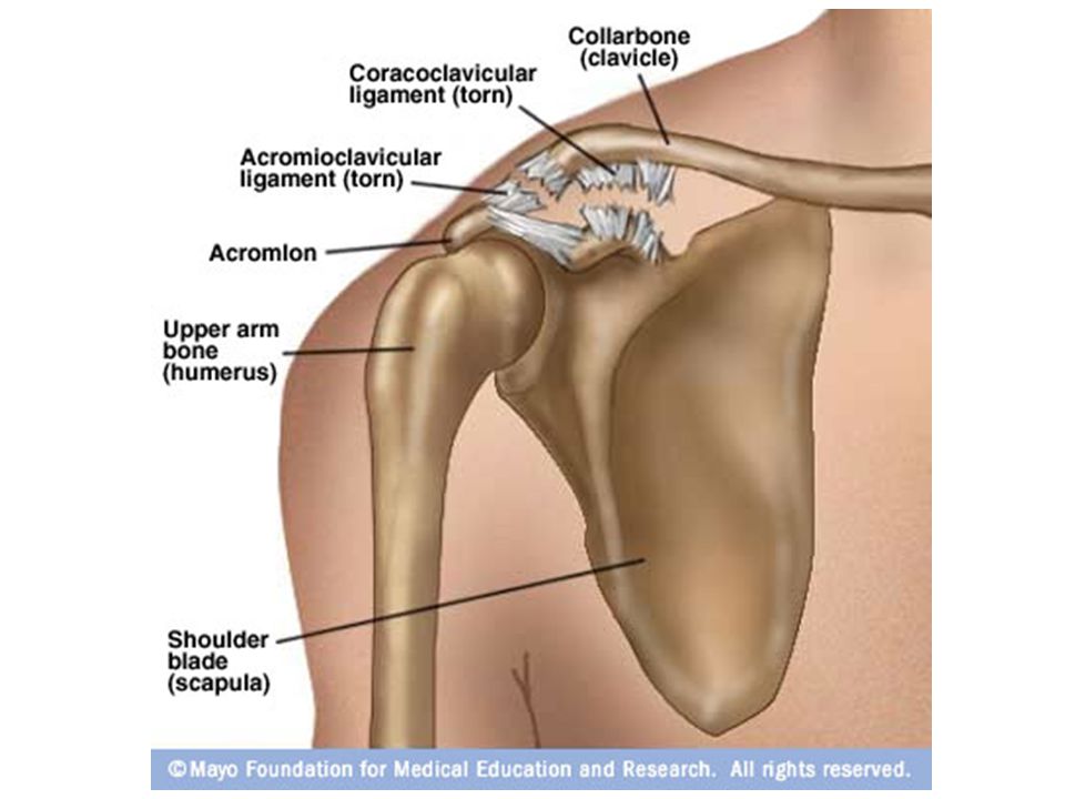
____________-Articulates with the __________
____________-Articulates with the __________. (This is where a “separated shoulder” occurs.) ACROMION CLAVICLE __________________ Articulates with the head of the _________________ to form a ball & socket joint. Glenoid Fossa humerus
 ACROMION. CLAVICLE. __________________ Articulates with the head of the _________________ to form a ball & socket joint. Glenoid Fossa. humerus.")
17
______________________________
Used for attachment to chest and arm muscles. Coracoid process ____________________- faces towards the ribs. Subscapular fossa

18
Separated shoulder 3. ____________________________- an injury where the joint between the lateral end of the clavicle and the acromion of the shoulder is affected (often happens with forceful trauma).
.")
20
Humerus C. ____________________- (upper arm bone) _____________articulates with the glenoid fossa of the scapula. HEAD __________________________ Where the humerus narrows. Anatomical neck _________________________- Attachment point for deltoid muscle. Deltoid Turberosity

21
______________ ___________- Articulates with the ulna when arm is bent. CORONOID FOSSA ____________ (lateral side) Articulates with the radius CAPITULUM ___________-(spool shaped) Articulates with the ulna. TROCHLEA
 Articulates with the ulna. TROCHLEA.")
22
_______________. Articulates with the olecranon process of the ulna.
Olecranon fossa

23
D. ____________________- (lower arm bone on
the little finger side) Ulna ULNA
 Ulna. ULNA.")
24
Ulna – full view _______________Articulates with the carpals of the wrist Styloid Process

25
Ulna – enlarged proximal end; lateral view
Trochlear notch _______________where the humerus fits _____________________ “point” of elbow Olecranon process _____________________ Where the radius fits. Radial Notch ________________________-articulate with coronoid fossa of humerus CORONOID PROCESS

26
E. ____________________- (lower arm on the
thumb side) Radius RADIUS
 Radius. RADIUS.")
27
_______________Articulates with the capitulum
_______________Articulates with the capitulum. (allows the hand to face “palms up”) head _______________ (commonly “fall” fracture by people over 50) Styloid process
 head. _______________ (commonly fall fracture by people over 50) Styloid process.")
28
1. ______________________________ - the head of the radius slides past or ruptures the ligament that forms a collar around the head of the radius (most common upper limb dislocation in children). Dislocation of the radial head

30
F. Carpals - _______________________________
8 bones in the wrist _______________2 rows of bones that forms a tunnel for nerves & tendons to pass through. CARPALS Carpal Tunnel Syndrome ______________________- when the nerves of this tunnel are compressed and cause numbness, tingling & weakening in the hand.

32
G. Metacarpals - ___________________________
5 bones that make up the palm of the hand ______________________ - Each metacarpal has a base (proximal), body & a head (distal). It is the _____________ of the metacarpal that makes _________________ when you clench your fist. METACARPALS head knuckles
, body & a head (distal). It is the _____________ of the metacarpal that makes _________________ when you clench your fist. METACARPALS. head. knuckles.")
33
H. Phalanges - ____________________________
14 bones of the fingers & thumb Proximal Phalanges

34
Middle Phalanges

35
Distal Phalange

36
I. Pollex - ____________________________
Specialized phalange that makes up the thumb Pollex – no middle phalange

37
IV. _________________________- both hip bones A
IV. _________________________- both hip bones A. Three functions of the pelvic girdle: 1. _________________ the vertebral column 2. _____________________________ of the pelvis (bladder, reproductive organs, rectum). 3. __________________________ to the axial skeleton. Pelvic Girdle supports Protects the organs Attaches lower limbs
. 3. __________________________ to the axial skeleton. Pelvic Girdle. supports. Protects the organs. Attaches lower limbs.")
38
B. Features of the Pelvic Girdle
Coxal ______________- the two “hip” bones that are made up of the: _______________ _______________________ which fuse together by age 23. Illium, ischium & pubis

39
_________________- joint that joins the two coxal bones.
Pubic Symphasis

40
_________________- divides the pelvis into an upper & lower portion.
Pelvic Brim

41
_________________- joint on the posterior side where the 2 coxal bones join the sacrum.
Sacroilliac joint

42
_________________everything above the pelvic brim
_________________everything above the pelvic brim. It is actually considered to be part of the ______________ & only contains the bladder when it is _________ and uterus during _____________. False Pelvis abdomen full pregnancy

43
_________________below the pelvic brim which surrounds the pelvic cavity
True Pelvis

44
_______________ upper opening of the true pelvis.
Pelvic Inlet

45
_______________ lower opening of the true pelvis.
Pelvic Outlet

46
C. ________________ - largest part of the coxal bone Illium
Anterior View Illiac crest

47
Lateral View of Coxal Bone
_________________________ - where the sciatic nerve (longest nerve in body) passes through. Greater sciatic notch
 passes through. Greater sciatic notch.")
48
D. ________________ - lower, posterior part of coxal bone. Ischium
Anterior View

49
E. ________________ - lower, anterior part of the coxal bone. Pubis
Anterior View

50
F. Features created by the joining of the bones of the coxal
F. Features created by the joining of the bones of the coxal. (lateral view) ____________- socket for the head of the femur formed by the joining of all 3 coxal bones. Acetabulum
 ____________- socket for the head of the femur formed by the joining of all 3 coxal bones. Acetabulum.")
51
__________________- largest foramen of the skeleton where nerves and vessels pass (formed by joining of ischium & pubis. Obturator Foramen

52
G. Structural differences between a male and female pelvis 1
G. Structural differences between a male and female pelvis 1. ____________________- of the female is larger and the _________________ is oriented so that a baby can fit through. 2. The angle made by the two ischium bones is a larger _______ shape in females, and narrower _________ shape in males. Pelvic Inlet Pelvic outlet U V

53
V. __________________ (the leg) A
V. __________________ (the leg) A. _________________(thigh bone) – the STRONGEST bone in the body Lower Limb Femur
 A. _________________(thigh bone) – the STRONGEST bone in the body Lower Limb. Femur.")
54
Greater Trochanter ____________ __________- projection on side of femur where muscles attach(also landmark for injections in the thigh). __________________- “ball” of the femur which articulates with the acetabulum. Head

55
__________________- articulates with the patella (“knee cap”).
Patellar Surface

56
____________ __________- Towards the outside of the leg. Articulates with the tibia. Lateral Condyle __________________- towards the inside of the leg. Articulates with the tibia. Medial Condyle

57
B. ___________________ (“knee cap”) – normally tracks(moves) up & down in a __________ between the femoral condyles. Patella groove

58
1. ___________________________- “runner’s knee” occurs when the patella tracks _________________ and causes pain. (due to running on the same side of the road that is naturally sloped). Patellofemoral Stress Syndrome laterally

60
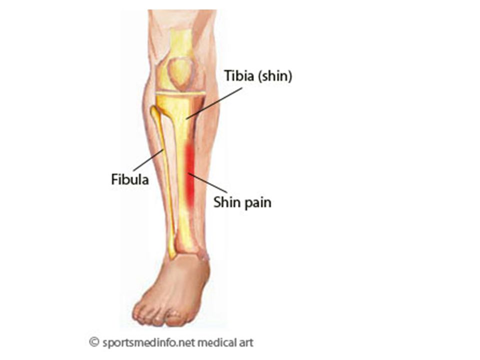
C. _________________(shin bone)
Tibia

61
________________________-
Articulates with the lateral condyle of the femur. Lateral condyle ______________________- Articulates with the medial condyle of the femur. Medial condyle

62
Medial Malleolus _____________________- Forms the prominence you feel on the medial ankle bone.

63
1. ______________- pain along the tibia that results from inflammation of the tibia’s ___________________ (usually caused by over-exertion of the calf muscles) Shin splints periosteum

65
D. ___________________- smaller bone next to the tibia in the lower leg.
Fibula

66
___________________-
Forms the prominence you feel on the lateral part of the ankle surface. Lateral malleolus

67
E. The FOOT & its functions 1
E. The FOOT & its functions 1. ______________- 7 ankle bones of the foot Tarsals ___________________- It is the only tarsal that articulates with the tibia & fibula. It initially bears the _________ of the entire body when walking. Talus weight

68
___________- Between the talus and cuniforms Navicular

69
___________- 3 bones between the navicular & metatarsals. cuniforms

70
___________- Between the calcaneous and metatarsals Cuboid

71
___________________-
(heel bone) It is the largest & strongest tarsal. _________ the weight is transferred to it from the __________ when walking. Calcaneous Half Talus
 It is the largest & strongest tarsal. _________ the weight is transferred to it from the __________ when walking. Calcaneous. Half. Talus.")
72
metatarsals 2. _______________________- 5 bones that are similar to the metacarpals that make up the “sole” of the foot. a. like the metacarpals, each have a __________ base, a body, and a ___________ head. proximal distal

73
3. __________________- 14 bones similar to the fingers in the hand.
phalanges Proximal phalanges

74
Middle Phalange (only 4)
Distal Phalanges

75
4. ___________________- specialized phalange that lacks a ______________ phalange.
Hallux middle

76
functions 5. The four ___________ of the arches foot: a. _________________ leverage when walking b. _________________ shock (by “giving when weight is applied & “springing back” afterwards). c. ____________________ body weight over parts of the foot. d. ____________________ the weight of the body. Acronym help: ______________ (think…..the “PADS” of your feet) P.rovides A.bsorbs D.istributes S.upports P. A. D. S.
. c. ____________________ body weight over parts of the foot. d. ____________________ the weight of the body. Acronym help: ______________ (think…..the PADS of your feet) P.rovides. A.bsorbs. D.istributes. S.upports. P. A. D. S.")
77
6. Three arches of the foot
Transverse arch ________________- Runs horizontally across the foot. Formed by the navicular, cuniforms, & bases of metatarsals.

78
Medial Longitudinal Arch
_________________________- Runs front to back of foot by the inside of the foot.

79
Lateral Longitudinal Arch
______________________- Runs front to back of foot by the outside of the foot.

81
7. __________________- abnormally low height of the medial longitudinal arch.
Flat footed

82
VI. Joints of the Skeletal System A
VI. Joints of the Skeletal System A. __________________- point of contact between ___________, between ________________________ or between ____________________. Joint bones Cartilage & bones Teeth & bones

83
B. Divisions of Medical Studies of Joints 1
B. Divisions of Medical Studies of Joints 1. ______________________- the study of joints. 2. ______________________- study of joint disease and related conditions Arthology Rheumatology

84
C. Movement of Joints 1. Range of motion: a
C. Movement of Joints 1. Range of motion: a. in general, the _______________ the distance between the articulating bone, more _________________ the range of motion for that joint. Ex - shorter restricted Skull bones- very close together no range of motion femur & tibia are farther apart large range of motion

85
2. Three Factors that determine joint flexibility: a
2. Three Factors that determine joint flexibility: a. ____________________ of the ligaments that bind the bones together. b. _____________________of articulating bones. c. _____________________tension of associated muscles and tendons. ACRONYM HELP: What’s your favorite math class at DPHS? __________________ F.lexibility S.hape T.ension F. S. T.

86
JOINTS D. Classes of Joints Fibrous Bones are held together by__________ connective tissue. Lack a ________________ cavity Three Types _____________ fibrous synovial suture syndesmosis gomphosis

87
JOINTS Synovial ____________ a synovial cavity Most ___________ Six Types ______________ contains movable planar condyloid hinge saddle pivot Ball & socket

88
JOINTS Cartilaginous Bones are held together by ______________. Also _________ a synovial cavity. Two Types ______________ cartilage lacks synchondris symphasis

89
1. FIBROUS JOINTS SUTURE immovable -They are ________________
-Found where__________ unite Skull bones

90
-Found in_____________________
SYNDESMOSIS (sin-dez-MŌ-sis) Slightly movable -They are ________________ -Found in_____________________ Tibia/fibula connection & sacrum/coxal connection
 Slightly movable. -They are ________________. -Found in_____________________. Tibia/fibula connection & sacrum/coxal connection.")
91
-They are ________________ -Found only in_________________
GOMPHOSIS (gom-FŌ-sis) immovable -They are ________________ -Found only in_________________ Tooth sockets
 immovable. -They are ________________. -Found only in_________________. Tooth sockets.")
92
2. SYNOVIAL JOINTS PLANAR Wrist & ankles Found in _________________
Btwn clavicle & sternum Btwn clavicle & scapula

93
- Found in ________________ Wrist and ankles
CONDYLOID (KON-di-loyd) - Found in ________________ Wrist and ankles
 - Found in ________________. Wrist and ankles.")
94
SADDLE - Found in ________________ Thumb only

95
HINGE - Found in __________________ Knee, elbow, ankle, & fingers

96
PIVOT - Found in __________________ Elbow & head to say “no”

97
BALL-&-SOCKET - Found in __________________ Shoulder & hip joints

98
a. ____________________- allows the joint to move freely.
Synovial cavity

99
b. specific joint structures of the knee:
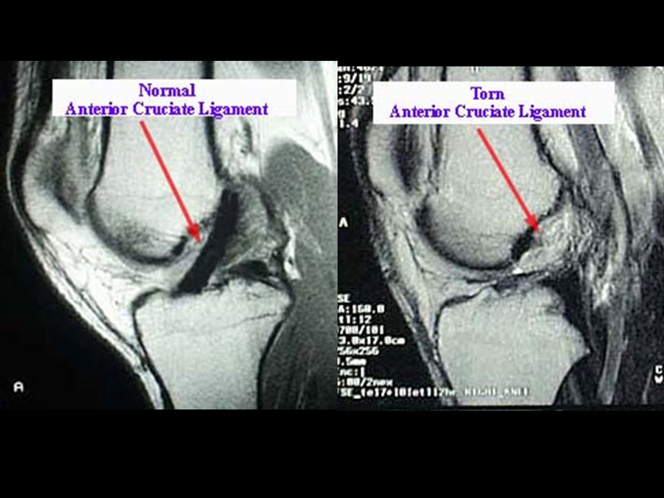
_________________________________________- Ligament that extends posteriorly & laterally from the tibia to the fibula. Anterior Cruciate Ligament (ACL)
")
100
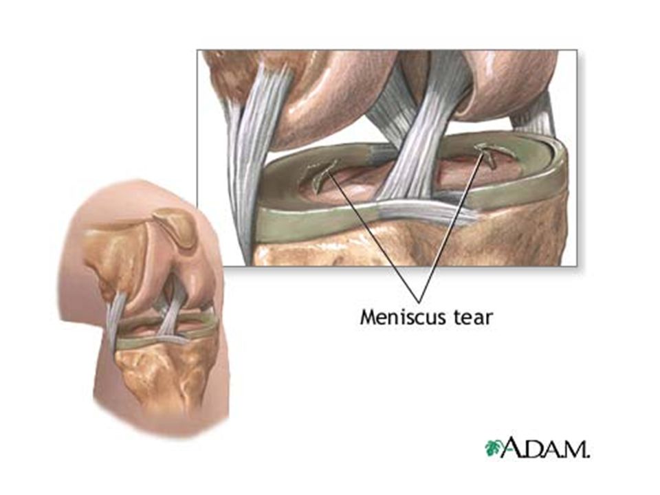
___________________________-
Found on the outside of the knee. Meniscus allow for a____________ fit between two different shaped bones. Lateral Meniscus tighter

101
Medial Meniscus ___________________________- Found on the inside of the knee.

102
c. Common injuries of the knee: 1
c. Common injuries of the knee: 1. _____________________- when the anterior cruciate ligament is stretched or torn (70% of all serious knee injuries). ACL injury
. ACL injury.")
105
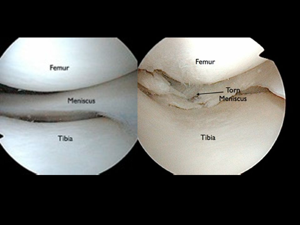
2. _______________________________ - when the lateral or medial meniscus is damaged. (If the damaged cartilage is not removed, it may lead to arthritis) Torn Cartilage of the knee

108
3. CARTILAGENOUS JOINTS immovable -They are ________________
SYNCHONDROSIS (sin-kon-DRŌ-sis) immovable -They are ________________ -Found in _____________ of elongating bones & between the ____________ & ____________ Growth plates Rib cage sternum
 immovable. -They are ________________. -Found in _____________ of elongating bones & between the ____________ & ____________. Growth plates. Rib cage sternum.")
109
-They are ________________
SYMPHASIS (sim-fi-sis) Slightly movable -They are ________________ -Found between the __________ bones and between ____________ pubis vertebrae
 Slightly movable. -They are ________________. -Found between the __________ bones and between ____________. pubis. vertebrae.")
110
E. Common Joint Diseases & Complications 1
E. Common Joint Diseases & Complications 1. _______________________- an autoimmune disease in which the immune system of the body attacks its own cartilage & joint linings. It is characterized by _______________ of the synovial cavity. Rheumatoid Arthritis (RA) swelling
 swelling.")
113
2. _________________________- degenerative joint disease characterized by deterioration of ________________ cartilage. (the “wear & tear” arthritis) Osteoarthritis articular

115
3. ________________- forcible wrenching or twisting of the ________________ of a joint.
Sprain ligaments

116
Strain 4. _____________________- stretched or partially torn _________________. (often occurs when a muscle contracts suddenly & powerfully) muscle

Similar presentations