Download presentation
Presentation is loading. Please wait.

1
Internal features of Heart Dr. Sama ul Haque Dr Rania Gabr

2
Objectives List the chambers of the heart. Describe the internal features of right atrium. Describe the internal features of the right ventricle. Discuss internal features of the left atrium. Discuss the internal features of the left ventricle. Differentiate between right and left ventricles. Define conducting system of the heart.

3
Chambers of Heart 1.Right Atrium 2.Right Ventricle 3.Left Atrium 4.Left Ventricle

4
Internal Structure of Heart

5
Right Atrium The right atrium consists of a main cavity and a small out pouching, the auricle. On the outside of heart at the junction between the right atrium and the right auricle is a vertical groove. This is called the sulcus terminalis, which on the inside forms a ridge, the crista terminalis.

6
Right Atrium Crista Terminalis Musculi pectinati (Pectinate muscles) 1.Rough anterior part. 2.Smooth posterior part. 1 2

7
Cavity of Right Atrium Crista terminalis divides right atrium into: 1- Anterior part: rough and trabeculated by bundles of muscle fibres (musculi pectinati). 2- Posterior part (sinus venarum): is smooth. The interatrial septum carries an oval depression called Fossa ovalis. The margin of this depression is called Anulus ovalis. The blood leaves right atrium to right ventricle via tricuspid valve.
: is smooth. The interatrial septum carries an oval depression called Fossa ovalis. The margin of this depression is called Anulus ovalis. The blood leaves right atrium to right ventricle via tricuspid valve..")
8
Right Ventricle

9
Right ventricle The right ventricle communicates with the right atrium through right atrioventricular orifice. It also communicates with the pulmonary trunk through the pulmonary orifice. Infundibulum

10
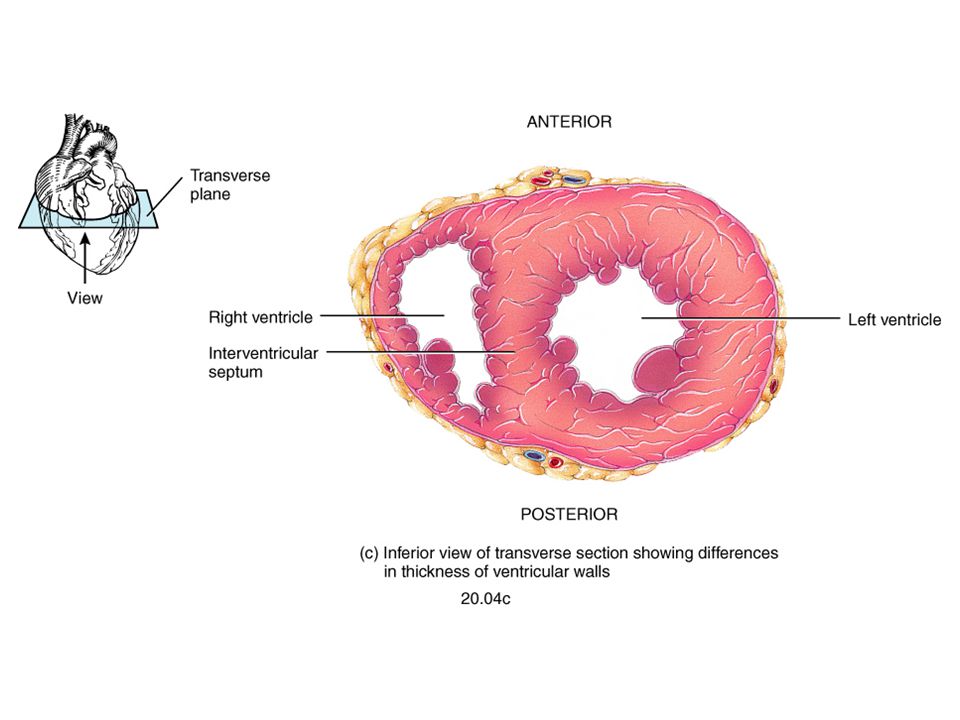
Cavity of right ventricle Its wall is thinner than that of left ventricle. Its wall contains rough muscular projections called trabeculae carneae. Tricuspid valve has three cusps: anterior, septal, and posterior Large projections arise from the walls called papillary muscles : Anterior papillary muscle Posterior papillary muscle Septal papillary muscle S P A trabeculae carnae.

11
Cavity of right ventricle Each papillary muscle is attached to the cusps of tricuspid valve by tendinous threads called chordae tendinae (Tendinous cords). The wall of infundibulum is smooth and contains no trabeculae. Interventricular septum is connected to anterior papillary muscle by a muscular band called moderator band ( the septomarginal trabecula ) Infundibulum
 Infundibulum.")
12
The conus arteriosus (infundibulum): is the cone-shaped portion of the right ventricle inferior to the opening of the pulmonary trunk. The pulmonary valve: consists of three semilunar cusps: anterior, right, and left. Right Ventricle Infundibulum

13
Right atrio-ventricular (tricuspid) orifice It is surrounded by a fibrous ring which gives attachment to the tricuspid valve. It has 3-cusps (anterior-posterior- septal or medial). The atrial surface of the cusps are smooth, while their ventricular surfaces give attachment to the chordae tendinae. A P M
. The atrial surface of the cusps are smooth, while their ventricular surfaces give attachment to the chordae tendinae. A P M.")
14
Pulmonary orifice Surrounded by a fibrous ring which gives attachment to the cusps of the pulmonary valve. The valve is formed of 3 semilunar cusps : one anterior only. No chordae tendineae or papillary muscles are attached to these cusps A P P

15
Right Ventricle

16
It communicates with the left ventricle through left atrioventricular orifice. It forms the greater part of base of heart. Its wall is smooth except for small musculi pectinati in the left auricle. Recieves 4 pulmonary veins which have no valves. Sends blood to left ventricle through the left atrioventricular orifice which is guarded by mitral valve. Left atrium of the heart Left atrium

17
Left Atrium Bicuspid valve: blood passes through it into left ventricle (mitral valve has two cusps).
.")
18
Left ventricle of the heart Its wall is thicker than that of right ventricle. It receives blood from left atrium through left atrio- ventricular orifice which is guarded by mitral valve. Its wall contains trabeculae carnae. Its wall contains 2 large papillary muscles (anterior & posterior). They are attached by chordae tendinae to cusps of mitral valve.
. They are attached by chordae tendinae to cusps of mitral valve..")
19
Left ventricle of the heart The blood leaves the left ventricle to the ascending aorta through the aortic orifice. The part of left ventricle leading to ascending aorta is called aortic vestibule. The wall of this part is fibrous and smooth. Aortic vestibule

20
Left Ventricle have thick muscular wall

21
Left atrio-ventricular (mitral) orifice Smaller than the right, admitting only tips of 2 fingers. Guarded by a mitral valve. Surrounded by a fibrous ring which gives attachment to the cusps of mitral valve. Mitral valve is composed of 2 cusps: Anterior cusp : lies anteriorly and to right. Posterior cusp : lies posteriorly and to left. The atrial surfaces of the cusps are smooth, while ventricular surfaces give attachment to chordae tendinae. A P

22
Aortic orifice Surrounded by a fibrous ring which gives attachment to the cusps of aortic valve. Aortic valve is formed of 3 semilunar cusps which are similar to those of pulmonary valve, but the position of the cusps differs being one posterior only. P A A

23
Valves of the Heart 1 2 3 4 1.Right Atrioventricular (Tricuspid) 2.Left Atrioventricular (Bicuspid or Mitral) 3.Pulmonary 4.Aortic
 2.Left Atrioventricular (Bicuspid or Mitral) 3.Pulmonary 4.Aortic")
24
Fibrous Skeleton of the Heart Dense collagenous fibrous tissue framework lies at the junction of the atria with the ventricles. Includes: 1-Tricuspid fibrous annulus: 2-Mitral fibrous annulus 3-Pulmonary fibrous annulus: 4-Aortic fibrous annulus Functions: 1-Supports the valves. 2-Electrically separetes atria from ventricles.

25
Valves of the Heart

26
Valves of Heart in Diastole (Viewed from base with atria removed)
")
27
Valves of Heart in Systole (Viewed from base with atria removed)
")
30
Direction of blood flow

31
Conducting system of Heart Sinu-Atrial Node (S-A Node) Atrio-ventricular Node (A-V Node) Atrio-ventricular Bundle Right and left Bundle branch Purkinje Fibers
 Atrio-ventricular Node (A-V Node) Atrio-ventricular Bundle Right and left Bundle branch Purkinje Fibers")
32
Conducting system of Heart S-A Node A-V Node

33
Conducting system of Heart PF

34
Conducting system of Heart

Similar presentations


 in thorax, in inferior mediastinum>")


