Download presentation
Presentation is loading. Please wait.

1
Unit 3 – The Integumentary System

2
The Integumentary System
Integument is skin Skin and its appendages make up the integumentary system Skin, hairs, nails, vessels, nerves, and glands A fatty layer (hypodermis) lies deep to it
 lies deep to it.")
3
The Integumentary System
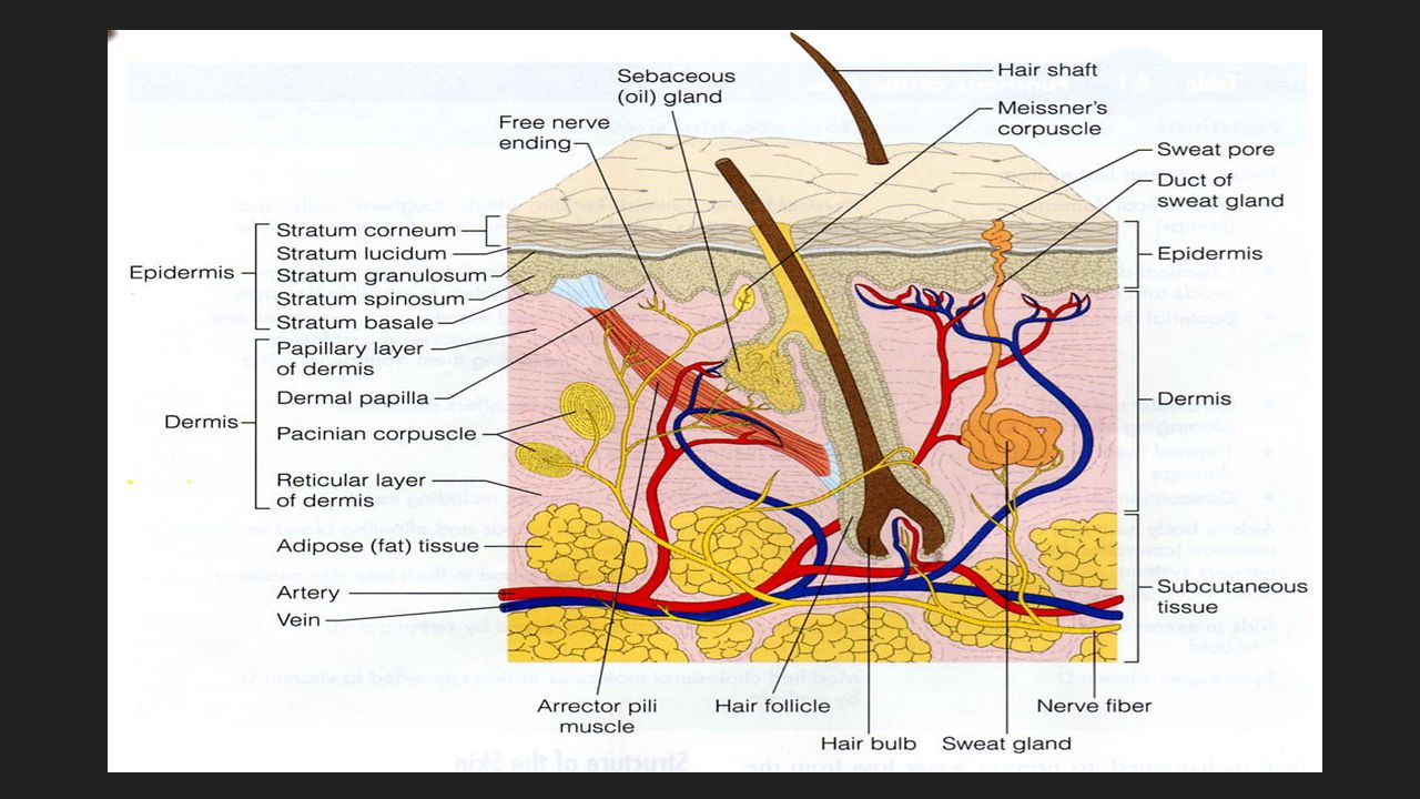
Two major components: Cutaneous Membrane Epidermis/Superficial Epithelium Dermis/Underlying Connective Tissue Accessory Structures Located in dermis Hair, nails, exocrine glands, blood vessels Sensory receptors for touch, pressure, temperature and pain Deep to the dermis, the loose connective tissue of the subcutaneous layer/superficial fascia/hypodermis separates the integument from the deep fascia around other organs

4
Functions of the Skin Protection Regulate Body Temperature
Covering to protect deeper tissues from dehydration, trauma, and germ invasion Regulate Body Temperature Controls heat loss Evaporation of water from the skin, in the form of perspiration Helps rid the body of excess heat Helps manufacture Vitamin D The sunshine vitamin Ultraviolet light on the skin is necessary for the first stages of vitamin D

5
Functions of the Skin Storage Absorption
Fat, glucose, water, and salt Absorption Can absorb certain medications and chemicals Screens out harmful ultraviolet radiation and eliminates wastes Site of many receptors and nerve endings for sensory information Touch, pressure, pain, and temperature

7
Layers of Skin Epidermis Dermis Subcutaneous Membrane Hypodermis

8
Epidermis Outer layer of the skin Renews itself ~ every 45 days

9
Epidermis – Cell Types Keratinocytes
Produce keratin waterproofing protein Originate in deeper layers & get pushed to surface Connected to each other by desmosomes & tight junctions Cell production & keratinization are accelerated in areas of friction Think callus thickened skin

10
Epidermis – Cell Types Melanocytes Produce melanin
Prevents DNA mutation from UV radiation UV increases melanin production Same number in everyone but different amount of pigment produced Accumulation of melanin results in freckles and moles

11
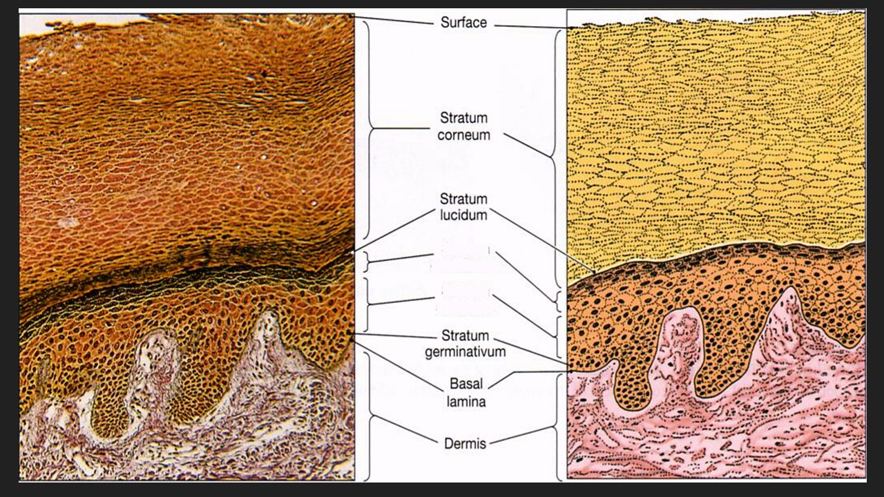
5 Layers of the Epidermis
In order from deep to superficial Stratum germinative (basale) Stratum spinosum Stratum granulosum Stratum lucidum Stratum corneum Takes days for a cell to move through all five levels
 Stratum spinosum. Stratum granulosum. Stratum lucidum. Stratum corneum. Takes days for a cell to move through all five levels.")
13
Stratum Germinative/Basale
Highly mitotic (goes through mitosis quickly) Produces new skin layer ~25% melanocytes
 Produces new skin layer. ~25% melanocytes.")
14
Stratum Spinosum Slightly mitotic – one of the daughter cells from the stratum germinativum is pushed into the stratum spinosum Consists of 8-10 layers of cells Contains Langerhans macrophages Stimulate a defense against: Microorganisms that manage to penetrate the superficial layers of the epidermis Superficial skin cancers

15
Stratum Granulosum Not mitotic but begin making keratin and keratohyalin Keratin = tough fibrous protein component of hair and nails Keratohyalin = forms dense granules that dehydrate the cell and aggregate cross-linking of the keratin fibers Also contains Langerhans cells Nuclei and other organelles disintegrate = Cell Death

16
Stratum Lucidum ONLY found in thicker epidermis – palms, soles, callus
Completely keratinized (and dead!) Contains closely packed, clear cells that contain gel-like substance eleiden
 Contains closely packed, clear cells that contain gel-like substance eleiden.")
17
Stratum Corneum Outermost layer – Exposed Skin
Also completely keratinized Dead cells Remain in this layer for two weeks before they are shed Tough, waterproofing protection

18
Dermis Middle layer of skin – your “hide” – like leather
Contains hair follicles, glands, nerves, vessels, and muscle

19
Layers of the Dermis Mainly strong, flexible connective tissue – 2 layers Papillary Layer Upper region Uneven and has fingerlike projections called dermal papillae that create fingerprints and are important for grip Contain capillaries, pain receptors (free nerve endings), and touch receptors called Meissner’s corpuscles Reticular Layer Deepest skin layer Contains blood vessels, adipose (fat) sweat and oil glands, and deep pressure receptors
, and touch receptors called Meissner’s corpuscles. Reticular Layer. Deepest skin layer. Contains blood vessels, adipose (fat) sweat and oil glands, and deep pressure receptors.")
20
Hypodermis Not usually part of the skin Also called subcutaneous layer
Site of subcutaneous injections – absorbed directly into the blood stream Anchors skin to underlying organs, bones, and muscles Shock absorption and insulation Composed mostly of adipose tissue Very vascular

21
Skin Color Skin color is determined by 3 factors:
3 Types of pigments present Melanin Brown, black, or yellow Carotene Orange-yellow pigment from some vegetables Vitamin A precursor – vitamin A forms retinal which is needed for sight Accumulates in adipose and stratum corneum cells Hemoglobin Red, oxygen-carrying pigment in erythrocytes More obviously detected in fair skin Blood circulation Stratum corneum thickness

22
Skin Color People who produce a lot of melanin have brown-toned skin
The crimson color of oxygen-rich hemoglobin gives the skin a rosy color When hemoglobin is poorly oxygenated, the skin appears blue – a condition called cyanosis Common during heart failure and severe breathing disorders

23
Skin Color Signals Disease States
Rubor Redness or erythema Embarrassment (Blushing) Fever Hypertension Inflammation Allergy
 Fever. Hypertension. Inflammation. Allergy.")
24
Skin Color Signals Disease States
Pallor or Blanching Emotional stress (fear, anger, and others) Pale skin may also signify anemia , low blood pressure, or impaired blood flow into the area Jaundice A yellow-case Liver disorder in which excess bile pigments is in the blood Bruises Sites where blood has escaped and has clotted in the tissue spaces Called hematomas Unusual bruising may signify a deficiency of vitamin C or hemophilia
 Pale skin may also signify anemia , low blood pressure, or impaired blood flow into the area. Jaundice. A yellow-case. Liver disorder in which excess bile pigments is in the blood. Bruises. Sites where blood has escaped and has clotted in the tissue spaces. Called hematomas. Unusual bruising may signify a deficiency of vitamin C or hemophilia.")
25
Hair Millions of hairs all over the body Guards head
Shields eyes (eyelashes) Keeps foreign particles out of the respiratory tract (nose hairs)
 Keeps foreign particles out of the respiratory tract (nose hairs)")
26
Hair A hair is produced by a hair follicle Structure of Hair
Shaft – protects skin Follicle – extends into dermis Root – lies within the follicle Bulb – growth zone at the inferior end of the follicle Sebaceous Gland – lubricates hair Arrector Pili Muscle – attached to follicle and contracts to move hair (growth or goosebumps)
")
27
Hair Growth Influenced by (in this order) Baldness (alopecia)
Nutrition – main influence Hormones Blood flow Baldness (alopecia) Male pattern baldness – sex-linked recessive genetic trait Thinning – can be caused by medications, nutrition, stress
 Male pattern baldness – sex-linked recessive genetic trait. Thinning – can be caused by medications, nutrition, stress.")
28
Hair Pigment Caused by proportions of 3 melanin types:
Dark Hair = true melanin Blonde & Red Hair = melanin with iron and sulfur Gray/White Hair = melanin replaced by air bubbles in shaft

29
Nails Scale-like modification of the epidermis Heavily keratinized
Stratum basale extends beneath the nail bed to form the nail matrix Responsible for growth (matrix region) Lack of pigment makes them colorless Lunula “little moon” – area of cell growth (white semicircle at base of nail) Cuticle – area of skin that covers base of nail
 Lack of pigment makes them colorless. Lunula little moon – area of cell growth (white semicircle at base of nail) Cuticle – area of skin that covers base of nail.")
31
Glands of the Body Cutaneous Glands Exocrine Glands 2 Groups:
All are exocrine glands Exocrine Glands Release secretions to surface via ducts 2 Groups: Sweat Glands Sebaceous Glands Both formed by stratum basale and push into dermis

32
Sweat Glands More than 2.5 million per person 2 Primary Types
Eccrine Glands Widely distributed in skin; abundant on palms, soles, and forehead Sweat composition: mostly water with a slightly acidic 4-6 pH Function: thermoregulation

33
Sweat Glands Apocrine Glands Ducts empty into hair follicles
Found mainly in anogenital and axillary region Begin to function at puberty due to hormones/pheromones Organic contents: fatty acids and proteins – can have a yellowish color that stains clothes Odor is from associated bacteria Cerminous Glands Modified apocrine gland Found in outer 1/3 of ear canal Produce ear wax to trap “invaders”

34
Sebaceous (Oil) Glands
All over except palms and soles of feet Produce oil for waterproofing Lubricant for skin and kills bacteria Most with ducts that empty into hair follicles Some open onto skin surface in lips, eyelids, genitalia Sebum (seb = grease) Mixture of oily substances and fragmented cells Glands are activated at puberty stimulated by hormones
 Mixture of oily substances and fragmented cells. Glands are activated at puberty stimulated by hormones.")
35
Sebaceous (Oil) Glands
Acne Active infection of sebaceous glands Can be mild or extremely severe Whitehead A sebaceous gland‘s duct becomes blocked by sebum Blackhead Accumulated material oxidized, dries, and darkens

36
Skin Diseases & Disorders
The most common skin disorders result from allergies or bacterial, viral, or fungal infections. Homeostatic imbalances of the skin

37
Common Skin Disorders Acne = disease of sebaceous glands
Alopecia = hair loss Tinea pedis = athletes foot Carbuncle = bacterial infection like a boil but subcutaneous Cyst = liquid filled sac Dermatitis = inflammation Eczema = non-contagiuous skin rash Impetigo = contagious bacterial infection causes eruption Moles = (nevi) tumors that are pigmented Pediculosis = lice Pruritis = itching without eruption Scabies = mites Shingles = (Herpes Zoster) virus causes blisters at nerve path
 tumors that are pigmented. Pediculosis = lice. Pruritis = itching without eruption. Scabies = mites. Shingles = (Herpes Zoster) virus causes blisters at nerve path.")
38
Contact Dermatitis Itching, redness, and swelling of the skin, &blistering. Caused by exposure of the skin to chemicals Ex: poison ivy Provokes an allergic response

39
Psoriasis Chronic condition
Reddened epidermal lesion-covered with dry, silvery scales When severe, may be disfiguring Cause unknown; may be hereditary in some cases Attacks often triggered by trauma, infection hormonal changes, and stress.

40
Athlete's Foot tinea pedis
Itchy, red, peeling skin between the toes, resulting from a fungal infection Athlete's Foot Tips From The APMA Avoid walking barefoot; use shower shoes Reduce perspiration by using talcum powder Wear light and airy shoes Wear socks that keep your feet dry, and change them frequently if you perspire heavily

41
Boils and Carbuncles Inflammation of hair follicles and sebaceous glands, Common on the dorsal neck Carbuncles are composite boils Typically caused by the bacterial infection (Staphylococcus aureus)
")
42
Cold Sores Fever blisters
Small fluid-filled blisters that itch and sting Caused by herpes simplex virus Virus localizes in a cutaneous nerve Remains dormant until activated by emotional upset, fever, or UV radiation Cold sores usually occur around the lips and in the oral mucosa of the mouth

43
Impetigo Pink, water-filled, raised lesions
Common around the mouth and nose Develop a yellow crust and eventually rupture Caused by a highly contagious staphylococcus infection Common in elementary school-aged children

44
Necrotizing Fasciitis
Severe type infection that involves the skin, subcutaneous fat, and muscle fascia Caused by several bacteria both aerobic and anaerobic The most severe kind is caused by a virulent streptococcus species Infection usually enters through the skin and releases toxins that: Directly kill tissue Interfere with blood flow to tissue Digest materials in tissue and allows bacteria to spread rapidly Cause widespread effects, i.e. shock

45
Necrotizing Fasciitis Symptoms
Infection begins as a small reddish painful spot or bump on the skin It quickly changes to a brown or purplish patch, the center of the wound will begin to turn black (dead cells) The wound will visibly expand in less that 1 hour Symptoms include sweating, chills, nausea, dizziness, profound weakness, and finally shock. Without treatment death occurs rapidly Many times the patient requires a surgeon to diagnose by culture of wound drainage
 The wound will visibly expand in less that 1 hour. Symptoms include sweating, chills, nausea, dizziness, profound weakness, and finally shock. Without treatment death occurs rapidly. Many times the patient requires a surgeon to diagnose by culture of wound drainage.")
46
Necrotizing Fasciitis Treatment
Powerful, broad spectrum anti-biotic administered IV immediately and immediate surgery required to open and drain infection and debride dead material Skin grafts are required after infection is cleared Infection in a limb and is not containable = amputation Prognosis Outcomes vary, depending on organism, rate of spread, susceptibility to antibiotics and how early infection is diagnosed Complications Sepsis, scarring and disfigurement, loss of limb, and death The disease untreated has 100% mortality

47
Basal Cell Carcinoma Least malignant Most common skin cancer
Cells of the stratum basale are altered so that they cannot form keratin & no longer honor the boundary between epidermis and dermis They proliferate, invading the dermis and subcutaneous tissue. Lesions occur most often on sun-exposed areas of the face Appear as shiny, dome-shaped nodules that later develop a central ulcer with a "pearly" beaded edge Relatively slow-growing Metastasis seldom occurs before it is noticed Full cure is the rule in 99 percent of cases where the lesion is removed surgically

48
Squamous Cell Carcinoma
Arises from the cells of the stratum spinosum The lesion appears as a scaly, reddened papule (small, rounded elevation) that gradually forms a shallow ulcer with a firm, raised border Scalp, ears, dorsum of the hands, and lower lip Grows rapidly Metastasizes to adjacent lymph nodes if not removed Believed to be sun-induced If it is caught early and removed surgically or by radiation therapy, the chance of complete cure is good
 that gradually forms a shallow ulcer with a firm, raised border. Scalp, ears, dorsum of the hands, and lower lip. Grows rapidly. Metastasizes to adjacent lymph nodes if not removed. Believed to be sun-induced. If it is caught early and removed surgically or by radiation therapy, the chance of complete cure is good.")
49
Malignant Melanoma Cancer of melanocytes
Accounts for 5 percent of skin cancers Incidence is increasing It is often deadly Melanoma can begin wherever there is pigment Appear spontaneously, but some develop from pigmented moles Appears as a spreading brown to black patch that metastasizes rapidly to surrounding lymph and blood vessels Chance for survival is about 50 percent Early detection helps – the American Cancer Society suggests that sun worshippers periodically examine their skin for new moles or pigmented spots

50
Malignant Melanoma Apply the ABCD rule for recognizing melanoma:
Asymmetry: the two sides of the pigmented spot or mole do not match. Border irregularity: the borders of the lesion are not smooth but exhibit indentations. Color: the pigmented spot contains areas of different colors (blacks, browns, tans, and sometimes blues and reds). Diameter: the spot is larger than 6 rum in diameter (the size of a pencil eraser) The usual therapy for malignant melanoma is wide surgical excision along with immunotherapy
. Diameter: the spot is larger than 6 rum in diameter (the size of a pencil eraser) The usual therapy for malignant melanoma is wide surgical excision along with immunotherapy.")
51
Burns Protein denaturation and cell death caused by heat, electricity, UV radiation (sunburn), or chemicals 2 main dangers: Dehydration Loss of fluids and electrolytes lead to Renal shutdown Circulatory shock Infection Skin (mechanical) barrier lost Immune system depresses
 barrier lost. Immune system depresses.")
52
Rules of Nines Way to determine extent of burns
Primary importance is to estimate fluids needed for rehydration Body is divided into 11 areas for quick estimation Each area represents about 9% This along with cause of burn helps determine the severity

53
First Degree Burns (Superficial Burns)
Only epidermis is damaged Local redness, swelling, and pain Usually heal in 2-3 days (short time period) with NO scarring
 with NO scarring.")
54
Second Degree Burns (Partial Thickness Burns)
Epidermis, dermis, and structures within dermis are damaged Appearance of blisters of any size Skin regeneration in 3-4 weeks with some scarring There is a danger of infection Very painful

55
Third Degree Burns (Full Thickness Burns)
Epidermis, dermis, hypodermis, and all structures within are completely destroyed Usually painless at site of burn due to destruction of sense receptors Burn is gray-white, tan, brown, black, or deep cherry red Surrounded by areas of 1st & 2nd degree burns that are painful Treatments are numerous but will involve skin grafting of some sort, fluid replacement, and debridement

56
Emergent Care Burning process stopped with removal of clothing & jewelry and covering affected area with cool water Increase blood volume with IV inserted in intact skin area Urinary catheter to monitor fluid output, indicates dehydration Intubation to secure an airway Vitals: BP, HR, BPM, Temp

57
Complications of Major Burns
Pulmonary injury; Stridor (whistling) with breathing Hypovolaemia – loss of plasma and decreased BP Hypothermia – with skin gone there is no thermoregulation Cardiac Arrhythmia – irregular heart beat Kidney Failure Death
 with breathing. Hypovolaemia – loss of plasma and decreased BP. Hypothermia – with skin gone there is no thermoregulation. Cardiac Arrhythmia – irregular heart beat. Kidney Failure. Death.")
58
When Burns Are Critical…
Any burn greater than 25% BSA Full or deep-partial-thickness burns greater than 10% BSA Burns complicated by a respiratory or airway injury Most burns involving the face, hands, feet or genitals Burns complicated by a fracture or major soft-tissue injury Electrical or deep-chemical burns Burns occurring in patients with serious pre-existing medical conditions

Similar presentations

 glands Sebaceous glands.>")


. + Skin Functions Protects deeper tissues from: Mechanical damage (bumps) Chemical damage (acids and bases) Bacterial damage.>")


