Download presentation
Presentation is loading. Please wait.

1
LORIE GOTTWALD, MD PROFESSOR AND CHIEF DIVISION OF DERMATOLOGY UNIVERSITY OF TOLEDO Dermatology for the Internist

3
Objectives Review common dermatological conditions recognizing uncommon variants; why are these important? Distinguish which cases to refer, and to whom List emerging therapies

4
Disclosures Speakers Bureau: Amgen, Centicor, Abbott, Galderma Consultant: Abbott

5
Outline Skin Cancers Non-Melanoma Skin Cancers (NMSC)--Basal and Squamous Cell Melanoma Psoriasis Manifestations of Systemic Disease
--Basal and Squamous Cell Melanoma Psoriasis Manifestations of Systemic Disease")
7
Skin Cancers Why? More than 3.5 million skin cancers in 2.5 million patients in U.S. annually Current estimates are that 1 in 5 Americans will develop skin cancer in their lifetime By 2015, in in 50 will develop melanoma Melanoma is the most common form of cancer for young adults aged 25-29, second most common for 15-29 On average, one person dies from melanoma every hour

8
Skin Cancers Why? Basal and squamous cell cancers have cure rates approaching 95% if detected and treated early If melanoma is detected early, 5 year survival rates approach 98% This decreases to 62% for regional disease, and 15% for distant spread In 2004, the direct cost associated with treating non- melanoma skin cancer was $1.5 billion in the U. S. The 2010 cost for treating melanoma in the U.S. was $2.36 billion

9
This is a huge burden!

10
Non-Melanoma Skin Cancers

14
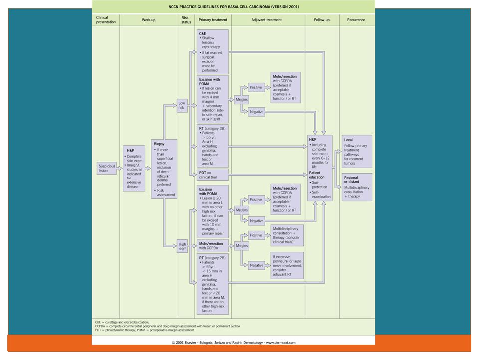
Basal Cell Carcinoma Classic pearly papule with central ulceration, telangectasia 26 variants exist: nodular, superficial, sclerosing, multifocal, pigmented, basosquamous, cystic et cetera May have history of radiation treatment—i.e. acne Comprises 80-85% of skin cancers; most common human cancer overall Usually slow-growing; derived from hair follicle/pilosebaceous unit Treatment via excision, 5-fluorouracil, dessication and currettage, Mohs surgery, cryosurgery, radiation therapy, chemical peels, laser, imiquimod, photodynamic therapy, chemotherapy --vismodegib

16
Squamous Cell Carcinoma Clinical appearance varies: Scaly, keratotic plaque; primary ulceration; exophytic, eroded plaque Often a history of tenderness of the lesion Precursor lesion: actinic keratosis/cheilitis; keratoacanthoma Sun exposed areas or prior burns/scars; check history for arsenic or radiation exposure, tobacco use Arises from keratinocytes Head and neck areas herald higher metastatic rate Treatment same as for basal cell carcinoma save not responsive to imiquimod/vismodegib

18
Management Caveats—When to Refer Histology BCC: Sclerosing/morpheaform; multifocal, micronodular, recurrent/within scar tissue—BAD signs SCC: Moderately to poorly differentiated; neurotropic, inflitrating; adenoid/adenosquamous/desmoplastic Size Anything greater than 20 mm or greater than 6 mm if in a high risk area Location Mask areas of face; genitalia; hands/feet; cosmetically sensitive Clinical Concomitant immunosuppression Chronic inflammation/ulceration/scarring Genetic disorders

20
Emerging Therapies: Basal Cell Vismodegib Small molecule inhibitor of the hedgehog signaling pathway Approved for the treatment of metastatic basal cell carcinoma or locally advanced disease that has recurred after surgery or for those who are not candidates for surgery or radiation therapy; recent approval for basal cell nevus syndrome 150 mg daily oral dosing 30% response rate in metastatic disease; 43% in locally advanced disease; mean duration 7.6 mos NEJM 2012; 366: 2171-2179 Significant side effects: muscle spasms, alopecia, dysgeusia, fatigue, weight loss—30% Cost--$90,000/yr Sequential intralesionial interferon therapy Mechanism of action similar to imiquimod Ingenol mebutate In clinical trials; currently only approved for actinic keratoses Inducer of cell death; exact mechanism of action unknown

21
Emerging Therapies: Squamous Cell Cituximab Monoclonal antibody directed against the human epidermal growth factor receptor has been approved in combination with radiation therapy for locally advanced head and neck disease Antiangiogenic therapies and tyrosinase kinase (gefitinib and erlotinib) in clinical trials Anti-vascular endothelial growth factor and human epidermal growth factor vandetanib in clinical trial Oral retinoid therapy Multiple studies in organ transplant, immunocompetent and – compromised, and high UV exposure patients Ingenol mebutate Clinical trials
 in clinical trials Anti-vascular endothelial growth factor and human epidermal growth factor vandetanib in clinical trial Oral retinoid therapy Multiple studies in organ transplant, immunocompetent and – compromised, and high UV exposure patients Ingenol mebutate Clinical trials")
22
Melanoma

25
Risk Factors Personal history of melanoma Family history of melanoma Atypical (>5) or multiple moles (>50) Fair skin type Light eyes Red-blonde hair Sunburn—Particularly prior to age 21 or intermittent Two outdoor summer jobs as a youth Giant congenital nevi (>15 cm) Genetic markers and/or syndromes
 or multiple moles (>50) Fair skin type Light eyes Red-blonde hair Sunburn—Particularly prior to age 21 or intermittent Two outdoor summer jobs as a youth Giant congenital nevi (>15 cm) Genetic markers and/or syndromes")
26
Risk Factors Higher income level Tanning Bed Use Tanning beds can emit UVA and UVB radiation at fifteen times the strength of the sun Int J Cancer 2007—meta-analysis of 19 studies RR 1.15 for melanoma with tanning bed use RR 1.75 if tanning began prior to age 35 No protective effect of tanning bed against damage from subsequent sun exposure

27
Definition Cancer of melanocytes, the pigment producing cells of the skin Melanocytes reside at the basal cell layer of the skin Neuroectodermal in origin Other common areas: retina, Organ of Corti, adrenal glands, leptomeninges Multiple variants: Superficial spreading, nodular, lentigo maligna melanoma, acral lentiginous, amelanotic, nevoid, malignant blue nevus, desmoplastic, clear cell sarcoma type, animal type Can be very difficult to classify both clinically and pathologically

28
Self-exam components ABCD (and sometimes E!) Asymmetry Border Irregularity Color Irregularity Diameter Extension or Elevation The “Ugly Duckling” sign Looking for the “outlying” mole Arch Dermatol 2008 Jan:144(1): 58-64
 Asymmetry Border Irregularity Color Irregularity Diameter Extension or Elevation The Ugly Duckling sign Looking for the outlying mole Arch Dermatol 2008 Jan:144(1): 58-64")
29
Assessment Breslow level Measurement of depth of invasion from granular cell layer down to last malignant cell Expressed in millimeters Ulceration Gross or microscopic Mitotic rate Histology—Number of dermal mitoses per mm2 (1 mm2=4.5 high power(40x) fileds Replaced Clark level for defining T1b subcategory N staging Micro- (a) or macro- (b) metastasis to one node broken out
 fileds Replaced Clark level for defining T1b subcategory N staging Micro- (a) or macro- (b) metastasis to one node broken out")
30
Treatment Excision Breslow level defines margins In situ—1/2 cm margins < or = 1mm—1 cm margins > 1mm to 2mm—1-2 cm margins > 2mm– 2 cm margins Consideration of SLN biopsy for lesions > 1mm thick or discordant Clark/Breslow Ulceration High mitotic rate

31
Survival curves by number of mitoses per millimeter squared. Thompson J F et al. JCO 2011;29:2199-2205 ©2011 by American Society of Clinical Oncology

32
Survival curves from the American Joint Committee on Cancer Melanoma Staging Database comparing (A) the different T categories and (B) the stage groupings for stages I and II melanoma. Balch C M et al. JCO 2009;27:6199-6206 ©2009 by American Society of Clinical Oncology

33
Treatment Sentinel lymph node biopsy Status of sentinel lymph node is most important prognostic indicator for disease-specific survival in patients with primary cutaneous melanoma Impact on overall survival remains unclear Not recommended for in situ or T1a Not certain of effect of adjuvant interferon therapy—promising 8-9% increased survival for stage III disease—ECOG 1684 trial Significant toxicity

34
Kaplan-Meier survival for patients undergoing successful lymphatic mapping and SLN biopsy stratified by SLN status. Gershenwald J E et al. JCO 1999;17:976-976 ©1999 by American Society of Clinical Oncology

37
Baseline laboratory tests and imaging studies are generally not recommended in asymptomatic patients with newly diagnosed primary melanoma of any thickness. No clear data regarding follow-up interval exist, but at least annual history and physical examination with attention to skin and lymph nodes is recommended. Regular clinical follow-up and interval patient selfexamination of skin and regional lymph nodes are most important means of detecting recurrent disease or new primary melanoma; findings from history and physical examination should direct need for further studies to detect local, regional, and distant metastasis. Surveillance laboratory tests and imaging studies in asymptomatic patients with melanoma have low yield for detection of metastatic disease and are associated with relatively high false-positive rates. J AM ACAD DERMATOL VOLUME 65, NUMBER 5 Bichakjian et al 1039 Recommendations

38
Management Caveats: When to Refer American Academy of Dermatology All patients with a prior diagnosis of melanoma should be seen by a Dermatologist annually Greater frequency if newly diagnosed based on the staging All first degree relatives should be screened Regular eye examination Multidisciplinary melanoma clinics based on patient, staging, recurrence Safest just to refer

39
Management Caveats When will you get in trouble? Resting on the laurels of a “negative” biopsy Resting on the laurels of time Not listening to patient Not undressing patient Not counseling patient or family Share the responsibility

40
New and Emerging Therapies Vemurafenib Inhibitor of mutated BRAF genes; 40-60% of melanomas carry this mutation—”mutation-specific” therapy 86% ages 20-30 22% aged 70+ Pivotal trial versus dacarbazine for stage IIIc or IV disease At 6 mos, survival 84% in vemurafenib group, 64% dacarbazine Median progression free survival in vemurafenib group 5.3 mos versus 1.6 with dacarbazine NEJM 2011; 364: 2507-2516 Overall still very poor prognostic rates Not a durable drug—resistance occurs; anti-sense therapy candidate? Targets bcl 2 gene expression Significant side effect of eruptive keratoacanthomata, squamous cell carcinomata—amongst others

41
New and Emerging Therapies Ipilimumab Inhibitor to CTLA4 (inhibits the inhibitor) 3mg/kg every three weeks/ 4 cycles Two studies: Ipilimumab and gp vaccine/ipilimumab alone/vaccine alone Hodi NEJM 2010; 363:711 10 mos drug and vaccine median survival 10.1 drug alone 6.4 vaccine alone Ipilimumab 10 mg/kg +dacarbazine v dacarbazine mono Robert NEJM 2011 Median 11.2 mos for both drugs 9.1 mos dacarbazine alone
 3mg/kg every three weeks/ 4 cycles Two studies: Ipilimumab and gp vaccine/ipilimumab alone/vaccine alone Hodi NEJM 2010; 363:711 10 mos drug and vaccine median survival 10.1 drug alone 6.4 vaccine alone Ipilimumab 10 mg/kg +dacarbazine v dacarbazine mono Robert NEJM 2011 Median 11.2 mos for both drugs 9.1 mos dacarbazine alone")
42
New and Emerging Therapies Ipilimumab: Significant side effects Colitis Cutaneous Stevens-Johnson/Toxic Epidermal Necrolysis Generalized eruptions Autoimmune hepatitis Endocrine abnormalities Thyroid Pituitary Demyleination Severe or fatal Sea in 10-15% of patients Cost $120,000 for a 70 kg person

43
Psoriasis

45
Why? Psoriasis affects 2.9 million people in the United States alone; 1.7 million seek treatment Average age of onset 28 years; 10% are under 10 years of age Skin lesions can antedate arthritic symptoms by ten years; 25% have arthritis 400 psoriasis related deaths each year Metabolic, treatment-related, suicide About 25% of patients have moderate to severe disease Body surface area measurements Psoriasis Area and Severity Index (PASI) scores
 scores.")
46
Psoriasis Psychological burden Increased obesity and higher incidence of smoking—Utah Psoriasis Initiative Higher incidence of diabetes, hypertension, hyperlipidemia, obesity and smoking—EADV Greater risk of myocardial infarction—JAMA 2006;296:1735-41 Independent of co-morbidities Increased risk of diabetes mellitus and likelihood of treatment for such Arch Dermatol 2012;148(9):995-1000 Independent of co-morbidities Inherent risk of lymphoma
: Independent of co-morbidities Inherent risk of lymphoma")
47
Psoriasis Co-Morbidities

50
Psoriasis can present as all of the following except: A. Erythroderma B. Isolated hand/foot disease C. Pustules D. Without cutaneous disease at all E. Only at the groin and axillae F. On the mucous membranes

52
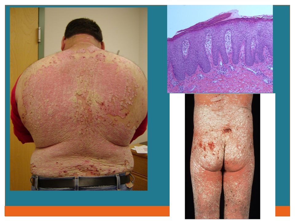
Types of Psoriasis Plaque Most common, 80% of all cases Raised, red scaly lesions Guttate Small, dot-like lesions Erythrodermic Intense redness, inflammation, some scaling Pustular Pus-filled lesions, some scaling; often localized to palms and soles Photos from the National Psoriasis Foundation

53
Types of Psoriasis Nail Pitting, onycholysis, “oil drop” changes Inverse Intertriginous areas Annular Circumferential and sepriginous Psoriatic arthritis

55
Therapies/Emerging Therapies Standard Topicals: Steroids, vitamin A and D derivatives, tar, anthralin Systemic: Methotrexate, Acitretin, Cyclosporine, Sulfasalazine’ Mycophenolate mofetil, hydroxyurea Phototherapy and chemophototherapy Biologic TNF-alpha blockers: Infliximab, etanercept, adalimumab, golimumab IL 12/23 blocker: Ustekinumab T cell blocker: Alefacept Watch for: Infection, heart failure, demyelinization Non-melanoma skin cancer, lupus, hematologic No increase in lymphoma to date with psoriasis use

56
Emerging Therapies Biologic TH 17 blockers Three finishing phase three trials Systemic Fumaric acid esterases

57
Management Caveats Referral Anyone with moderate to severe disease Suspected psoriatic arthritis Erythroderma Difficult presentation—inverse, pustular Screening questions Co-morbidities well documented Accountability for management

59
(THERE ARE WELL OVER A HUNDRED) Manifestations of Internal Disease
 Manifestations of Internal Disease")
60
What is your diagnosis? A. Stasis dermatitis B. Cellulitis C. Necrobiosis lipoidica diabeticorum D. Psoriasis

61
Stasis Dermatitis Inflammatory changes of the lower extremities in association with edema Peripheral vascular disease/venous insufficiency/congestive heart failure Acute stages/exacerbations associated with bright erythema; chronic cases with progressive lichenification, pigmentation Frequently mistaken for cellulitis, vasculitis, venous thrombosis, diabetic dermopathy

62
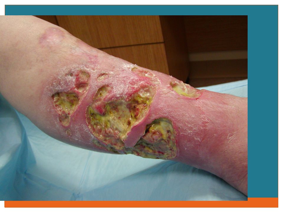
Management of this lesion should include: A. Wide surgical debridement B. Oral Vitamin C supplementation C. IV antibiotics D. Oral steroids

64
Pyoderma Gangrenosum Erythematous nodules progressing to pustules and eventually ulcers; heal with characteristic “cribriform” scarring Adults aged 40-60; seen in association with ulcerative colitis, rheumatoid arthritis, monoclonal gammopathies, hepatitis, leukemia Neutrophilic infiltration of the skin leads to abscesses and breakdown

65
Pyoderma Gangrenosum Bullous variant recognized, associated with hematologic disorders Differential diagnosis Venous ulcers/vascular disease Infectious processes Sweet’s syndrome Erythema nodosum Treatment via immunosuppressives and local care; biologics

67
What should we screen for in this condition? A. ANA B. CCP or RA C. Blood glucose D. Zinc deficiency

68
C. Blood glucose

69
Necrobiosis Lipoidica Diabeticorum Well-circumscribed, yellow-brown plaques with an erythematous border Progress to atrophic or slcerotic patches Lower extremities>>upper extremities; women>>men Strong association with the development of diabetes mellitus Differential diagnosis Dermatitidies, Erythema nodosum, Infection

71
You see these lesions on a patient’s ankle— where else should you look?

72
Lichen Planus Pruritic, purple, polygonal papules and plaques Overlying reticulate scale—”Wickham’s striae” Widespread or localized Hypertrophic variant recognized—predominately lower extremities; lichen planus pemphigoides bullous variant Skin and mucous membranes; nails show “angel wing” deformity; scarring hair loss Oral forms can rarely lead to squamous cell carcinoma

73
Lichen Planus Can see as an overlap with lupus erythematosus Unknown etiology; association with Hepatitis C, medications including non-steroidals, beta-blockers, thiazides, antimalarials, penicillamine, others Multiplicity of forms makes differential extensive

76
What should you do for this patient? A. Perform a biopsy for H and E staining B. Begin treatment for psoriasis C. Check an RPR D. Counsel not to worry—this is a self limited viral infection

77
A. Perform a biopsy

78
Sarcoidosis Granulomatous disease of uncertain etiology Violaceous papules and plaques; favor African- American women Great mimicker—verrucous, ulcerative, hypopigmented variants; can arise in scars Centro-facial involvement heralds higher incidence of pulmonary and bony disease

79
Sarcoidosis Must do systemic evaluation after confirmatory biopsy Alterations in cell-mediated immune responses Rare overlap syndrome with lymphoma Corticosteroids/chloroquine mainstay; biologics increasing in favor but may be controversial

80
This patient is experiencing progressive thickening of the skin with a loss of flexion/extension. What is a key part of her history? A. Experienced a tick bite B. Has renal failure C. Has had MRIs with exposure to Gadolinium D. Is ANCA positive E. B and C

81
E. B and C above

82
Nephrogenic Systemic Fibrosis Fibrosis or hardening of the skin and internal organs suggestive of, but distinct from, scleroderma or scleromyxedema Seen exclusively in patients with renal failure/insufficiency with or without hemodialysis Length or cause of kidney disease not relevant

83
Nephrogenic Systemic Fibrosis Diagnosis made by histopatholgy in conjunction with clinical setting Pathophysiology not understood Improvement in renal function can lead to some improvement in clinical disease, but not consistent No single effective treatment modality All anecdotal/case report

84
Nephrogenic Systemic Fibrosis Incidence equal between males, females Affects all ages, though middle-aged more common No ethnic predilection Can be widespread and fulminant in 5% of cases Extensive skin hardening can lead to contractures and rapid progression to wheelchair status

85
Nephrogenic Systemic Fibrosis Higher incidence in patients that have undergone surgical procedures—i.e. fistula placement, or have a history of clotting—question relation to imaging needs Recent association with use of Gadolinium- containing contrast agents in MRI/MRA studies has led to an FDA cautionary announcement

86
This is not ringworm...!

87
Granuloma Annulare Serpiginous, annular plaques with peripheral expansion Lightly erythematous to flesh-colored; no epidermal change Pediatric age group favored Frequently at sites of trauma Singular or eruptive forms May spontaneously involute

88
Granuloma Annulare Association with diabetes mellitus New association with dyslipidemia Arch Dermatol 2012;148 1131-7 Differential diagnosis Sarcoidosis Lichen planus Tinea corporis Amyloidosis Treatment success highly variable; steroids mainstay

Similar presentations