Download presentation
Presentation is loading. Please wait.

8
Primary lesions are de-novo lesions while secondary are either a sequence of the natural history of the disease or a modification of the primary lesion which can be either due to drug use or iatrogenic by itching. Dermatological test of choice is skin biopsy and it is done whenever we are in doubt.

15
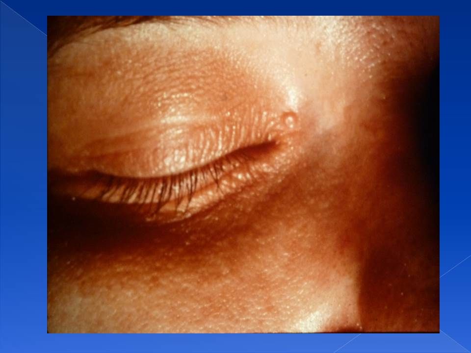
Milia Its an epidermal inclusion cyst. Simple benign and asymptomatic.
Unknown cause. Seen as papules, in the next picture they are over the cheeks and nose. Papules contain keratinized material. Resolves spontaneously within days.

16
Milia

17
Ebstein pearls It is an epidermal inclusion cysts, it affects mucous membrane and most commonly the mouth. When it ruptures it involutes spontaneously.

18
Ranula It is a congenital mucous retention thin cyst, less commonly traumatic. It ruptures spontaneously and needs no surgical intervention.

19
Ranula

20
Erythema Toxicum Neonatorum
It is a macular generalized patchy red rash. Benign and self limiting. Ruptures and resolves spontaneously. Therefore reassure the patient. Patient does not look sick and has no fever. Macules are the commonest presentation, but can also present with papules and vesicles. It is an esinophilic eruptions. Scraping taken from the lesion will show esinophils aggregate. The next picture also shows infantile gynaecomastia, which is physiological due to the maternal hormones. It can also produce milk. It resolves spontaneously when the estrogen levels go down in the blood. Do not squeeze it or touch it because you might induce mastitis.

21
Erythema Toxicum Neonatorum

22
Congenital Macular rash that appears on the back of the neck, glabella and the upper eyelid. Its an ictatic salmon like rash. It disappears with time especially if above the clavicle or on the upper eyelid. It might also persists for years and then get covered by hair.

23
Mongolian spots It occurs anywhere but mostly in the lower back or buttocks. It is a bluish- greenish discoloration that are caused by the arrest of melanocyte in the dermis. It is seen in Mongolian race, but it has nothing to do with down syndrome. Might disappear with time. Common in our society and dark-skinned people, but less common in Caucasians.

24
Mongolian spots

25
Hemangiomas It is an abnormal proliferation of the blood vessels.
It might involve the viscera or the respiratory tract. It appears as papules and increase in size. Two types: strawberry and capillary hemangiomas. Strawberry hemangioma: rapidly enlarge within the first two years, and disappears around school age. Therefore it only need reassurance of the patient. It is more benign than the capillary form. When the strawberry hemangioma enlarges, it can cause bleeding, disfigurement or ulceration. When complications occur, you treat with phototherapy, laser systemic steroids or interferon. Capillary hemangioma: it most commonly affect the area supplied by the ophthalmic division of the trigeminal nerve. It is usually associated with other symptoms. It never disappear by itself might lead to glucoma (one eye is bigger than the other).
.")
26
Strawberry hemangioma

27
Strawberry hemangioma

28
Ulcerated enlarged strawberry hemangioma

29
Capillary hemangioma with glaucoma

30
Sturge-Weber syndrome: leptomeningioma with contra-lateral hemiplegia, mental retardation and capillary hemangioma.

31
Kabel Kanani syndrome they have hemi-hypertrophy on the side of the hemangioma.

32
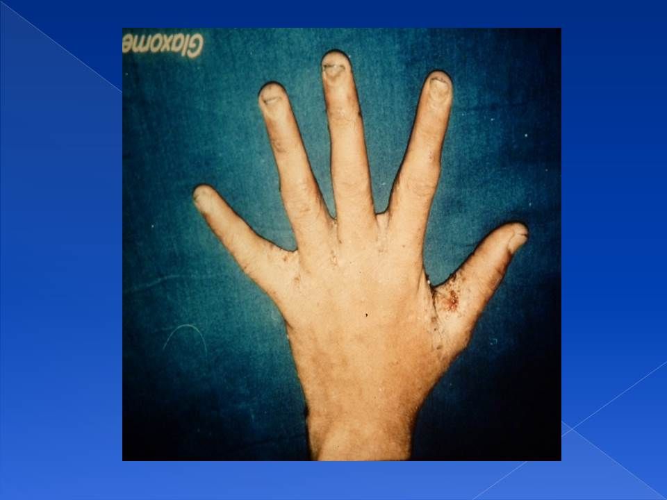
Epidermolysis Bullosa: whenever the skin is touched it sloughs out
Epidermolysis Bullosa: whenever the skin is touched it sloughs out. There is no specific treatment, you need to only prevent infection, and when infection occurs treat it.

33
Collodion baby AKA Icthyosis Congenita: the skin is very tight the baby can not move or breath. It is a form of icthyosis. The skin will get shed with time but he will continue to have icthyosis, requires supportive manegemnt.

34
Nevi They are abnormal cells in an abnormal locations.
Black hairy nevus is AKA giant or trunk nevus. It might covers the whole trunk. The larger it is the more it is likely to convert into malignant melanoma (it increases the risk 10x normal). All the previous images are congenital skin lesions.
. All the previous images are congenital skin lesions.")
35
Black Giant Nevus

36
Infantile Eczema- Atopic Dermatitis
Its not present at birth. It is an inflammatory immunological disorder. There is activation of the mast cells (increased IgE) causing the release of histamine that attacks the skin. Unknown cause. It waxes and wanes (intermittent), it might stay for several years. Usually seen on the flexures. it causes itching, which will result in secondary lesions as: liquenifacation, ulceration and excoriation. It requires intermittent long term treatment. Most common complication is by secondary bacterial infection by staph or strept because the skin barrier is broken. It can also be complicated by eczema herpiticus where herpes virus invade abnormal skin.
 causing the release of histamine that attacks the skin. Unknown cause. It waxes and wanes (intermittent), it might stay for several years. Usually seen on the flexures. it causes itching, which will result in secondary lesions as: liquenifacation, ulceration and excoriation. It requires intermittent long term treatment. Most common complication is by secondary bacterial infection by staph or strept because the skin barrier is broken. It can also be complicated by eczema herpiticus where herpes virus invade abnormal skin.")
37
Atopic dermatitis

38
Atopic dermatitis

39
Atopic Dermatitis

41
Seborrheic Dermatitis
Scaly eythematous oily skin lesions. Has nothing to do with seborrheic glands. More common in extreme ages. Involves skin creases as the axilla, face and scalp (cradle scalp-hallmark). More benign, more superficial and less itchy than atopic dermatitis. Might disappear and will not reappear until later in life (60-70) years. Treated by removing the scales, wash properly with shampoo, we might sometimes give mild steroids or salicylic acids or keratolytic drugs.
. More benign, more superficial and less itchy than atopic dermatitis. Might disappear and will not reappear until later in life (60-70) years. Treated by removing the scales, wash properly with shampoo, we might sometimes give mild steroids or salicylic acids or keratolytic drugs.")
42
seborrheic dermatitis

43
seborrheic dermatitis

44
seborrheic dermatitis

45
Cradle scalp: a hallmark for seborrheic dermatitis.

46
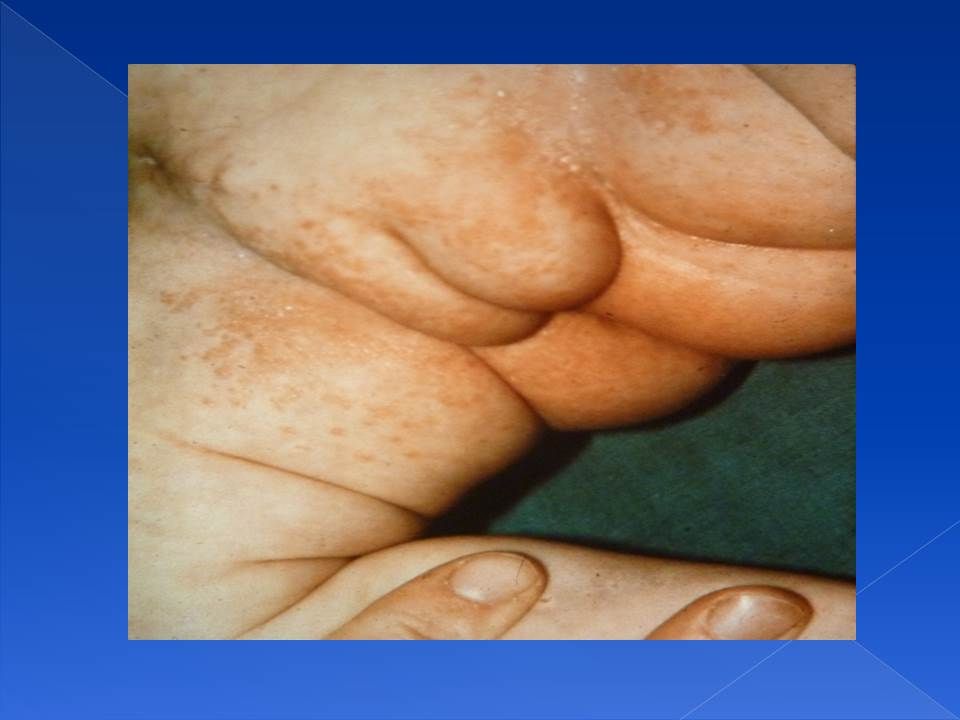
Contact Dermatitis Its irritant dermatitis AKA diaper dermatitis.
It spares creases. Its due to irritation from urine, stool and hot humid environment (as in the diaper). It might be complicated with fungal infection. In this case the creases will be involved and you might see vesicles. Whenever suspected look and the mouth for oral thrush.
. It might be complicated with fungal infection. In this case the creases will be involved and you might see vesicles. Whenever suspected look and the mouth for oral thrush.")
47
Contact Dermatitis (sparing the creases)
")
49
Fungal candidal infection

50
Oral thrush by candida

52
Bullous impetigo: caused by staph and sometimes by strept, it involves the face and limbs. It causes honey-like crusted skin infection. The bullas might rupture causing severe pain. Treated by systemic antibiotics and other supportive measures.

53
Scalded skin syndrome: its is an acute infection caused by exotoxin secreted from staph aurus. Whenever you touch the skin it comes out (? Phenomena). It is treated by antibiotics and supportive measures. It responds to treatment unlike the inherited epidermolysis bullosa.
. It is treated by antibiotics and supportive measures. It responds to treatment unlike the inherited epidermolysis bullosa..")
54
Molluscum Contagiosum: it is caused by POX DNA virus, human is the only resorvoir. There will be thick wall vesicles with indurations and umbilication. Its asymptomatic and not itchy. It is self-limiting disease, it usually disappears alone but might need scraping and if multiple cryotherapy.

56
Herpetic gingivo-stomatitis: painful ulcers that are caused by HSV type 1 if affecting the oral mucosa, or HSV type 2 if it affected the genetalia. The patient will look sick and will be febrile. it needs supportive treatment and if the patient is immuno-deficient give systemic acylovir.

57
Vericilla zoster: painful vesicular eruptions that involves a certain dermatomal area. It is caused by the activation of a dormant virus. In children it is less painful than adults. The child is infective and might transmit the disease (chicken pox) to other contacts, therefore he must be isolated. if it is severe give analgesics and acylovir.
 to other contacts, therefore he must be isolated. if it is severe give analgesics and acylovir..")
58
Scabies: it is caused by mite Sarcoptes scabiei
Scabies: it is caused by mite Sarcoptes scabiei. It causes severe itching and ulceration, positive family history of itching. Itching is the hallmark of the disease.

59
Fungal infection: the skin lesion is rounded and superficial has an active periphery and an inactive pale center. It can affect any part of the body as the trunk (tinea corpus), scalp (tinea capitis), foot (tinea pedis). It is treated by topical antifungal if possible or systemic griseofalvin if multiple.
, scalp (tinea capitis), foot (tinea pedis). It is treated by topical antifungal if possible or systemic griseofalvin if multiple..")
62
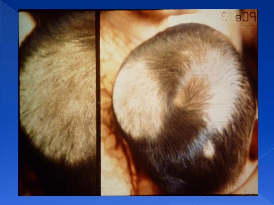
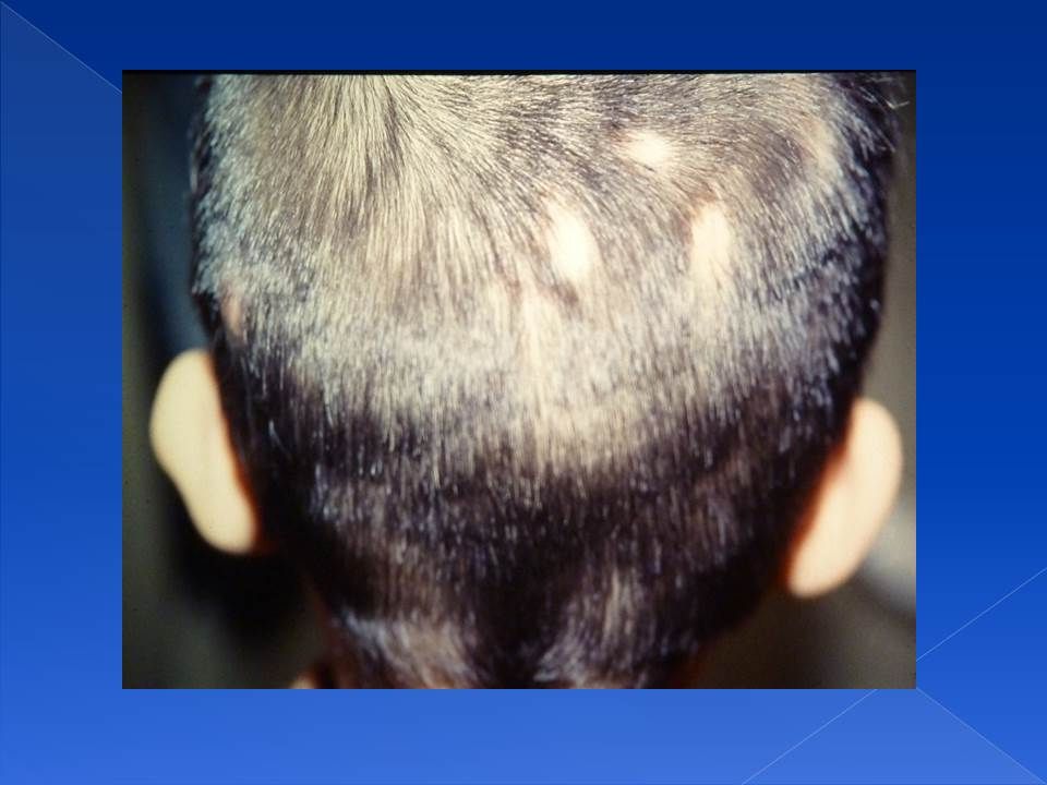
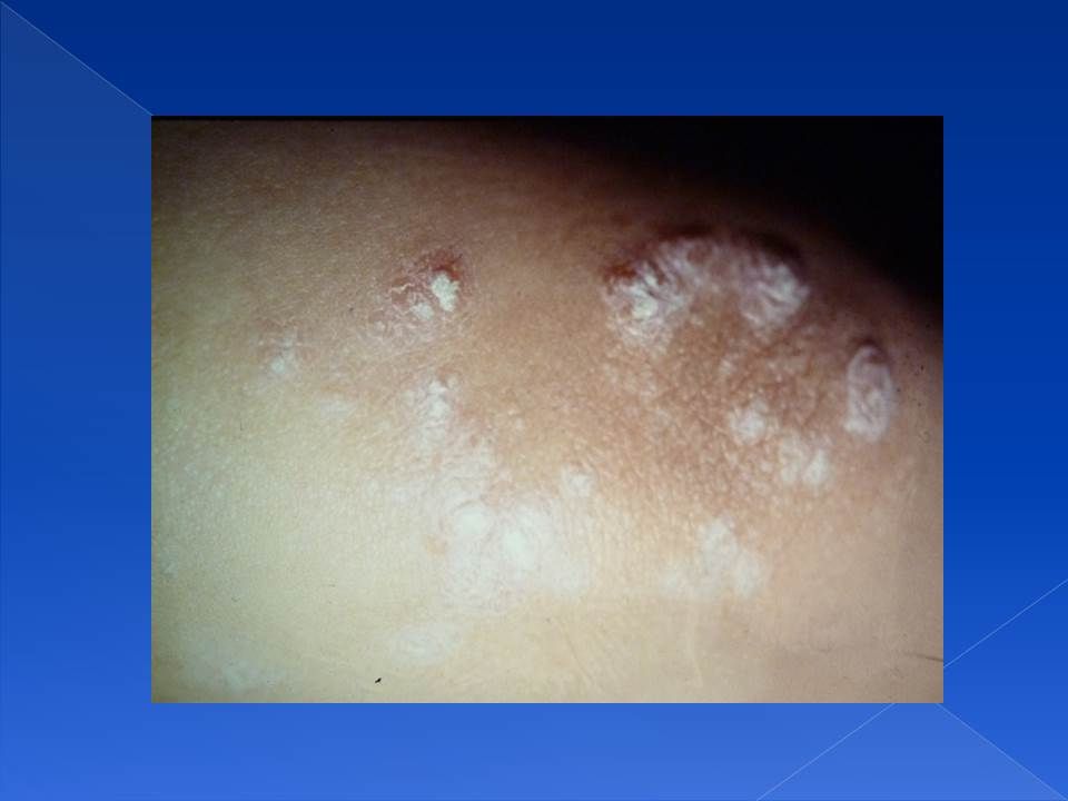
Alopecia Alopecia areata is an autoimmune diseas, should be investigated for other autoimmune endocrinopathies. The lesion is clean. Tinea capitis: dirty lesion that might ulcerate and ooze and has a bad smell. It is treated by steroids and antifungal, steroid, and sometimes antibiotics.

64
Tinea capitis

65
Alopecia areata

67
This picture is pediculosis
This picture is pediculosis. in pediculosis the nets cant be removed while dandruff it can fall off without the hair coming out.

68
Cutaneous leishmaniasis: It is caused by the parasite Leishmania Tropica or Donovani, the sand fly is the vector. It affects exposed skin. The lesion is not very itchy. It is a chronic disease treated by antimonial medications.

69
Pityriasis Rosea: it’s a scaly hyper-pigmented lesion
Pityriasis Rosea: it’s a scaly hyper-pigmented lesion. It affects adolescent age group more common in boys. It is mildly itchy and is a self limiting disease. If it was severely itchy we give steroids.

70
Pityriasis Rosea

71
Psoriasis: scaly patchis that bleeds upon removal known as auspitz’s sign. Guttate psoriasis: is milder form seen in children after throat , it causes guttate spots.

Similar presentations


.>")


