Download presentation
Presentation is loading. Please wait.

1
Dr.MOHAMED NASR Lecturer Of Dermatology & Venereology Zagazig University SYPHILIS

2
SYPHILIS Syphilis is a chronic infectious sexually transmitted disease capable of involving every structure of the body.

3
Causative organism: Treponema pallidum is a spirochaete, with the following characters: Slender, motile, and regular spiral organism. 6-15 microns in length, 0.25 micron in width and formed of 8-24 coils. Cannot be stained by ordinary bacterial stains and appears pale (hence the name pallidum).
..")
5
Diagnosis of syphilis: 1- Clinical examination. 2- Dark-ground examination. 3- Serological tests. 4- Other investigations.

6
Serological tests of syphilis: A- Reagin tests or standard tests of syphilis (S.T.S) or non-specific tests. B- Specific tests.

7
A- Reagin tests or S.T.S.: In the course of syphilis, an antibody (reagin) appears in the serum of the patients. This reagin may be demonstrated by using non- treponemal antigen, i.e. antigen not extracted from treponema pallidum. There are 2 types of S.T.S.

8
1-Complement fixation test: Wasserman reaction (W.R.): The reagin unites with the detecting antigen in the presence of complement. The union is invisible and fixation of complement could be detected by using another antigen –antibody system (indicator system; sheep's RBCs+sheep'S RBCs antibody). If haemolysis occurs this means the complement is not fixed during the first reaction i.e the test is negative. If no haemolysis ocurrs this means that the complement is consumed and fixed in the first antigen antibody reaction, i.e positive result.
. If haemolysis occurs this means the complement is not fixed during the first reaction i.e the test is negative. If no haemolysis ocurrs this means that the complement is consumed and fixed in the first antigen antibody reaction, i.e positive result..")
9
2-Flocculation or precipitation tests: a- Venereal Disease Research Laboratory Test (VDRL): the antigen –antibody reaction gives visible flocculation with no need for an indicator system. This can be done in a test tube or on a glass slide. b- Kahn's test is also a flocculation test but is less reliable than VDRL test.

10
The S.T.S. tests are: Negative in the first stage (15-30 days after the chancre has appeared) and become positive after that period in 50% of cases and then increasing gradually till the onset of secondary stage. Positive in 100% of cases in the secondary stage. Positive in 70-90% of cases of the tertiary stage.
 and become positive after that period in 50% of cases and then increasing gradually till the onset of secondary stage. Positive in 100% of cases in the secondary stage. Positive in 70-90% of cases of the tertiary stage..")
11
B-Specific tests for syphilis: Specific antibodies against Treponema pallidum are found in the serum of syphilitic patients and could be demonstrated by using treponemal antigens. These tests include:

12
1-Treponema Pallidum Immobilization Test (T.P.I.): Specific antitreponema antibodies are found in the serum of the patient. They can immobilize T. pallidum when patient's serum is put on living spirochetes suspension. The test is positive when 70% or more of living T. pallidum are immobilized.

13
2-Fluorescent Treponemal Antibody Test (F.T.A.): A diluted serum of the patient is added to a dried drop of suspension of dead T.pallidum fixed on a glass slide. If the serum contains the antibody it will adhere and coat the treponemes. This could be detected by adding a fluorescent substance and examining microscopically.

14
3-Treponema Pallidum Hemoagglutination Test (T.P.H.A.): Sheep erythrocytes which are sensitized to Treponema and treated with tannic acid are added to the patient's serum. Haemagglutination of the erythrocytes means positive results due to the presence of anti- Treponema antibody in the serum.

15
Other investigations: 1. Cerebrospinal fluid (C.S.F.): Examination to exclude or to diagnose neurosyphilis. 2. Radiological examination: For diagnosis of late acquired syphilis and congenital syphilis. 3. Biopsy: a- Chronic inflammation with infiltration of lymphocytes and plasma cells. b- Endarteritis obliterans of the blood vessels. c- Fibroblastic activity in the periphery of the lesion.
: Examination to exclude or to diagnose neurosyphilis. 2. Radiological examination: For diagnosis of late acquired syphilis and congenital syphilis. 3. Biopsy: a- Chronic inflammation with infiltration of lymphocytes and plasma cells. b- Endarteritis obliterans of the blood vessels. c- Fibroblastic activity in the periphery of the lesion..")
16
ACQUIRED SYPHILIS Classification: a. Early infectious phase (first 2 years of the infection): 1. Primary stage (3-9 weeks). 2. Secondary stage (3-9 months). 3. Recurrent stage (relapse). 4. Early latent stage (9 months - 2 years). b. Late non-infectious phase (after 2 years of the infection): 1- Late latent stage. 2- Tertiary stage. 3- Cardiovascular and neurosyphilis.
. 2. Secondary stage (3-9 months). 3. Recurrent stage (relapse). 4. Early latent stage (9 months - 2 years). b. Late non-infectious phase (after 2 years of the infection): 1- Late latent stage. 2- Tertiary stage. 3- Cardiovascular and neurosyphilis..")
17
Mode of infection: a. Direct: 1. Sexual intercourse with infected partner. 2. Close contact with a syphilitic person e.g. kissing. 3. Touching a syphilitic lesion, e.g. doctors, nurses. 4. Blood transfusion from a syphilitic donor. b. Indirect 1. Using infected materials, e.g. cups, spoons....etc. 2. Unsterilized medical instruments, e.g. needles.

18
A. EARLY INFECTIOUS PHASE I. PRIMARY STAGE OF SYPHILIS Incubation period: 9-90 days, (average 2-4 weeks). a. Chancre It is the first tissue reaction to treponemal infection. It is an ulcer with the following characters: 1- Single but occasionally multiple. 2-Raised border, clean dull-red granulating floor and indurated base. 3- It oozes serum which dry giving yellowish gray crust and contains treponemas. 4- Not painful not tender unless secondarily infected. 5- Does not bleed easily on manipulation due to end arteritis obliterans. It occurs on the genital areas (95%). It may rarely occur on extragenital areas (5%). The ulcer heals, if untreated leaving a thin, atrophic and non- contractile scar.
. a. Chancre It is the first tissue reaction to treponemal infection. It is an ulcer with the following characters: 1- Single but occasionally multiple. 2-Raised border, clean dull-red granulating floor and indurated base. 3- It oozes serum which dry giving yellowish gray crust and contains treponemas. 4- Not painful not tender unless secondarily infected. 5- Does not bleed easily on manipulation due to end arteritis obliterans. It occurs on the genital areas (95%). It may rarely occur on extragenital areas (5%). The ulcer heals, if untreated leaving a thin, atrophic and non- contractile scar..")
20
Special types of chancre: 1. Kissing chancre: on contagious surfaces e.g scrotum, thigh and lips. 2. Condom chancre: at the root of the penis of those wearing condoms. 3. Concealed chancre: intracervical, intrameatal or intraanal. 4. Chancre redux: recurrence of chancre at the site of healed chancre, during the secondary stage. 5. Pseudochancre redux: is a gumma that appears at the site of original chancre.

21
b. Regional lymph glands Enlarged, firm rubbery and not matted. Painless and not tender unless secondarily infected. The enlargement is bilateral in genital chancre and unilateral in extragenital one.

22
Fate of primary syphilis: 30% of cases: the disease burns itself out. 30% of cases: become serologically positive for life. 20% of cases: pass to benign tertiary syphilis. 20% of cases: pass to malignant syphilis.

23
Diagnosis: 1-Clinical examination. 2-Dark-ground examination. 3-Serological tests: they are negative early in this stage (sero-negative) and later become positive.
 and later become positive..")
24
Differential diagnosis

25
a. Genital ulcer: 1- Scabies. 2- Fixed drug eruption. 3- Herpes genitalis; grouped painful vesicles on an erythematous base. 4- Soft sore (chancroid). 5. Lymphogranuloma Venereum: the ulcer is small and transient. 6. Granuloma inguinale: The genital ulcer may simulate chancre but the lymph glands are not enlarged. 7. Malignant ulcer: the ulcer is indurated with rolled everted edge and may be fixed to the underlying structure. Lymph nodes enlargement late and they are stony hard. 8. Herpes Zoster: unilateral Grouped painful vesicles on an erythematous base.
. 5. Lymphogranuloma Venereum: the ulcer is small and transient. 6. Granuloma inguinale: The genital ulcer may simulate chancre but the lymph glands are not enlarged. 7. Malignant ulcer: the ulcer is indurated with rolled everted edge and may be fixed to the underlying structure. Lymph nodes enlargement late and they are stony hard. 8. Herpes Zoster: unilateral Grouped painful vesicles on an erythematous base..")
26
b. Extragenital chancre: On the lips or tongue: 1. Primary tuberculous lesion. 2. Herpes simplex with secondary pyogenic infection. 3. Aphthous stomatitis. On the nipple: 1. Simple fissuring. 2. Paget's disease. On the anus: 1. Anal fissure. 2. Thrombosed external piles.

28
II. SECONDARY STAGE OF SYPHILIS It occurs in untreated patients, this stage starts usually 6-8 weeks after the appearance of chancre. It is characterized by: 1. Healing or healed chancre. 2. Skin rash. 3. Generalized lymphadenopathy. 4. Mucous membrane lesions. 5. Condylomata lata. 6. Other signs.

29
1. Healing or healed chancre: The chancre may still be present or a scar may be formed indicating its previous existence.

30
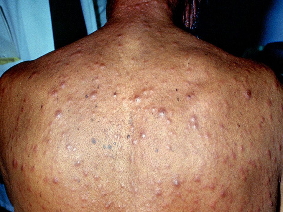
2. Skin Rash: a. Widely spread, bilateral, symmetrical and more prominent on the flexor surfaces. b. Rounded, dull red in colour and polymorphic but never vesiculates. No itching. c. The rash is chronic and may persist for weeks or months if untreated.

32
Four main types of rash occur: 1. Macular syphilid: Rose pink in colour, discrete lesions distributed mainly on the chest, back, abdomen and flexural surfaces 2. Papular syphilid: According to their distribution they may get many descriptive names: a. Corona veneris: line of papules on the forehead just below the hair line. b. Follicular syphilid: follicular papules related to the hair follicles. These follicular papules when occurring on the scalp may lead to irregular alopecia (moth-eaten alopecia). 3. Papulo-squamous syphilid: These are papules with scaling which resulted from diminution of the blood supply due to endarteritis obliterans. 4. Pustular syphilid: The papules may undergo deep central necrosis giving the appearance of a pustule.
. 3. Papulo-squamous syphilid: These are papules with scaling which resulted from diminution of the blood supply due to endarteritis obliterans. 4. Pustular syphilid: The papules may undergo deep central necrosis giving the appearance of a pustule..")
33
3. Generalized lymphadenopathy: - Non-painful, rubbery and discrete enlargement of lymph glands are present in 50% of cases.

34
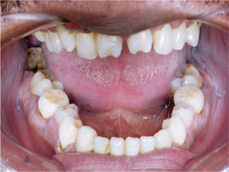
4. Mucous membrane lesions: The characteristic lesion is the mucous patch which is painless, grayish white patch edged by a dull red border. These patches may be present on buccal mucosa, tongue, larynx and soft palate leaving erosion which is, sometimes, called snail-track ulcer.

36
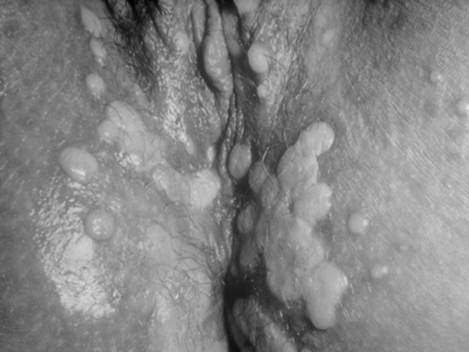
5. Condylomata lata: These are papules, flat-toped, dull red or grayish white (due to necrosis), with broad base and are very infectious (the most infective syphilitic lesions).
, with broad base and are very infectious (the most infective syphilitic lesions)..")
39
6. Other signs of secondary stage: a. Hepatitis with mild jaundice. b. Uveitis and choroido-retinitis. c. Periostitis of long bones. d. Onychia and paronychia. e. Acute or subacute meningitis.

40
Diagnosis of secondary stage: 1. History and clinical examination. 2. Dark-ground examination from the moist lesions. 3. Serological tests for syphilis are 100% positive. 4. C.S.F examination: changes are found only in 10- 20% of cases (not routinely examined in secondary stage).
..")
41
III. STAGE OF RECURRENCE (RELAPSE) a. By relapse we mean return of the disease (clinically or serologically) after its disappearance with or without treatment. b. This usually occurs in the first two years of infection.
 after its disappearance with or without treatment. b. This usually occurs in the first two years of infection..")
42
IV. EARLY LATENT SYPHILIS At this stage, the patient is completely free of signs and symptoms with positive blood tests for syphilis. This stage ends by the beginning of the third year of infection.

43
B. LATE NON INFECTIOUS SYPHILIS This phase includes 3 stages: 1. Late latent sage. 2. Tertiary stage. 3. Cardiovascular and neurosyphilis.

44
1. LATE LATENT STAGE This is the continuation of the early latent stage. The disease in late latent stage may be discovered by chance, e.g. during investigations done before marriage, ante-natal care, before joining military or governmental services. The diagnosis of this stage depends upon positive S.T.S. and should be confirmed by one of the specific tests.

45
2. TERTIARY STAGE This stage starts usually 3-10 years after infection. The characteristic lesion of this stage is the gumma which is a localized swelling, single or multiple, varying in size from a pin-head to a bigger mass several centimeters in diameter.

47
The gumma is characterized by: a. Pathologically: 1. Central necrosis (due to endarteritis obliterans) surrounded by: granulation tissue infiltrated by plasma cells, lymphocytes and fibroblasts. 2. No treponemas are present.
 surrounded by: granulation tissue infiltrated by plasma cells, lymphocytes and fibroblasts. 2. No treponemas are present..")
48
b. Clinically: 1. Asymmetrical and painless. 2. Single or in isolated groups. 3. Destructive (forming punched out ulcer) 4. Slow healing (forming thin atrophic scar). 5. It may affect: Covering structures of the body, e.g. skin, mucous membranes and subcutaneous tissues. Supporting structures of the body, e.g. bones, joints, muscles and ligaments. Internal organs, e.g. stomach, intestines, liver, spleen, lung and testis.
 4. Slow healing (forming thin atrophic scar). 5. It may affect: Covering structures of the body, e.g. skin, mucous membranes and subcutaneous tissues. Supporting structures of the body, e.g. bones, joints, muscles and ligaments. Internal organs, e.g. stomach, intestines, liver, spleen, lung and testis..")
49
The gumma involving the skin is either: 1. Nodular. 2. Squamous or psoriasiform lesion; a nodule with waxy scaling. It may occur on the palms and soles leading to unilateral hyperkeratosis. 3. Subcutaneous; which may be ulcerated, giving gummatous ulcer which is: punched out with vertical wall and polycyclic borders, not painful with no regional lymph glandular enlargement.

50
The gumma of the mucous membrane When it ulcerates it may destruct the underlying structures: 1. In the nasal septum it leads to saddle nose. 2. In the soft palate it ends by destruction and perforation 3. In the tongue it may give one of the following pictures: a. Macroglossia: due to diffuse gummatous infiltration. b. Superficial glossitis: red smooth glazed area due to destruction of the tongue papillae. c. Chronic interstitial glossitis: the tongue may be smaller than normal. d. Leucoplakia: irregular white patch induced by chronic irritation.

52
Diagnosis of Tertiary stage: a.Clinically: Any residual stigmata of early syphilis should be looked for. b.Serologically: S.T.S. is positive in 90% of cases (if negative specific tests should be done).
..")
53
CONGENITAL SYPHILIS Congenital syphilis is acquired by placental transmission of infection from the mother. Congenital syphilis can be prevented by detection and treatment of infected mothers.

54
The outcome of prengancy: During early stages of infection, there are large numbers of T.pallidum with the following effects: a. Abortion. b. Stillbirth. c. Early neonatal death. If infection occurs late in pregnancy or is milder; the following may occur: a. Premature born child. b. Some children are born apparently healthy and signs may be delayed for days, weeks or months.

55
I. EARLY CONGENITAL SYPHILIS In this phase the manifestations, occur in the first 2 years of life. They are similar to those of secondary stage of acquired syphilis. There is no primary stage in congenital syphilis as the infection is blood-borne.

56
1. Skin: a. Bullous eruption mainly on the palms and soles. b. Rashes like those in the secondary stage of acquired syphilis. c. Condylomata lata in the napkin area. d. Fissuring of the angles of the mouth and wrinkling of the skin of the face due to weight loss (old man look). e. Brownish yellow patches on the skin (café au lait tint).
. e. Brownish yellow patches on the skin (café au lait tint)..")
57
2. In the mucous membrane: (mucous patches) a. The lips, mouth and throat. b. The mucoperiosteum of the nasal cavity giving rise to syphilitic rhinitis. c. The larynx leading to thin or aphonic cry.

58
3. Lymphadenitis.

59
4. Bones: a. In the first six months : Osteochondritis of the long bones. b. After 6 months: Periostitis of the long bones. c. After one year: Osteoperiostitis of the phalanges (syphilitic dactylitis).
..")
60
5. Viscera: a. Enlarged liver and spleen. b. Lungs (Pneumonia Alba). c. Meningitis
. c. Meningitis")
61
Diagnosis of early congenital syphilis: 1- Dark-ground examination. 2- Serological tests.

62
II. LATE CONGENITAL SYPHILIS After the first 2 years of life.

63
1. Interstitial keratitis (the commonest): Circumcorneal vascularization of the sclera, which then extends to the deep layers of the cornea.
: Circumcorneal vascularization of the sclera, which then extends to the deep layers of the cornea..")
64
2. Gummatous osteoperiostitis of the bones: Thickening of the middle third of tibia (Sabre tibia). Bony swellings of the skull (Parot's nodes). Destruction of the nasal septum with depression of the lower part of the nose. Swelling (not painful) of big joints, especially the knee joint (Clutton's joint).
. Destruction of the nasal septum with depression of the lower part of the nose. Swelling (not painful) of big joints, especially the knee joint (Clutton s joint)..")
66
3. Auditory nerve affection. 4. Skin and mucous membrane affection: Similar to those in late acquired state. 5. C.N.S. affection.

67
III. THE STIGMATA These include the scars and deformities of previous syphilitic lesions and remain for life.

68
1. Stigmata of early congenital syphilis: a. Saddle nose: Depression of the bridge of the nose due to decreased growth of the nasal bones and septum which is the end result of syphilitic rhinitis. b. Hutchinson's Teeth: Small, separated and screw driver like central incisors of the permanent teeth due to affection of tooth buds late in pregnancy. c. Syphilitic Rhagades: Scars radiating from the angles of the mouth due to papular eruption with maceration and secondary infection in the mouth angles.

72
2. Stigmata of late conegnital syphilis: a. Sabre tibia: Due to osteoperiostitis in the tibia with new bone formation. b. Perforation: Perforation of nasal septum and soft palate. c. Ghost vessels: Network of empty vessels deep in the cornea end result of interstitital keratitis. d. Frontal bossing: Broad frontal region due to periosteal nodes in the frontal bone. e. Optic atrophy: A part of C.N.S. affection.

73
N.B.: Hutchinson's triad: Hutchinson's teeth + Interstitial keratitis + 8th nerve deafness.

74
Diagnosis of late congenital syphilis: 1. Clinical manifestations 2. S.T.S. are positive in 80-90% of cases and when negative specific tests should be done.

75
TREATMENT OF SYPHILIS Penicillin is the drug of choice as: 1-Procaine penicillin G (2% aluminum monostearate was added to prolong its action (P.A.M) but this is historical and not used now) 2-Benzathine penicillin G.
 but this is historical and not used now) 2-Benzathine penicillin G.")
76
TREATMENT REGIMEN STAGE OF SYPHILIS Aqueous procaine penicillin GBenzathine penicillin G 600.000 units IM daily for 15 days 2.4 million Units IM once. Early syphilis (primary, secondary, early latent). 600.000 units IM daily for 15 days 2.4 million units IM once/week for 3 weeks Late latent and late benign syphilis 600.000 units IM daily for 20 days -Cardiovascular syphilis 600.000 units IM daily for 20 days -Neurosyphilis 450.000 units /kg IM divided on 10 daysNot recommended, as it is painful.Congenital syphilis
 units IM daily for 15 days 2.4 million units IM once/week for 3 weeks Late latent and late benign syphilis units IM daily for 20 days -Cardiovascular syphilis units IM daily for 20 days -Neurosyphilis units /kg IM divided on 10 daysNot recommended, as it is painful.Congenital syphilis.")
77
COMPLICATIONS OF TREATMENT 1. Jarisch-Herxheimer reaction (J-H): Following the first injection of penicillin by 2-12 hours as the patient may get fever and flushing with influenza like attack. The syphilitic lesion becomes inflamed and intensified. This reaction usually disappears the next day. The cause of this reaction is unknown but it is said that it may be a hypersensitivity reaction to the products of the treponemes destroyed by penicillin i.e it is not a drug reaction. It is an all or none type of reaction regardless the type or dose of penicillin
: Following the first injection of penicillin by 2-12 hours as the patient may get fever and flushing with influenza like attack. The syphilitic lesion becomes inflamed and intensified. This reaction usually disappears the next day. The cause of this reaction is unknown but it is said that it may be a hypersensitivity reaction to the products of the treponemes destroyed by penicillin i.e it is not a drug reaction. It is an all or none type of reaction regardless the type or dose of penicillin.")
78
2. Penicillin hypersensitivity: a- Anaphylactic shock (immediate). b- Skin rash (delayed) e.g. urticaria. If the patient is allergic to penicillin, other drugs should be used: a. Tetracyclines: 2 grams daily for 15-20 days. b. Erythromycin: 2 grams daily for 15-20 days. c. Cephaloridine. 2 grams daily for 10-15 days. d.quinolones
 e.g. urticaria. If the patient is allergic to penicillin, other drugs should be used: a. Tetracyclines: 2 grams daily for days. b. Erythromycin: 2 grams daily for days. c. Cephaloridine. 2 grams daily for days. d.quinolones.")
79
3. Therapeutic paradox: It is defined as worsening of the disease after antisyphilitic therapy resulting from excessive scarring produced by too rapid destruction of treponemes and healing of syphilitic lesions by fibrosis. This fibrosis leads to distortion of the surrounding structures. It is most notable in patients with aortic disease.

80
THANK YOU

Similar presentations


>")


>")


