Download presentation
Presentation is loading. Please wait.

1
Digestive System Chapter 17

2
Undernourished – diet deficient in calories
Malnourished – diet is missing one or more essential nutrients 4 classes of essential nutrients: 1. Essential amino acids 2. Essential fatty acids 3. Vitamins Minerals

4
Obesity in the US % of obese people has doubled in 20 years 30% in US
Another 35% are overweight 15% of children and adolescents are overweight Obesity is a factor in over 300,000 deaths/year

7
Food Processing Ingestion – act of eating (1st stage)
Digestion – process of breaking down into molecules small enough for the body to absorb (2nd stage) Enzymatic Hydrolysis Absorption – take-up of small molecules by cells (3rd stage) Elimination – of undigested material (4th stage)
 Enzymatic Hydrolysis. Absorption – take-up of small molecules by cells (3rd stage) Elimination – of undigested material (4th stage)")
8
Alimentary Canals Tubular part of the digestive tract from the mouth to the anus - mouth, pharynx, esophagus, stomach, small intestine, large intestine, and anal canal - accessory organs of the digestive system; salivary glands, liver, gall bladder, pancreas (these are not a part of the alimentary canal)
")
9
Movements through the alimentary canal
Segmentation – alternating contraction and relaxing of smooth muscle ex: muscular contraction of muscle every 20 seconds Peristalsis – a wavelike motion in which a ring of contraction occurs in the wall of the tube ex: pushes food down the esophagus

10
Layers of the Wall of the Alimentary Canal
Mucosa – epithelium, connective tissue, smooth muscle protection, secretion, absorption Submucosa – loose connective tissue, blood and lymph vessels, nerves Nourishment of tissues, transports absorbed material Muscular Layer – smooth muscle fibers Movements of the tube and its contents Serosa – epithelium, connective tissue Protection, lubrication

11
Mouth, pharynx and esophagus initiate food processing
Oral Cavity – saliva (mucin, buffers, antibacterial agents) Digestion of carbohydrates Bolus Pharynx – the throat, leads to esophagus Glottis and epiglottis Esophagus – peristalsis pushes bolus Salivary amylase continues digestion of starch
 Digestion of carbohydrates. Bolus. Pharynx – the throat, leads to esophagus. Glottis and epiglottis. Esophagus – peristalsis pushes bolus. Salivary amylase continues digestion of starch.")
12
Salivary Glands Serous cells produce watery fluid with salivary amylase Mucous cells produce thick liquid called mucus 1. Parotid Glands: largest salivary glands, secrete clear, watery fluid with salivary amylase (Stenson’s duct) Submandibular Glands: floor of mouth, ducts open near lingual frenulum, secrete serous and mucus fluids (Wharton’s duct) Sublingual Glands: inferior to tongue, produce thick, stringy mucus (Rivinus’s duct)
 Submandibular Glands: floor of mouth, ducts open near lingual frenulum, secrete serous and mucus fluids (Wharton’s duct) Sublingual Glands: inferior to tongue, produce thick, stringy mucus (Rivinus’s duct)")
13
The Teeth 20 primary (deciduous) 32 secondary (permanent)
Hardest structures in body 2 portions – crown and root Enamel covers crown Cementum (bone-like material) and periodontal ligament surrounds root
 and periodontal ligament surrounds root.")
14
The Mouth Tongue Lingual frenulum Uvula Palate

15
Connects nasal and oral cavity with larynx and esophagus 3 parts:
Pharynx Connects nasal and oral cavity with larynx and esophagus 3 parts: Nasopharynx Oropharynx Laryngopharynx

16
Esophagus Passageway for food from the pharynx to the stomach
Penetrates the diaphragm through the esophageal hiatus Esophageal sphincter (cardiac sphincter) Hiatal Hernia – stomach protruding through weakened area in diaphragm and the esophageal sphincter
 Hiatal Hernia – stomach protruding through weakened area in diaphragm and the esophageal sphincter.")
19
The Stomach Parts: Cardia, Fundus, Body, Pyloric Antrum
Contents mixed every 20 seconds – an acid chyme is formed. Hunger pains result when an empty stomach churns Openings at both ends - Cardiac sphincter (esophagus to stomach) and pyloric sphincter (stomach to small intestine) Acid chyme is produced Stomach wall not adapted for absorption
 and pyloric sphincter (stomach to small intestine) Acid chyme is produced. Stomach wall not adapted for absorption.")
21
Gastric Secretions Mucous Membrane – inner lining with many gastric glands 3 cell types: mucous cell mucous chief cell digestive enzymes parietal cell HCl Gastric Juice – pepsinogen, pepsin, HCl, mucus, intrinsic factor

22
Gastric Secretion Cephalic Phase (30% - 50%)
sight, smell, taste, and thought of food triggers gastric juice secretion Gastric Phase (40% - 50%) Food in stomach Gastrin released gastric juice released pH approaches 1.5 gastrin secretion stops HCl is released Intestinal Phase (5%) Begins when food leaves stomach Intestinal gastrin released increases gastric juice secretion Fats and proteins in intestine stimulate cholecystokinin decreases gastric motility. Fats also increase intestinal somatostatin inhibits gastric juice
 Food in stomach. Gastrin released gastric juice released. pH approaches 1.5 gastrin secretion stops. HCl is released. Intestinal Phase (5%) Begins when food leaves stomach. Intestinal gastrin released increases gastric juice secretion. Fats and proteins in intestine stimulate cholecystokinin decreases gastric motility. Fats also increase intestinal somatostatin inhibits gastric juice.")
23
ALKALINE TIDE ENTEROGASTRIC REFLEX Stomach secretes HCl
Gets H+ from blood Bicarbonate released into blood Blood conc. of bicarbonate increases Urine excretes excess bicarbonate ENTEROGASTRIC REFLEX Entero = small intestine Gastric = stomach - Food in duodenum stretches walls Reflex: - decreased peristalsis in stomach - intestinal filling slows Regulates rate at which chyme leaves the stomach

24
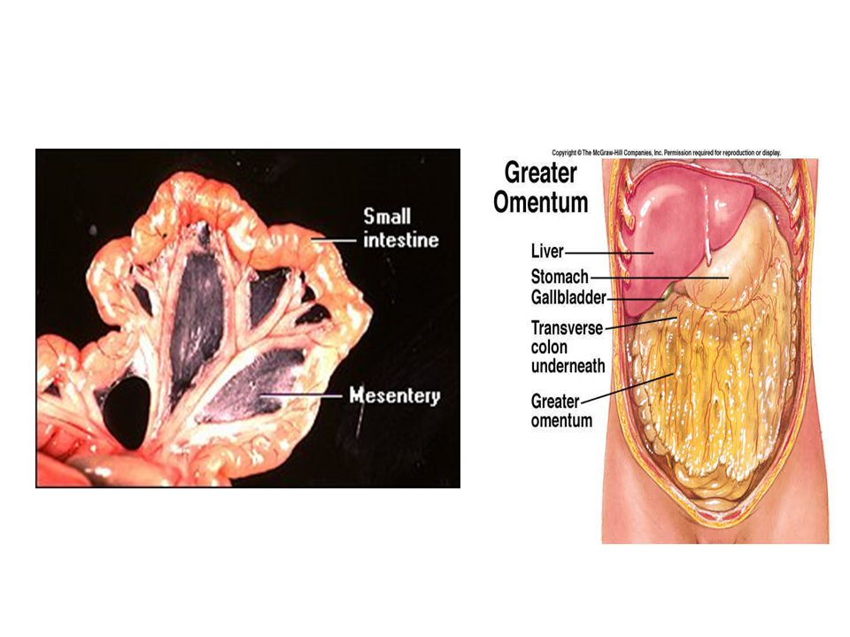
Small Intestine Major organ of digestion and absorption
20 feet in length (5.5 – 6.0 meters) 3 regions: 1. Duodenum – shortest section 2. Jejunum – 2/5 of intestine 3. Ileum Mesentery – fold of the peritoneum Greater Omentum – a drap over the stomach and small intestine
 3 regions: 1. Duodenum – shortest section. 2. Jejunum – 2/5 of intestine. 3. Ileum. Mesentery – fold of the peritoneum. Greater Omentum – a drap over the stomach and small intestine.")
26
3 Main Functions of Small Intestine
Completes digestion Absorbs digestive products Transports remaining residue to Large Intestine

27
Structure of the Small Intestine
Intestinal villi – most numerous in duodenum and proximal jejunum Microvilli – brush border Goblet cells and Brunner’s Glands secrete mucous Cell lining is replaced every 3-6 days through mitosis – Cellular Turnover - Feces is 25% dead intestinal cells Plicae Circulares – circular folds of the mucosa (helps increase surface area)
")
28
Structure of Intestinal Wall

29
Intestinal Enzymes Peptidases Sucrase, Maltase, Lactase
Intestinal Lipase Enterokinase

30
Large Intestine Reabsorption of water – 90% of water that enters alimentary canal (along with small intestine), preparation of feces, and bacteria that produce Vitamin K, biotin, folic acid, and methane 12 – 24 hours to travel length Rich flora of mostly harmless bacteria (E. coli) Ileocecal valve prevents movement back into small intestine Terminal portion is the rectum – waste exits the anal sphincter through voluntary control
, preparation of feces, and bacteria that produce Vitamin K, biotin, folic acid, and methane. 12 – 24 hours to travel length. Rich flora of mostly harmless bacteria (E. coli) Ileocecal valve prevents movement back into small intestine. Terminal portion is the rectum – waste exits the anal sphincter through voluntary control.")
31
Parts of Large Intestine
Cecum – beginning, pouchlike structure, hangs inferior to ileocecal valve appendix – no digestive function Colon: - ascending, Transverse, Descending, Sigmoid colon Rectum Anal Canal

33
Wall lacks villi and plicae circularis
Teniae coli – 3 distinct bands of muscle fibers which exert tension and create a series of pouches (haustra) No digestive functions Has Goblet cells – mucous secretions Absorption is limited to water and electrolytes Intestinal flora – break down some cellulose, and help produce vitamins such as K, B12, thiamine and riboflavin Feces – water, electrolytes, mucus, and bacteria. Bile gives its color
 No digestive functions. Has Goblet cells – mucous secretions. Absorption is limited to water and electrolytes. Intestinal flora – break down some cellulose, and help produce vitamins such as K, B12, thiamine and riboflavin. Feces – water, electrolytes, mucus, and bacteria. Bile gives its color.")
34
The Pancreas Pancreatic acinar cells form clusters called acini – produce pancreatic juice Pancreatic duct – extends length of pancreas into duodenum Hepatopancreatic ampulla (ampulla of Vater) – pancreatic and bile ducts Hepatopancreatic sphincter (sphincter of Oddi) – band of smooth muscle that surrounds the ampulla
 – pancreatic and bile ducts. Hepatopancreatic sphincter (sphincter of Oddi) – band of smooth muscle that surrounds the ampulla.")
35
Pancreatic Juice Enzymes that digest carbs, fats, proteins, and nucleic acids Pancreatic amylase Pancreatic Lipase Trypsin Chymotrypsin Carboxypeptidase Nucleases A peptide hormone – SECRETIN – stimulates pancreas to secrete bicarbonate when acid chyme enters the stomach. Secretin is released from duodenal mucous membrane Cholecystokinin stimulates pancreas to release pancreatic juice

36
The Liver Largest internal organ 4 lobes
Falciform Ligament – separates right and left lobes Lobes are separated into hepatic lobules – the functional unit of the liver - hepatic cells around a central vein - hepatic sinusoids surround cells - hepatic portal vein - carries blood from digestive tract to liver - hepatic artery carries oxygenated blood to liver Kupffer cells – line hepatic sinusoids and phagocytize

37
Liver Functions Carbohydrate metabolism – responds to insulin and glucagon Lipid metabolism – oxidizes fatty acids Synthesizes lipoproteins, phospholipds, and cholesterol Protein metabolism – deaminate amino acids, forming urea Storage of glycogen, iron, vitamins A,D, B12 Destruction of damages RBC’s Removal of toxic substances from blood

38
Gall Bladder Connected to cystic duct which joins the common hepatic duct to form bile duct Stores bile between meals, concentrates bile by reabsorbing water, and releases bile into duodenum Bile duct – forms from union of common hepatic duct and cystic duct Bile salts break fat globules into droplets – emulsification Lipases are then able to digest fats more effectively Bile also helps in the absorption of fat soluble vitamins

39
Digestion in Herbivores
Must digest cellulose, but mammals don’t produce cellulase. Large teeth grind the cellulose 4-chambered stomach that contains protozoans and bacteria that breakdown cellulose Ruminants – mammals (order Artiodactyla) that digest plant-based food. Cattle, goats, sheep, giraffes, deer, etc.
 that digest plant-based food. Cattle, goats, sheep, giraffes, deer, etc.")
40
Energy Content of Food Fat = 9 kcal/gram Protein = 4 kcal/gram
Carbohydrates = 4 kcal/gram

Similar presentations


>")


