Download presentation
Presentation is loading. Please wait.

1
Richard G. Lembach, M.D. Professor of Ophthalmology The Ohio State University Medical Center 10

2
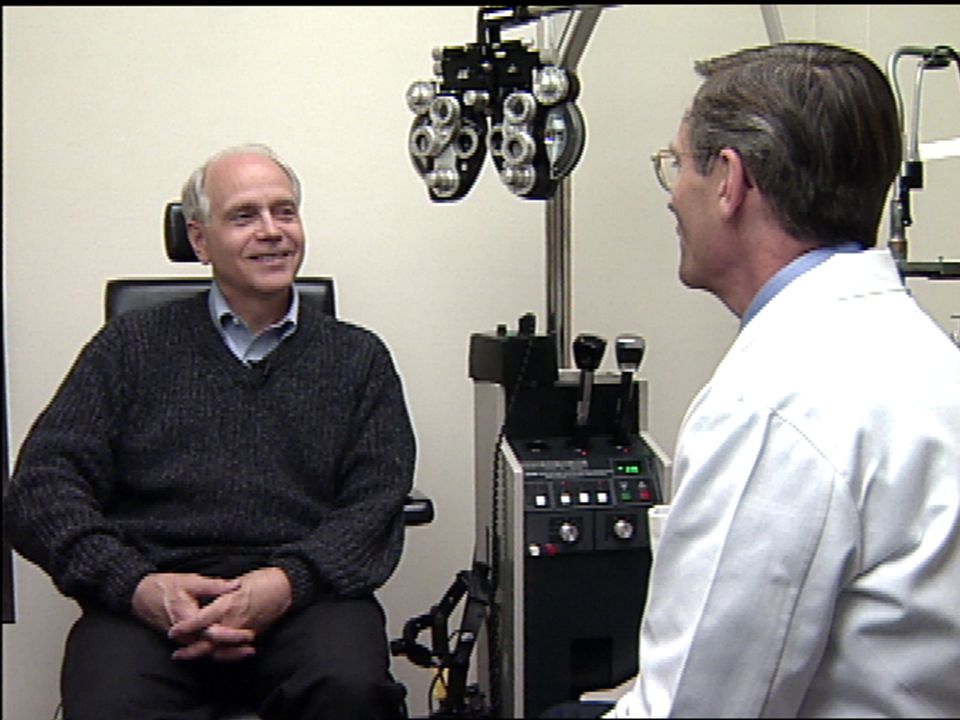
Profile Dr. Cunin Practicing endodontist 60 year old man Has worn glasses since 12 years Tried rigid contact lenses and found them uncomfortable Dr. Cunin Practicing endodontist 60 year old man Has worn glasses since 12 years Tried rigid contact lenses and found them uncomfortable

3
Profile Dr. Cunin After consultation in office and his sister-in-laws success with lasik surgery, he elected to undergo lasik procedure He now enjoys perfect distance vision of 20 / 16 in both eyes unaided but does wear reading glasses for near vision 2a Dr. Cunin After consultation in office and his sister-in-laws success with lasik surgery, he elected to undergo lasik procedure He now enjoys perfect distance vision of 20 / 16 in both eyes unaided but does wear reading glasses for near vision 2a

5
Japan, 1950 SatoJapan, 1950 Sato Columbia, 1960 BarraquerColumbia, 1960 Barraquer Russia, Fyodorov (RK)Russia, Fyodorov (RK) USA, 1983 Trokel (PRK)USA, 1983 Trokel (PRK) Crete, 1988 Pallikaris ( LASIK)Crete, 1988 Pallikaris ( LASIK) Japan, 1950 SatoJapan, 1950 Sato Columbia, 1960 BarraquerColumbia, 1960 Barraquer Russia, Fyodorov (RK)Russia, Fyodorov (RK) USA, 1983 Trokel (PRK)USA, 1983 Trokel (PRK) Crete, 1988 Pallikaris ( LASIK)Crete, 1988 Pallikaris ( LASIK) History of Refractive Surgery
Russia, Fyodorov (RK) USA, 1983 Trokel (PRK)USA, 1983 Trokel (PRK) Crete, 1988 Pallikaris ( LASIK)Crete, 1988 Pallikaris ( LASIK) Japan, 1950 SatoJapan, 1950 Sato Columbia, 1960 BarraquerColumbia, 1960 Barraquer Russia, Fyodorov (RK)Russia, Fyodorov (RK) USA, 1983 Trokel (PRK)USA, 1983 Trokel (PRK) Crete, 1988 Pallikaris ( LASIK)Crete, 1988 Pallikaris ( LASIK) History of Refractive Surgery")
6
Myopia – NearsightedMyopia – Nearsighted - 0.5 to 14 Diopters- 0.5 to 14 Diopters Myopia – NearsightedMyopia – Nearsighted - 0.5 to 14 Diopters- 0.5 to 14 Diopters Refractive Errors of Vision

7
Hyperopia – FarsightedHyperopia – Farsighted +1.0 to + 6.0 Diopters+1.0 to + 6.0 Diopters Hyperopia – FarsightedHyperopia – Farsighted +1.0 to + 6.0 Diopters+1.0 to + 6.0 Diopters Refractive Errors of Vision 6

8
Astigmatism - Visual focus in one axisAstigmatism - Visual focus in one axis - 0.5 to 5.0 Diopters- 0.5 to 5.0 Diopters Astigmatism - Visual focus in one axisAstigmatism - Visual focus in one axis - 0.5 to 5.0 Diopters- 0.5 to 5.0 Diopters Refractive Errors of Vision

9
Presbyopia - Loss ofPresbyopia - Loss of accommodation (age related) accommodation (age related) MonovisionMonovision Presbyopia - Loss ofPresbyopia - Loss of accommodation (age related) accommodation (age related) MonovisionMonovision Refractive Errors of Vision
 accommodation (age related) MonovisionMonovision Presbyopia - Loss ofPresbyopia - Loss of accommodation (age related) accommodation (age related) MonovisionMonovision Refractive Errors of Vision")
10
Spectacles - Limited field of vision,Spectacles - Limited field of vision, cosmetic limitation, safety issues cosmetic limitation, safety issues Contact Lenses - Allergy, comfort,Contact Lenses - Allergy, comfort, astigmatism, microbial keratitis, cost astigmatism, microbial keratitis, cost Spectacles - Limited field of vision,Spectacles - Limited field of vision, cosmetic limitation, safety issues cosmetic limitation, safety issues Contact Lenses - Allergy, comfort,Contact Lenses - Allergy, comfort, astigmatism, microbial keratitis, cost astigmatism, microbial keratitis, cost Non-Surgical Methods of Correcting Refractive Error 9

11
David Castellano, M.D. Assistant Professor of Ophthalmology The Ohio State University Medical Center

12
Incisional: radial keratotomy,Incisional: radial keratotomy, astigmatic keratotomy astigmatic keratotomy 11 Incisional: radial keratotomy,Incisional: radial keratotomy, astigmatic keratotomy astigmatic keratotomy 11 Surgical Options

13
Intrastromal rings (INTACS) < 5 dioptersIntrastromal rings (INTACS) < 5 diopters Surgical Options
 < 5 dioptersIntrastromal rings (INTACS) < 5 diopters Surgical Options")
14
Excimer laserExcimer laser “Cold” laser UV light range 193 nm“Cold” laser UV light range 193 nm Excimer = Excited dimer (argon fluoride)Excimer = Excited dimer (argon fluoride) Excimer laserExcimer laser “Cold” laser UV light range 193 nm“Cold” laser UV light range 193 nm Excimer = Excited dimer (argon fluoride)Excimer = Excited dimer (argon fluoride) Surgical Options
Excimer = Excited dimer (argon fluoride) Excimer laserExcimer laser Cold laser UV light range 193 nm Cold laser UV light range 193 nm Excimer = Excited dimer (argon fluoride)Excimer = Excited dimer (argon fluoride) Surgical Options")
15
The excimer laser can be used to remove a shaped area of the corneaThe excimer laser can be used to remove a shaped area of the cornea Submicron precisionSubmicron precision Minimal collateral damageMinimal collateral damage Minimal thermal energy effectMinimal thermal energy effect The excimer laser can be used to remove a shaped area of the corneaThe excimer laser can be used to remove a shaped area of the cornea Submicron precisionSubmicron precision Minimal collateral damageMinimal collateral damage Minimal thermal energy effectMinimal thermal energy effect Excimer Laser

16
Ablation patterns - myopia Excimer Laser

17
Ablation patterns - hyperopia Excimer Laser

18
Ablation patterns - astigmatism Excimer Laser 1515

19
PRK (Photorefractive Keratectomy)PRK (Photorefractive Keratectomy) – remove corneal epithelium – remove corneal epithelium LASIK (Laser Assisted In SituLASIK (Laser Assisted In Situ Keratomileusis) – Create corneal flap Keratomileusis) – Create corneal flap PRK (Photorefractive Keratectomy)PRK (Photorefractive Keratectomy) – remove corneal epithelium – remove corneal epithelium LASIK (Laser Assisted In SituLASIK (Laser Assisted In Situ Keratomileusis) – Create corneal flap Keratomileusis) – Create corneal flap Laser Surgical Options
PRK (Photorefractive Keratectomy) – remove corneal epithelium – remove corneal epithelium LASIK (Laser Assisted In SituLASIK (Laser Assisted In Situ Keratomileusis) – Create corneal flap Keratomileusis) – Create corneal flap PRK (Photorefractive Keratectomy)PRK (Photorefractive Keratectomy) – remove corneal epithelium – remove corneal epithelium LASIK (Laser Assisted In SituLASIK (Laser Assisted In Situ Keratomileusis) – Create corneal flap Keratomileusis) – Create corneal flap Laser Surgical Options")
20
PRK (Photorefractive Keratectomy) – remove corneal epitheliumPRK (Photorefractive Keratectomy) – remove corneal epithelium LASIK (Laser Assisted In SituLASIK (Laser Assisted In Situ Keratomileusis) – create corneal flap Keratomileusis) – create corneal flap PRK (Photorefractive Keratectomy) – remove corneal epitheliumPRK (Photorefractive Keratectomy) – remove corneal epithelium LASIK (Laser Assisted In SituLASIK (Laser Assisted In Situ Keratomileusis) – create corneal flap Keratomileusis) – create corneal flap Laser Surgical Options 1717
 – remove corneal epitheliumPRK (Photorefractive Keratectomy) – remove corneal epithelium LASIK (Laser Assisted In SituLASIK (Laser Assisted In Situ Keratomileusis) – create corneal flap Keratomileusis) – create corneal flap PRK (Photorefractive Keratectomy) – remove corneal epitheliumPRK (Photorefractive Keratectomy) – remove corneal epithelium LASIK (Laser Assisted In SituLASIK (Laser Assisted In Situ Keratomileusis) – create corneal flap Keratomileusis) – create corneal flap Laser Surgical Options 1717")
21
Corneal Anatomy

22
Realistic expectations: 20/40 or betterRealistic expectations: 20/40 or better without correction (95%) without correction (95%) Need for reading glasses after age 40Need for reading glasses after age 40 Occupational needs (police, fire,Occupational needs (police, fire, professional sports) professional sports) Personal needs (sports, travel)Personal needs (sports, travel) Realistic expectations: 20/40 or betterRealistic expectations: 20/40 or better without correction (95%) without correction (95%) Need for reading glasses after age 40Need for reading glasses after age 40 Occupational needs (police, fire,Occupational needs (police, fire, professional sports) professional sports) Personal needs (sports, travel)Personal needs (sports, travel) Candidates for Refractive Surgery
 without correction (95%) Need for reading glasses after age 40Need for reading glasses after age 40 Occupational needs (police, fire,Occupational needs (police, fire, professional sports) professional sports) Personal needs (sports, travel)Personal needs (sports, travel) Realistic expectations: 20/40 or betterRealistic expectations: 20/40 or better without correction (95%) without correction (95%) Need for reading glasses after age 40Need for reading glasses after age 40 Occupational needs (police, fire,Occupational needs (police, fire, professional sports) professional sports) Personal needs (sports, travel)Personal needs (sports, travel) Candidates for Refractive Surgery")
23
Myopia <6.00 dioptersMyopia <6.00 diopters Low surgical riskLow surgical risk (no flap complications) (no flap complications) Lower costLower cost Myopia <6.00 dioptersMyopia <6.00 diopters Low surgical riskLow surgical risk (no flap complications) (no flap complications) Lower costLower cost Advantages of PRK
 (no flap complications) Lower costLower cost Myopia <6.00 dioptersMyopia <6.00 diopters Low surgical riskLow surgical risk (no flap complications) (no flap complications) Lower costLower cost Advantages of PRK")
24
DiscomfortDiscomfort Slow recoverySlow recovery Increased medications (>3 D)Increased medications (>3 D) Need for bandage soft contact lensNeed for bandage soft contact lens Usually monocular surgeryUsually monocular surgery Regression of correction more commonRegression of correction more common DiscomfortDiscomfort Slow recoverySlow recovery Increased medications (>3 D)Increased medications (>3 D) Need for bandage soft contact lensNeed for bandage soft contact lens Usually monocular surgeryUsually monocular surgery Regression of correction more commonRegression of correction more common Disadvantages of PRK 2121
Increased medications (>3 D) Need for bandage soft contact lensNeed for bandage soft contact lens Usually monocular surgeryUsually monocular surgery Regression of correction more commonRegression of correction more common DiscomfortDiscomfort Slow recoverySlow recovery Increased medications (>3 D)Increased medications (>3 D) Need for bandage soft contact lensNeed for bandage soft contact lens Usually monocular surgeryUsually monocular surgery Regression of correction more commonRegression of correction more common Disadvantages of PRK 2121")
25
Minimal discomfortMinimal discomfort Rapid visual recoveryRapid visual recovery Bilateral surgery possibleBilateral surgery possible Higher amount of myopic correction (-1.00 to -14.00 D myopia)Higher amount of myopic correction (-1.00 to -14.00 D myopia) Minimal discomfortMinimal discomfort Rapid visual recoveryRapid visual recovery Bilateral surgery possibleBilateral surgery possible Higher amount of myopic correction (-1.00 to -14.00 D myopia)Higher amount of myopic correction (-1.00 to -14.00 D myopia) Advantages of LASIK
Higher amount of myopic correction (-1.00 to D myopia) Minimal discomfortMinimal discomfort Rapid visual recoveryRapid visual recovery Bilateral surgery possibleBilateral surgery possible Higher amount of myopic correction (-1.00 to D myopia)Higher amount of myopic correction (-1.00 to D myopia) Advantages of LASIK")
26
Flap complications (decentration, button hole, partial or free cap, cap striae)Flap complications (decentration, button hole, partial or free cap, cap striae) Interface inclusionsInterface inclusions Interface inflammation (SOS)Interface inflammation (SOS) Steeper learning curveSteeper learning curve Flap complications (decentration, button hole, partial or free cap, cap striae)Flap complications (decentration, button hole, partial or free cap, cap striae) Interface inclusionsInterface inclusions Interface inflammation (SOS)Interface inflammation (SOS) Steeper learning curveSteeper learning curve Disadvantages of LASIK
Flap complications (decentration, button hole, partial or free cap, cap striae) Interface inclusionsInterface inclusions Interface inflammation (SOS)Interface inflammation (SOS) Steeper learning curveSteeper learning curve Flap complications (decentration, button hole, partial or free cap, cap striae)Flap complications (decentration, button hole, partial or free cap, cap striae) Interface inclusionsInterface inclusions Interface inflammation (SOS)Interface inflammation (SOS) Steeper learning curveSteeper learning curve Disadvantages of LASIK")
27
Profile Harvey 47 year old male Preoperatively Refractive error in: right eye was 4 left eye was 4.25 Treatment Surgery for monovision 2a Harvey 47 year old male Preoperatively Refractive error in: right eye was 4 left eye was 4.25 Treatment Surgery for monovision 2a

28
Profile Harvey Postoperatively Right eye 20 / 20 minus Left eye 20 / 40 Near vision left eye is J-1 2a Harvey Postoperatively Right eye 20 / 20 minus Left eye 20 / 40 Near vision left eye is J-1 2a

30
Unrealistic expectations (must have 20/20 result or be glasses independent)Unrealistic expectations (must have 20/20 result or be glasses independent) Expect vision better than with glasses or contact lensesExpect vision better than with glasses or contact lenses Unrealistic expectations (must have 20/20 result or be glasses independent)Unrealistic expectations (must have 20/20 result or be glasses independent) Expect vision better than with glasses or contact lensesExpect vision better than with glasses or contact lenses ContraindicationsContraindications
Unrealistic expectations (must have 20/20 result or be glasses independent) Expect vision better than with glasses or contact lensesExpect vision better than with glasses or contact lenses Unrealistic expectations (must have 20/20 result or be glasses independent)Unrealistic expectations (must have 20/20 result or be glasses independent) Expect vision better than with glasses or contact lensesExpect vision better than with glasses or contact lenses ContraindicationsContraindications")
31
Refractive error outside laser parametersRefractive error outside laser parameters Corneal pathology, scars, keratoconusCorneal pathology, scars, keratoconus Thin cornea (LASIK)Thin cornea (LASIK) Keratitis siccaKeratitis sicca Collagen vascular diseaseCollagen vascular disease PregnancyPregnancy Refractive error outside laser parametersRefractive error outside laser parameters Corneal pathology, scars, keratoconusCorneal pathology, scars, keratoconus Thin cornea (LASIK)Thin cornea (LASIK) Keratitis siccaKeratitis sicca Collagen vascular diseaseCollagen vascular disease PregnancyPregnancy ContraindicationsContraindications
Thin cornea (LASIK) Keratitis siccaKeratitis sicca Collagen vascular diseaseCollagen vascular disease PregnancyPregnancy Refractive error outside laser parametersRefractive error outside laser parameters Corneal pathology, scars, keratoconusCorneal pathology, scars, keratoconus Thin cornea (LASIK)Thin cornea (LASIK) Keratitis siccaKeratitis sicca Collagen vascular diseaseCollagen vascular disease PregnancyPregnancy ContraindicationsContraindications")
32
Muscle balance without correctionMuscle balance without correction Bright and low illumination ofBright and low illumination of pupil - size pupil - size Lids, aperture, lid fold, blepharitisLids, aperture, lid fold, blepharitis Muscle balance without correctionMuscle balance without correction Bright and low illumination ofBright and low illumination of pupil - size pupil - size Lids, aperture, lid fold, blepharitisLids, aperture, lid fold, blepharitis Pre-Operative Evaluation: External Exam 2828

33
Lids - meibomian glands, debris on lashes, follicles, concretionsLids - meibomian glands, debris on lashes, follicles, concretions Tear film - lakes, height, debrisTear film - lakes, height, debris Cornea - pannus, scarsCornea - pannus, scars Lens - opacity, nuclear sclerosisLens - opacity, nuclear sclerosis Intraocular pressureIntraocular pressure Lids - meibomian glands, debris on lashes, follicles, concretionsLids - meibomian glands, debris on lashes, follicles, concretions Tear film - lakes, height, debrisTear film - lakes, height, debris Cornea - pannus, scarsCornea - pannus, scars Lens - opacity, nuclear sclerosisLens - opacity, nuclear sclerosis Intraocular pressureIntraocular pressure Pre-Operative Evaluation: Slit Lamp

34
Optic discOptic disc MaculaMacula VesselsVessels Peripheral retina (lattice, holes)Peripheral retina (lattice, holes) Optic discOptic disc MaculaMacula VesselsVessels Peripheral retina (lattice, holes)Peripheral retina (lattice, holes) Pre-Operative Evaluation: Fundus
Peripheral retina (lattice, holes) Optic discOptic disc MaculaMacula VesselsVessels Peripheral retina (lattice, holes)Peripheral retina (lattice, holes) Pre-Operative Evaluation: Fundus")
35
Discontinue contact lens wearDiscontinue contact lens wear Stable refractionStable refraction Corneal topographyCorneal topography Corneal thicknessCorneal thickness Pupil sizePupil size Discontinue contact lens wearDiscontinue contact lens wear Stable refractionStable refraction Corneal topographyCorneal topography Corneal thicknessCorneal thickness Pupil sizePupil size Pre-Operative Evaluation: Refractive

36
Stable refraction, mapsStable refraction, maps No make-up day of surgeryNo make-up day of surgery Mark axis of astigmatism at slit lampMark axis of astigmatism at slit lamp Topical anesthesiaTopical anesthesia Stable refraction, mapsStable refraction, maps No make-up day of surgeryNo make-up day of surgery Mark axis of astigmatism at slit lampMark axis of astigmatism at slit lamp Topical anesthesiaTopical anesthesia Surgical Technique - PRK

37
Remove epithelium (laser or mechanical)Remove epithelium (laser or mechanical) Laser correction to stromaLaser correction to stroma Contact lensContact lens Remove epithelium (laser or mechanical)Remove epithelium (laser or mechanical) Laser correction to stromaLaser correction to stroma Contact lensContact lens Surgical Technique - PRK
Remove epithelium (laser or mechanical) Laser correction to stromaLaser correction to stroma Contact lensContact lens Remove epithelium (laser or mechanical)Remove epithelium (laser or mechanical) Laser correction to stromaLaser correction to stroma Contact lensContact lens Surgical Technique - PRK")
38
Drape lash marginsDrape lash margins Lid speculumLid speculum Mark corneaMark cornea Apply suction ringApply suction ring Check intraocular pressure (>60mmHg)Check intraocular pressure (>60mmHg) Make flap with microkeratomeMake flap with microkeratome Drape lash marginsDrape lash margins Lid speculumLid speculum Mark corneaMark cornea Apply suction ringApply suction ring Check intraocular pressure (>60mmHg)Check intraocular pressure (>60mmHg) Make flap with microkeratomeMake flap with microkeratome Surgical Technique - LASIK
Check intraocular pressure (>60mmHg) Make flap with microkeratomeMake flap with microkeratome Drape lash marginsDrape lash margins Lid speculumLid speculum Mark corneaMark cornea Apply suction ringApply suction ring Check intraocular pressure (>60mmHg)Check intraocular pressure (>60mmHg) Make flap with microkeratomeMake flap with microkeratome Surgical Technique - LASIK")
39
Lift flapLift flap Laser correction to stromaLaser correction to stroma Reposition flapReposition flap Irrigate interface post keratectomyIrrigate interface post keratectomy Dry gutterDry gutter Examine at slit lamp for striae or debrisExamine at slit lamp for striae or debris Lift flapLift flap Laser correction to stromaLaser correction to stroma Reposition flapReposition flap Irrigate interface post keratectomyIrrigate interface post keratectomy Dry gutterDry gutter Examine at slit lamp for striae or debrisExamine at slit lamp for striae or debris Surgical Technique - LASIK

40
Late regressionLate regression Induced astigmatismInduced astigmatism Central islandsCentral islands DecentrationDecentration Loss of best corrected visionLoss of best corrected vision Late regressionLate regression Induced astigmatismInduced astigmatism Central islandsCentral islands DecentrationDecentration Loss of best corrected visionLoss of best corrected vision Complications of Laser Surgery

41
InfectionInfection Decreased corneal sensationDecreased corneal sensation Recurrent erosion (PRK)Recurrent erosion (PRK) Late haze (PRK)Late haze (PRK) Flap complications (LASIK)Flap complications (LASIK) Interface syndromes (LASIK)Interface syndromes (LASIK) InfectionInfection Decreased corneal sensationDecreased corneal sensation Recurrent erosion (PRK)Recurrent erosion (PRK) Late haze (PRK)Late haze (PRK) Flap complications (LASIK)Flap complications (LASIK) Interface syndromes (LASIK)Interface syndromes (LASIK) Complications of Laser Surgery
Recurrent erosion (PRK) Late haze (PRK)Late haze (PRK) Flap complications (LASIK)Flap complications (LASIK) Interface syndromes (LASIK)Interface syndromes (LASIK) InfectionInfection Decreased corneal sensationDecreased corneal sensation Recurrent erosion (PRK)Recurrent erosion (PRK) Late haze (PRK)Late haze (PRK) Flap complications (LASIK)Flap complications (LASIK) Interface syndromes (LASIK)Interface syndromes (LASIK) Complications of Laser Surgery")
42
GlareGlare HaloHalo Multiple imagesMultiple images GhostingGhosting Night visionNight vision GlareGlare HaloHalo Multiple imagesMultiple images GhostingGhosting Night visionNight vision Complications of Laser Surgery - Quality of Vision

43
20/40 or better at 1 year - 98.8%20/40 or better at 1 year - 98.8% 20/20 or better at 1 year - 80.5%20/20 or better at 1 year - 80.5% Loss of 2 or more lines of BCV at 1 year - 1.2%Loss of 2 or more lines of BCV at 1 year - 1.2% Glare at 1 year - 2.4%Glare at 1 year - 2.4% Halos at 1 year - 2.4%Halos at 1 year - 2.4% 20/40 or better at 1 year - 98.8%20/40 or better at 1 year - 98.8% 20/20 or better at 1 year - 80.5%20/20 or better at 1 year - 80.5% Loss of 2 or more lines of BCV at 1 year - 1.2%Loss of 2 or more lines of BCV at 1 year - 1.2% Glare at 1 year - 2.4%Glare at 1 year - 2.4% Halos at 1 year - 2.4%Halos at 1 year - 2.4% Results – PRK

44
20/40 or better at 6 months - 97%20/40 or better at 6 months - 97% 20/20 or better at 6 months - 58.6%20/20 or better at 6 months - 58.6% Loss of 2 or more lines of BCV at 6 months - 0.5%Loss of 2 or more lines of BCV at 6 months - 0.5% 20/40 or better at 6 months - 97%20/40 or better at 6 months - 97% 20/20 or better at 6 months - 58.6%20/20 or better at 6 months - 58.6% Loss of 2 or more lines of BCV at 6 months - 0.5%Loss of 2 or more lines of BCV at 6 months - 0.5% Results – LASIK

45
Scanning Laser (LADAR)Scanning Laser (LADAR) Flying spotFlying spot TopolinkTopolink Stromal ablationStromal ablation Wavefront technologyWavefront technology Scanning Laser (LADAR)Scanning Laser (LADAR) Flying spotFlying spot TopolinkTopolink Stromal ablationStromal ablation Wavefront technologyWavefront technology Future Laser Development
Scanning Laser (LADAR) Flying spotFlying spot TopolinkTopolink Stromal ablationStromal ablation Wavefront technologyWavefront technology Scanning Laser (LADAR)Scanning Laser (LADAR) Flying spotFlying spot TopolinkTopolink Stromal ablationStromal ablation Wavefront technologyWavefront technology Future Laser Development")
Similar presentations


thus effecting baseline evaluation.>")


