Download presentation
Presentation is loading. Please wait.

1
SISTEMA DIGESTIVO

3
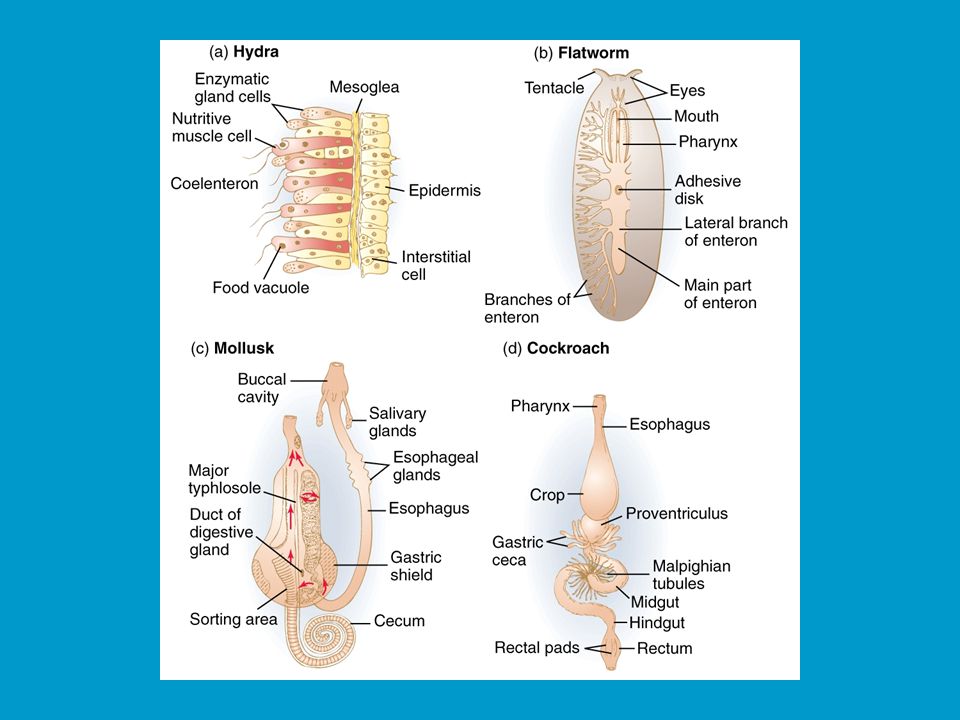
SISTEMA DIGESTIVO: bioreactores

4
SISTEMA DIGESTIVO Es un tubo abierto: extensión del medio ambiente! E S absorción Digestión: mecánica (trituración) química (enzimas hidrolíticas)
 química (enzimas hidrolíticas)")
5
Sistema de Tubos y Esfínteres
Direccionalidad Compartimentalización Almacenamiento.

6
EL TRACTO DIGESTIVO

7
Anatomía del sistema digestivo
Digestive tract Alimentary tract or canal GI tract Accessory organs Primarily glands Regions Mouth or oral cavity Pharynx Esophagus Stomach Small intestine Large intestine Anus

8
Cavidad oral Mouth or oral cavity Lips (labia) and cheeks
Vestibule: Space between lips or cheeks and alveolar processes Oral cavity proper Lips (labia) and cheeks Palate: Oral cavity roof Hard and soft Palatine tonsils Tongue: Involved in speech, taste, mastication, swallowing
 and cheeks. Palate: Oral cavity roof. Hard and soft. Palatine tonsils. Tongue: Involved in speech, taste, mastication, swallowing.")
9
Dientes

10
Dientes Two sets Types Primary, deciduous, milk: Childhood
Permanent or secondary: Adult (32) Types Incisors, canine, premolar and molars
 Types. Incisors, canine, premolar and molars.")
11
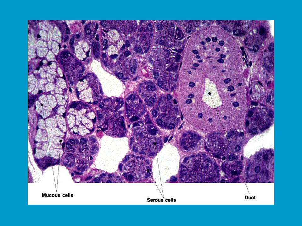
Glándulas salivales Produce saliva Three pairs
Prevents bacterial infection Lubrication Contains salivary amylase Breaks down starch Three pairs Parotid: Largest Submandibular Sublingual: Smallest

13
Producción de saliva % agua HCO3- , ph 6.5 moco, enzimas

14
SECRECIONES SALIVALES 1. Secreción serosa: amilasa salival:
hidrólisis a-1-4 polisacáridos Secreción mucosa: lubrica y protege. IgA y lisozima: antimicrobiana

15
Reflejo de deglución Fase voluntaria Fase refleja Fase refleja
Bolo alimenticio desde boca a faringe Fase refleja Apertura del esófago, cierre de laringe Fase refleja Transporte por esófago

16
Histología del tracto digestivo

17
Peritoneo y Mesenterios
Peritoneum Visceral: Covers organs Parietal: Covers interior surface of body wall Retroperitoneal: Behind peritoneum as kidneys, pancreas, duodenum Mesenteries Routes which vessels and nerves pass from body wall to organs Greater omentum Lesser omentum

18
Estómago Openings Regions Gastroesophageal: To esophagus
Pyloric: To duodenum Regions Cardiac Fundus Body Pyloric

19
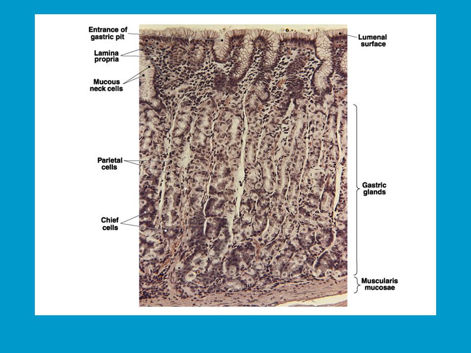
Histología del estómago
Layers Serosa or visceral peritoneum: Outermost Muscularis: Three layers Outer longitudinal Middle circular Inner oblique Submucosa Mucosa

22
Gastric pits and glands:
Contain cells Surface mucous: Mucus Mucous neck: Mucus Parietal: Hydrochloric acid and intrinsic factor Chief: Pepsinogen Endocrine: Regulatory hormones

23
Secreción de HCl en células parietales

24
Barrera mucus – HCO3-

25
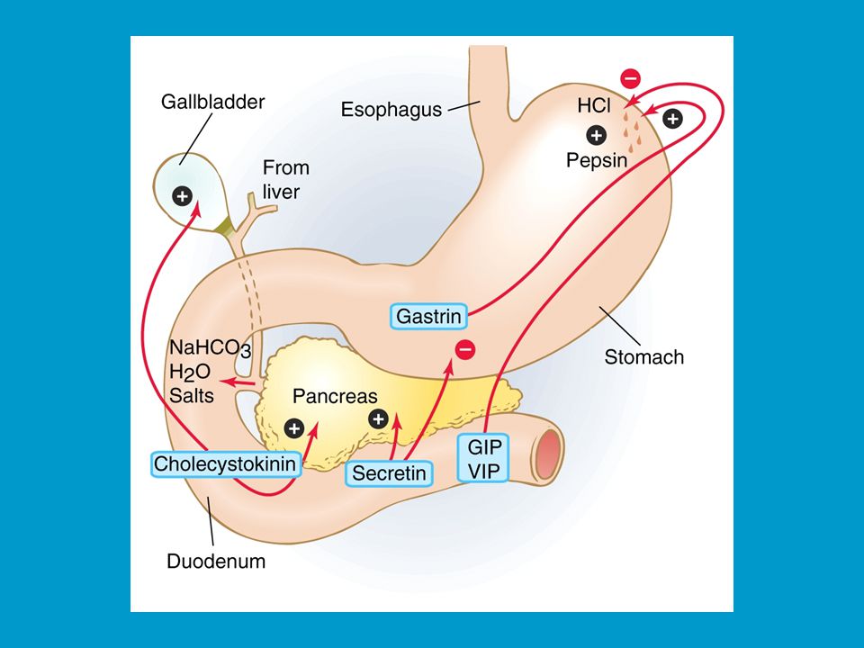
Fases de la secreción gástrica

26
FASE CEFÁLICA - percepción sensorial del alimento

27
FASE GÁSTRICA - presencia del alimento

28
Ondas de mezcla

29
FASE INTESTINAL VIP Gastrin

31
Duodeno y Pancreas

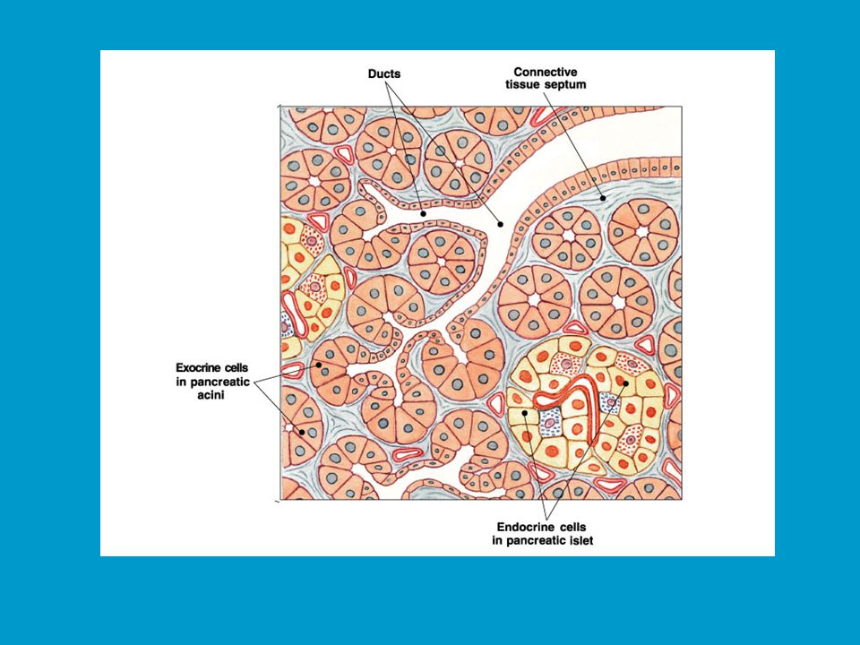
32
Pancreas Anatomy Secretions Endocrine Pancreatic juice (exocrine)
Pancreatic islets produce insulin and glucagon Exocrine Acini produce digestive enzymes and HCO3- Regions: Head, body, tail Secretions Pancreatic juice (exocrine) Trypsin Chymotrypsin Carboxypeptidase Pancreatic amylase Pancreatic lipases Elastase Nucleases (DNA, RNA) HCO3-, H20
 Trypsin. Chymotrypsin. Carboxypeptidase. Pancreatic amylase. Pancreatic lipases. Elastase. Nucleases (DNA, RNA) HCO3-, H20.")
35
Secreción de HCO3-

36
Enzimas Trypsinogen Chymotrysinogen Carboxypeptidases Pro-elastase
Phospholipase pancreatic lipase Pancreatic amylase Pancreatic juice is composed of two secretory products critical to proper digestion: digestive enzymes and bicarbonate. The enzymes are synthesized and secreted from the exocrine ascinar cells, whereas bicarbonate is secreted from the epithelial cells lining small pancreatic ducts. Digestive Enzymes The pancreas secretes a magnificent battery of enzymes that collectively have the capacity to reduce virtually all digestible macromolecules into forms that are capable of, or nearly capable of being absorbed. Three major groups of enzymes are critical to efficient digestion: Proteases Digestion of proteins is initiated by pepsin in the stomach, but the bulk of protein digestion is due to the pancreatic proteases. Several proteases are synthesized in the pancreas and secreted into the lumen of the small intestine. The two major pancreatic proteases are trypsin and chymotrypsin, which are synthesized and packaged into secretory vesicles as an the inactive proenzymes trypsinogen and chymotrypsinogen. As you might anticipate, proteases are rather dangerous enzymes to have in cells, and packaging of an inactive precursor is a way for the cells to safely handle these enzymes. The secretory vesicles also contain a trypsin inhibitor which serves as an additional safeguard should some of the trypsinogen be activated to trypsin; following exocytosis this inhibitor is diluted out and becomes ineffective - the pin is out of the grenade. Once trypsinogen and chymotrypsinogen are released into the lumen of the small intestine, they must be converted into their active forms in order to digest proteins. Trypsinogen is activated by the enzyme enterokinase, which is embedded in the intestinal mucosa. Once trypsin is formed it activates chymotrypsinogen, as well as additional molecules of trypsinogen. The net result is a rather explosive appearance of active protease once the pancreatic secretions reach the small intestine. Trypsin and chymotrypsin digest proteins into peptides and peptides into smaller peptides, but they cannot digest proteins and peptides to single amino acids. Some of the other proteases from the pancreas, for instance carboxypeptidase, have that ability, but the final digestion of peptides into amino acids is largely the effect of peptidases in small intestinal epithelial cells. More on this later. Pancreatic Lipase The major form of dietary fat is triglyceride, or neutral lipid. A triglyceride molecule cannot be directly absorbed across the intestinal mucosa. Rather, it must first be digested into a 2-monoglyceride and two free fatty acids. The enzyme that performs this hydrolysis is pancreatic lipase, which is delivered into the lumen of the gut as a constituent of pancreatic juice. Sufficient quantities of bile salts must also be present in the lumen of the intestine in order for lipase to efficiently digest dietary triglyceride and for the resulting fatty acids and monoglyceride to be absorbed. This means that normal digestion and absorption of dietary fat is critically dependent on secretions from both the pancreas and liver. Pancreatic lipase has recently been in the limelight as a target for management of obesity. The drug orlistat (Xenical) is a pancreatic lipase inhibitor that interferes with digestion of triglyceride and thereby reduces absorption of dietary fat. Clinical trials support the contention that inhibiting lipase can lead to significant reductions in body weight in some patients. Amylase The major dietary carbohydrate for many species is starch, a storage form of glucose in plants. Amylase is the enzyme that hydrolyses starch to maltose (a glucose-glucose disaccharide), as well as the trisaccharide maltotriose and small branchpoints fragments called limit dextrins. The major source of amylase in all species is pancreatic secretions, although amylase is also present in saliva of some animals, including humans. Other Pancreatic Enzymes In addition to the proteases, lipase and amylase, the pancreas produces a host of other digestive enzymes, including ribonuclease, deoxyribonuclease, gelatinase and elastase. Bicarbonate and Water Epithelial cells in pancreatic ducts are the source of the bicarbonate and water secreted by the pancreas. The mechanism underlying bicarbonate secretion is essentially the same as for acid secretion parietal cells and is dependent on the enzyme carbonic anhydrase. In pancreatic duct cells, the bicarbonate is secreted into the lumen of the duct and hence into pancreatic juice.
 is a pancreatic lipase inhibitor that interferes with digestion of triglyceride and thereby reduces absorption of dietary fat. Clinical trials support the contention that inhibiting lipase can lead to significant reductions in body weight in some patients. Amylase. The major dietary carbohydrate for many species is starch, a storage form of glucose in plants. Amylase is the enzyme that hydrolyses starch to maltose (a glucose-glucose disaccharide), as well as the trisaccharide maltotriose and small branchpoints fragments called limit dextrins. The major source of amylase in all species is pancreatic secretions, although amylase is also present in saliva of some animals, including humans. Other Pancreatic Enzymes. In addition to the proteases, lipase and amylase, the pancreas produces a host of other digestive enzymes, including ribonuclease, deoxyribonuclease, gelatinase and elastase. Bicarbonate and Water. Epithelial cells in pancreatic ducts are the source of the bicarbonate and water secreted by the pancreas. The mechanism underlying bicarbonate secretion is essentially the same as for acid secretion parietal cells and is dependent on the enzyme carbonic anhydrase. In pancreatic duct cells, the bicarbonate is secreted into the lumen of the duct and hence into pancreatic juice.")
37
Hígado Lobes Ducts Major: Left and right Minor: Caudate and quadrate
Common hepatic Cystic From gallbladder Common bile Joins pancreatic duct at hepatopancreatic ampulla

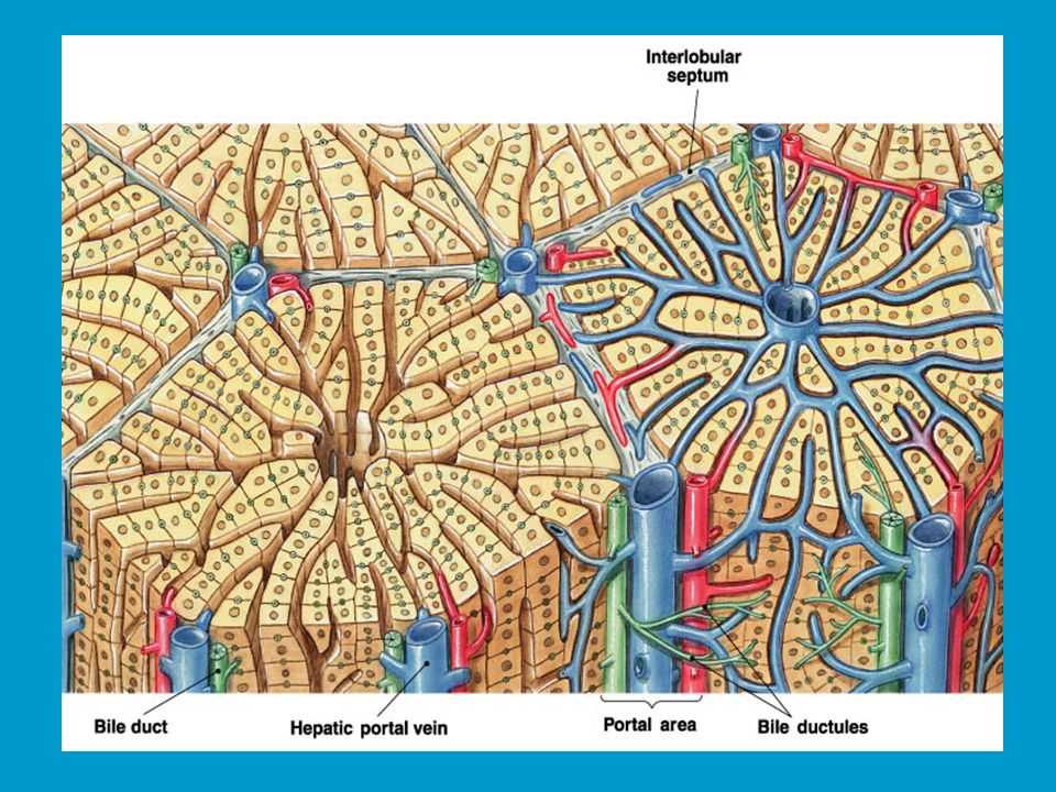
39
Histología del hígado triada portal Figure 24.20a, b

40
Funciones del hígado Bile production Storage Aminoacid synthesis
Salts emulsify fats, contain pigments as bilirubin Storage Glycogen, fat, vitamins, copper and iron Aminoacid synthesis Cholesterol and tryglicerid synthesis Gluconeogénesis, glucogénesis, glucogenólisis Hepatocytes remove ammonia and convert to urea Phagocytosis Kupffer cells phagocytize worn-out and dying red and white blood cells, some bacteria Plasma proteins and hemostatic factors Albumins, globulins, fibrinogen, heparin, K vitamin Detoxification and drug metabolism Hormonal secretion: trombopoietin, angotensinogen

41
…each day around 600 ml of bile is produced…
Bilis …each day around 600 ml of bile is produced… Bile acid Phospholipids Cholesterol Bilirubin Waste products Electrolytes Mucin HCO3-

42
Conductos

43
Intestino delgado Site of greatest amount of digestion and absorption
Divisions Duodenum Jejunum Ileum: Peyer’s patches or lymph nodules Modifications Circular folds or plicae circulares, villi, lacteal, microvilli Cells of mucosa Absorptive, goblet, granular, endocrine

44
Secreciones del int. delgado
Mucus and HCO3- Protects against digestive enzymes and stomach acids Enzymes Disaccharidases Peptidases Nucleotidases, nucleosidases Enteroquinase: tripsinógeno tripsina Duodenal glands (Brünner) Stimulated by vagus nerve, secretin, chemical or tactile irritation of duodenal mucosa
 Stimulated by vagus nerve, secretin, chemical or tactile irritation of duodenal mucosa.")
45
Duodeno llegan: quimo ácido, jugos pancreáticos, bilis

46
Absorción intestinal

47
Monosacáridos

48
Lípidos

49
Lipoproteinas Types Chylomicrons VLDL LDL HDL Enter lymph
Transports cholesterol to cells HDL Transports cholesterol from cells to liver

50
Intestino grueso Extends from ileocecal junction to anus
Consists of cecum, colon, rectum, anal canal Movements sluggish (18-24 hours)
")
51
Intestino grueso: Absorción de H2O y Defecación
Figure 21-27: Anatomy of the large intestine

52
Large Intestine Cecum Colon Rectum Anal canal
Blind sac, vermiform appendix attached Colon Ascending, transverse, descending, sigmoid Rectum Straight muscular tube Anal canal Internal anal sphincter (smooth muscle) External anal sphincter (skeletal muscle)
 External anal sphincter (skeletal muscle)")
53
Histology of Large Intestine

54
Figure 21-28: NaCl reabsorption by colonocytes
Water and electrolyte secretion &/or absorption Bacterial fermentation of HC Bacterial synthesis of Vit. K , B. Bacterial degradation of bile acids and esterols Absortion of lactate & butyrate Prevents infections and immune alterations Figure 21-28: NaCl reabsorption by colonocytes

55
Figure 21-29: NaCl secretion by colonic crypt cells

56
Toxina colérica ADP-ribosilación de Ga
G-GTP AC cAMP PKA p-CFTR PKA

57
Secreciones Mucus provides protection Pumps
Parasympathetic stimulation increases rate of goblet cell secretion Pumps Exchange of bicarbonate ions for chloride ions Exchange of sodium ions for hydrogen ions Bacterial actions produce gases called flatus

58
Reflejos en Colon y Recto

59
Secreciones del tubo digestivo

60
Enzimas del tubo digestivo

61
Regulación nerviosa y hormonal
Nervous regulation Involves enteric nervous system Types of neurons: sensory, motor, interneurons Coordinates peristalsis and regulates local reflexes Chemical regulation Production of hormones Gastrin, secretin Production of paracrine chemicals Histamine Help local reflexes in ENS control digestive environments as pH levels

63
Regulating Digestion: CNS and Enteric Nervous System (ENS)
Figure 21-9: The enteric nervous system

64
Hormonas gastrointestinales

Similar presentations


![Anatomy Practical [PHL 212]](/14/4428258/big_thumb.jpg "Anatomy Practical [PHL 212]>")


