Download presentation
Presentation is loading. Please wait.

1
Pharmaceutical Biotechnology
7. Product analysis Dr. Tarek El-Bashiti Assoc. Prof. of Biotechnology

2
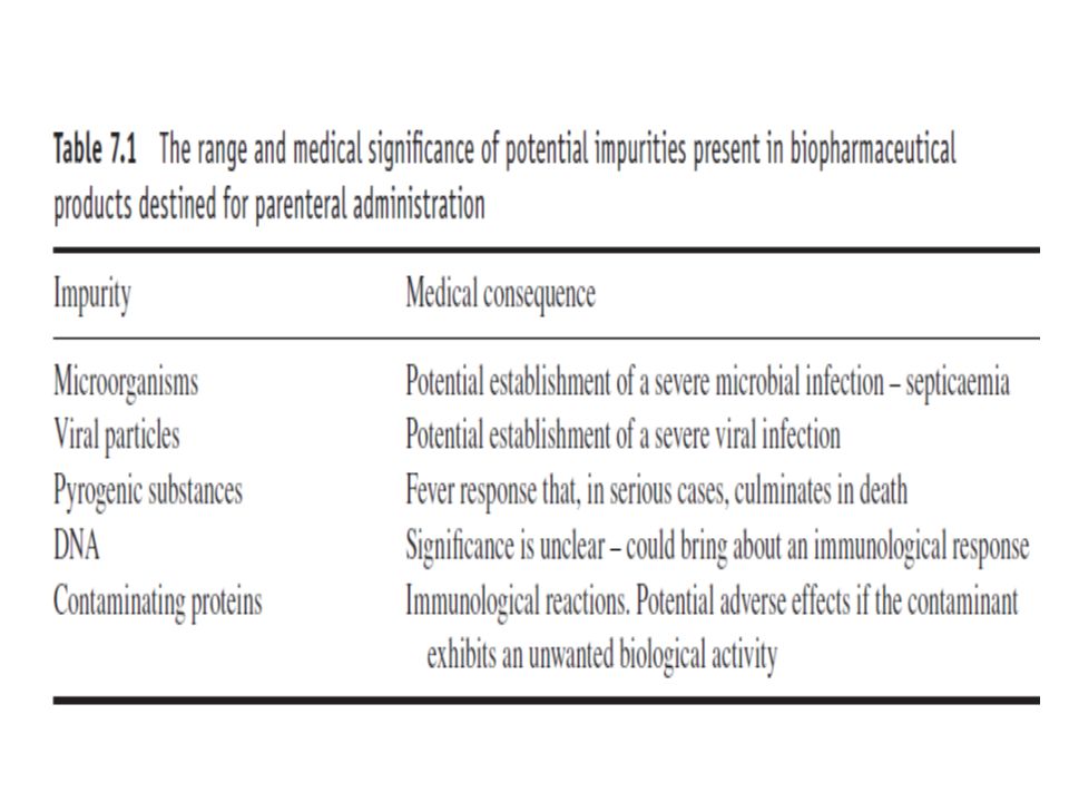
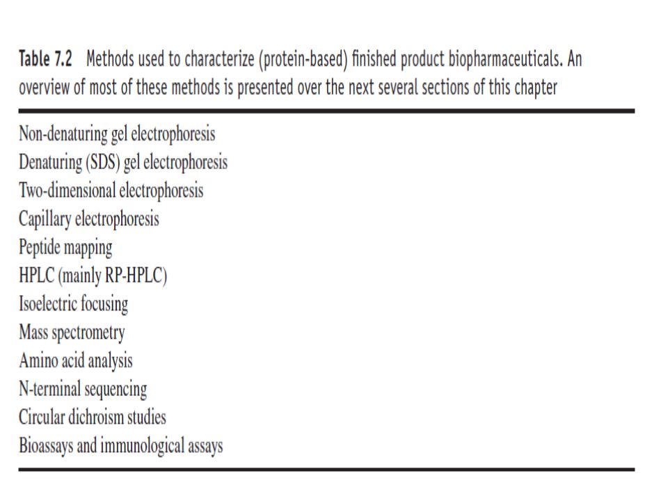
Introduction All pharmaceutical finished products undergo rigorous QC testing in order to confirm their conformance to predetermined specifications. Potency testing is of obvious importance, ensuring that the drug will be efficacious when administered to the patient. A prominent aspect of safety testing entails analysis of product for the presence of various potential contaminants (Fig. 7.1). An overview of the range of finished-product tests of recombinant protein biopharmaceuticals is outlined below.
. An overview of the range of finished-product tests of recombinant protein biopharmaceuticals is outlined below.")
4
Protein-based contaminants
Most of the chromatographic steps undertaken during downstream processing are specifically included to separate the protein of interest from additional contaminant proteins. Proteins may be introduced during upstream or downstream processing. For example, animal cell culture media are typically supplemented with bovine serum/foetal calf serum (2–25 per cent), or with a defined cocktail of various regulatory proteins required to maintain and stimulate growth of these cells.
, or with a defined cocktail of various regulatory proteins required to maintain and stimulate growth of these cells.")
5
Downstream processing of intracellular microbial proteins often requires the addition of endonuleases to the cell homogenate to degrade the large quantity of DNA liberated upon cellular disruption. The clinical significance of protein-based impurities relates to (a) their potential biological activities and (b) their antigenicity. Whereas some contaminants may display no undesirable biological activity, others may exhibit activities deleterious to either the product itself (e.g. proteases that could modify/degrade the product) or the recipient patient (e.g. the presence of contaminating toxins)
 their potential biological activities and. (b) their antigenicity. Whereas some contaminants may display no undesirable biological activity, others may exhibit activities deleterious to either the product itself (e.g. proteases that could modify/degrade the product) or the recipient patient (e.g. the presence of contaminating toxins)")
6
Their inherent immunogenicity also renders likely and immunological reaction against protein based impurities upon product administration to the recipient patient. Although the product itself is likely to be non-immunogenic (usually being coded for by a human gene), contaminant proteins will be endogenous to the host cell, and hence foreign to the human body. This is particularly likely if a requirement exists for ongoing, repeat product administration (e.g. administration of recombinant insulin) Immunological activation of this type could also potentially (and more seriously) have a sensitizing effect on the recipient against the actual protein product.
, contaminant proteins will be endogenous to the host cell, and hence foreign to the human body. This is particularly likely if a requirement exists for ongoing, repeat product administration (e.g. administration of recombinant insulin) Immunological activation of this type could also potentially (and more seriously) have a sensitizing effect on the recipient against the actual protein product.")
7
In addition to distinct gene products, modified forms of the protein of interest are also considered impurities, rendering desirable their removal from the product stream. Modified product ‘impurities’ may compromise the product in a number of ways, e.g.:- biologically inactive forms of the product will reduce overall product potency; some modified product forms remain biologically active, but exhibit modified pharmacokinetic characteristics (i.e. timing and duration of drug action); modified product forms may be immunogenic.
; modified product forms may be immunogenic.")
8
Removal of altered forms of the protein of interest from the product stream
Modification of any protein will generally alter some aspect of its physicochemical characteristics. This facilitates removal of the modified form by standard chromatographic techniques during downstream processing. Most downstream procedures for protein-based biopharmaceuticals include both gel-filtration and ion-exchange steps.

9
Aggregated forms of the product will be effectively removed by gel filtration (because they now exhibit a molecular mass greater by several orders of magnitude than the native product). This technique will also remove extensively proteolysed forms or glycoprotein variants of the product. Disulfide bond formation, partial denaturation and limited proteolysis can also alter the shape and surface charge of proteins, facilitating their removal from the product by ion exchange or other techniques, such as hydrophobic interaction chromatography (Fig. 7.2).
.")
11
Product potency Any biopharmaceutical must obviously conform to final product potency specifications. Such specifications are usually expressed in terms of ‘units of activity’ per vial of product (or per therapeutic dose, or per milligram of product). Bioassays represent the most relevant potency-determining assay, as they directly assess the biological activity of the biopharmaceutical. Bioassay involves applying a known quantity of the substance to be assayed to a biological system that responds in some way to this applied stimulus.
. Bioassays represent the most relevant potency-determining assay, as they directly assess the biological activity of the biopharmaceutical. Bioassay involves applying a known quantity of the substance to be assayed to a biological system that responds in some way to this applied stimulus.")
12
The response is measured quantitatively, allowing an activity value to be assigned to the substance being assayed. An example of a straightforward bioassay is the traditional assay method for antibiotics (disc or well diffusion methods). The biological system used can be whole animals, specific organs or tissue types, or individual mammalian cells in culture. Bioassays of related substances can be quite similar in design. Specific growth factors, for example, stimulate the accelerated growth of specific animal cell lines.
. The biological system used can be whole animals, specific organs or tissue types, or individual mammalian cells in culture. Bioassays of related substances can be quite similar in design. Specific growth factors, for example, stimulate the accelerated growth of specific animal cell lines.")
13
Relevant bioassays can be undertaken by incubation of the growth-factor-containing sample with a culture of the relevant sensitive cells and radiolabelled nucleotide precursors. After an appropriate time period, the level of radioactivity incorporated into the DNA of the cells is measured. This is a measure of the bioactivity of the growth factor. The most popular bioassay of EPO involves a mouse-based bioassay (EPO stimulates red blood cell production, making it useful in the treatment of certain forms of anaemia). Basically, the EPO-containing sample is administered to mice along with radioactive iron (57Fe).
. Basically, the EPO-containing sample is administered to mice along with radioactive iron (57Fe).")
14
Subsequent measurement of the rate of incorporation of radioactivity into proliferating red blood cells is undertaken. Although bioassays directly assess product potency (i.e. activity), they suffer from a number of drawbacks, including: 1. Lack of precision. The complex nature of any biological system, be it an entire animal or individual cell, often results in the responses observed being influenced by factors such as metabolic status of individual cells, or (in the case of whole animals) subclinical infections, stress levels induced by human handling, etc.
, they suffer from a number of drawbacks, including: 1. Lack of precision. The complex nature of any biological system, be it an entire animal or individual cell, often results in the responses observed being influenced by factors such as metabolic status of individual cells, or (in the case of whole animals) subclinical infections, stress levels induced by human handling, etc.")
15
2. Time. Most bioassays take days, and in some cases week, to run.
3. Cost. Most bioassay systems, in particular those involving whole animals, are extremely expensive to undertake. Because of such difficulties alternative assays have been investigated, and sometimes are used in conjunction with, or instead of, bioassays. The most popular alternative assay system is the immunoassay. Immunoassays employ monoclonal or polyclonal antibody preparations to detect and quantify the product. The specificity of antibody–antigen interaction ensures good assay precision.

16
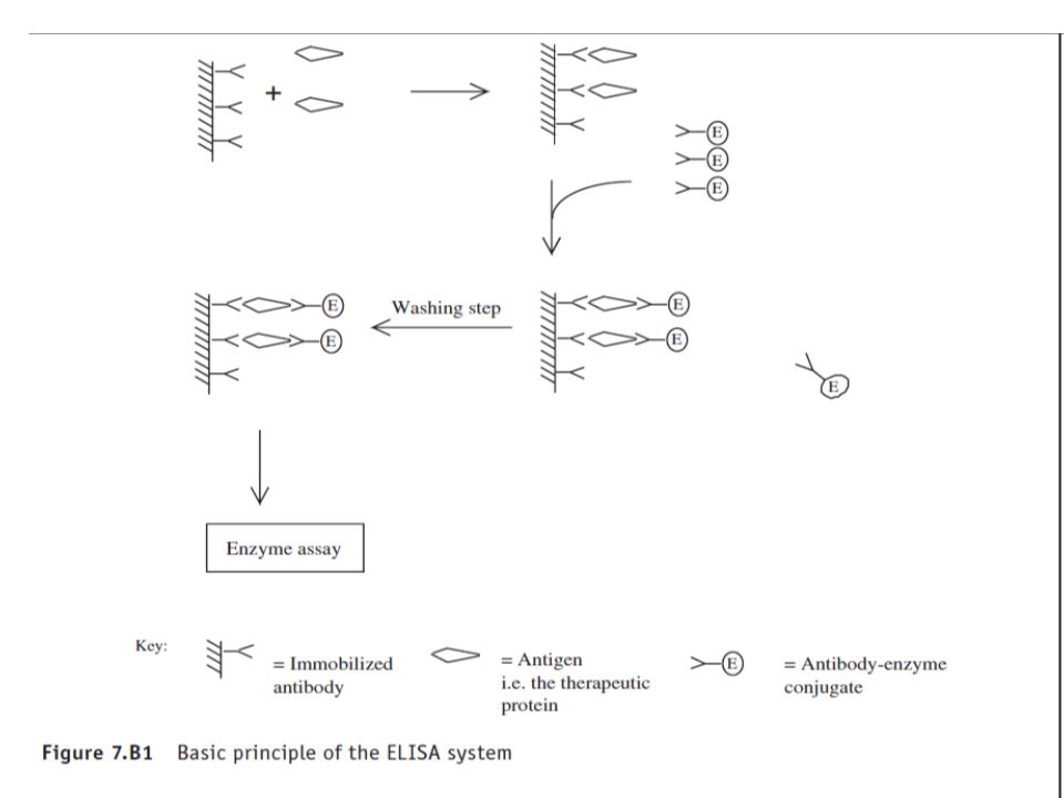
The use of conjugated radiolabels (RIA) or enzymes (EIA) to allow detection of antigen–antibody binding renders such assays very sensitive. Furthermore, when compared with a bioassay, immunoassays are rapid (undertaken in minutes to hours), inexpensive, and straightforward to undertake. In most such systems, the antibody is immobilized on the internal walls of the wells in a multi-well microtitre plate, which therefore serves as collection of reaction mini-test tubes. One of the most popular EIA systems currently in use is that of the ELISA (Fig 7.B1) In this form it is also often referred to as the double antibody sandwich technique.
, inexpensive, and straightforward to undertake. In most such systems, the antibody is immobilized on the internal walls of the wells in a multi-well microtitre plate, which therefore serves as collection of reaction mini-test tubes. One of the most popular EIA systems currently in use is that of the ELISA (Fig 7.B1) In this form it is also often referred to as the double antibody sandwich technique.")
18
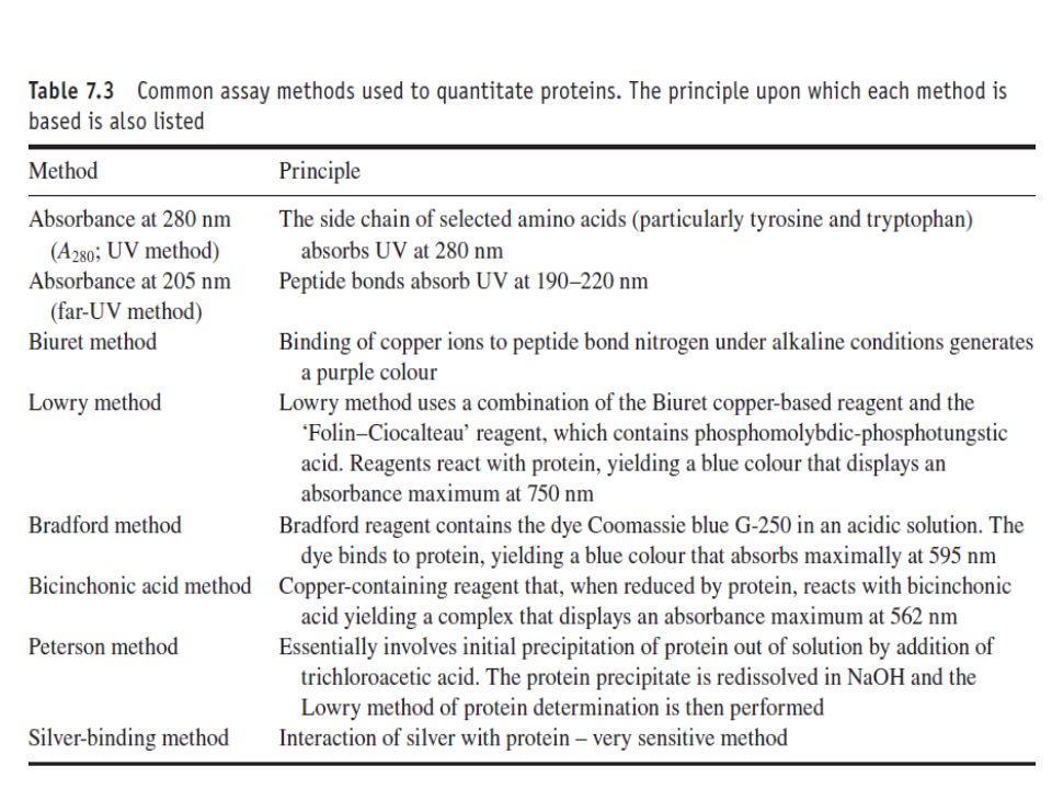
Determination of protein concentration
Quantification of total protein in the final product represents another standard analysis undertaken by QC. A number of different protein assays may be potentially employed (Table 7.3). Detection and quantification of protein by measuring absorbency at 280 nm is perhaps the simplest such method. This approach is based on the fact that the side chains of the amino acids tyrosine and tryptophan absorb at this wavelength.
. Detection and quantification of protein by measuring absorbency at 280 nm is perhaps the simplest such method. This approach is based on the fact that the side chains of the amino acids tyrosine and tryptophan absorb at this wavelength.")
19
The method is popular, as it is fast, easy to perform and is non-destructive to the sample.
However, it is a relatively insensitive technique, and identical concentrations of different proteins will yield different absorbance values if their content of tyrosine and tryptophan vary to any significant extent. Hence, this method is rarely used to determine the protein concentration of the final product, but it is routinely used during downstream processing to detect protein elution off chromatographic columns, and hence track the purification process.

21
Detection of protein-based product impurities:-
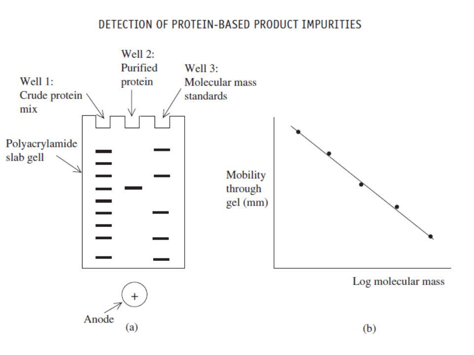
SDS polyacrylamide gel electrophoresis (SDS-PAGE) represents the most commonly used analytical technique in the assessment of final product purity (Figure 7.1). It provides high-resolution separation of polypeptides on the basis of their molecular mass. Bands containing as little as 100 ng of protein can be visualized by staining the gel with dyes such as Coomassie blue. Subsequent gel analysis by scanning laser densitometry allows quantitative determination of the protein content of each band.
 represents the most commonly used analytical technique in the assessment of final product purity (Figure 7.1). It provides high-resolution separation of polypeptides on the basis of their molecular mass. Bands containing as little as 100 ng of protein can be visualized by staining the gel with dyes such as Coomassie blue. Subsequent gel analysis by scanning laser densitometry allows quantitative determination of the protein content of each band.")
22
The use of silver-based stains increases the detection sensitivity up to 100 fold, with individual bands containing as little as 1ng of protein usually staining well. However, because silver binds to protein non-stoichiometrically, quantitative studies using densitometry cannot be undertaken. SDS-PAGE is normally run under reducing conditions. Addition of a reducing agent such as β-mercaptoethanol or dithiothreitol (DTT) disrupts interchain (and intrachain) disulfide linkages. Individual polypeptides held together via disulfide linkages in oligomeric proteins will thus separate from each other on the basis of their molecular mass.
 disrupts interchain (and intrachain) disulfide linkages. Individual polypeptides held together via disulfide linkages in oligomeric proteins will thus separate from each other on the basis of their molecular mass.")
23
The presence of bands additional to those equating to the protein product generally represent protein contaminants. Such contaminants may be unrelated to the product or may be variants of the product itself (e.g. differentially glycosylated variants, proteolytic fragment, etc.). Further characterization may include western blot analysis. This involves eluting the protein bands from the electrophoretic gel onto a nitrocellulose filter. The filter can then be probed using antibodies raised against the product. Binding of the antibody to the ‘contaminant’ bands suggests that they are variants of the product.
. Further characterization may include western blot analysis. This involves eluting the protein bands from the electrophoretic gel onto a nitrocellulose filter. The filter can then be probed using antibodies raised against the product. Binding of the antibody to the ‘contaminant’ bands suggests that they are variants of the product.")
25
One concern relating to SDS-PAGE-based purity analysis is that contaminants of the same molecular mass as the product will go undetected as they will comigrate with it. Two-dimensional electrophoretic analysis would overcome this eventuality in most instances. The most commonly utilized method entails separation of proteins by isoelectric focusing (see below) in the first dimension, with separation in the second dimension being undertaken in the presence of SDS, thus promoting band separation on the basis of protein size.
 in the first dimension, with separation in the second dimension being undertaken in the presence of SDS, thus promoting band separation on the basis of protein size.")
26
Isoelectric focusing entails setting up a pH gradient along the length of an electrophoretic gel.
Applied proteins will migrate under the influence of an electric field until they reach a point in the gel at which the pH equals the protein’s isoelectric point pI (the pH at which the protein exhibits no overall net charge; only species with a net charge will move under the influence of an electric field). Isoelectric focusing thus separates proteins on the basis of charge characteristics.
. Isoelectric focusing thus separates proteins on the basis of charge characteristics.")
27
This technique is also utilized in the biopharmaceutical industry to determine product homogeneity.
Homogeneity is best indicated by the appearance in the gel of a single protein band, exhibiting the predicted pI value. Isoelectric focusing also finds application in analysing the stability of biopharmaceuticals over the course of their shelf life.

28
Capillary electrophoresis:-
Separation is based upon different rates of protein migration upon application of an electric field. This separation occurs within a capillary tube with a diameter of 20–50 μm and be up to a 1 m long. This, in turn, allows operation at a higher current density, thus speeding up the rate of migration through the capillary. Sample analysis can be undertaken in 15–30 min, and on-line detection at the end of the column allows automatic detection and quantification of eluting bands. The speed, sensitivity, high degree of automation and ability to quantitate protein bands directly render this system ideal for biopharmaceutical analysis.

29
High-performance liquid chromatography:-
Most of the chromatographic strategies used to separate proteins under ‘low pressure’ (e.g. gel filtration, ion exchange, etc.) can be adapted to operate under high pressure. Reverse-phase-, size-exclusion- and, to a lesser extent, ion-exchange-based HPLC chromatography systems are now used in the analysis of a range of biopharmaceutical preparations. On-line detectors (usually a UV monitor set at 220 or 280 nm) allows automated detection and quantification of eluting bands.
 can be adapted to operate under high pressure. Reverse-phase-, size-exclusion- and, to a lesser extent, ion-exchange-based HPLC chromatography systems are now used in the analysis of a range of biopharmaceutical preparations. On-line detectors (usually a UV monitor set at 220 or 280 nm) allows automated detection and quantification of eluting bands.")
30
HPLC is characterized by a number of features that render it an attractive analytical tool. These include: excellent fractionation speeds (often just minutes per sample); superior peak resolution; high degree of automation (including data analysis); ready commercial availability of various sophisticated systems
; superior peak resolution; high degree of automation (including data analysis); ready commercial availability of various sophisticated systems.")
31
Mass spectrometry:- It is now possible to determine the molecular mass of many proteins to within an accuracy of +/-0.01 per cent. A protein variant missing a single amino acid residue can easily be distinguished from the native protein in many instances.

32
Immunological approaches to detection of contaminants:-
Most recombinant biopharmaceuticals are produced in microbial or mammalian cell lines. Thus, although the product is derived from a human gene, all product-unrelated contaminants will be derived from the producer organism. These non-self proteins are likely to be highly immunogenic in humans, rendering their removal from the product stream especially important.

33
Immunoassays have found widespread application in detecting and quantifying product impurities.
These assays are extremely specific and very sensitive, often detecting target antigen down to parts per million levels. Many immunoassays are available commercially, and companies exist that will rapidly develop tailor-made immunoassay systems for biopharmaceutical analysis.

34
Application of the analytical techniques discussed thus far focuses upon detection of proteinaceous impurities. A variety of additional tests are undertaken that focus upon the active substance itself. Tests performed to verify the product identity include amino acid analysis, peptide mapping, N-terminal sequencing and spectrophotometric analyses.

35
Amino acid analysis:- Amino acid analysis remains a characterization technique undertaken in many laboratories, in particular if the product is a peptide or small polypeptide (molecular mass ≤10 kDa.). The strategy is simple: Determine the range and quantity of amino acids present in the product and compare the results obtained with the expected (theoretical) values. The results should be comparable.
. The strategy is simple: Determine the range and quantity of amino acids present in the product and compare the results obtained with the expected (theoretical) values. The results should be comparable.")
36
Although this technique is relatively straightforward and automated amino acid analysers are commercially available, it is subject to a number of disadvantages that limits its usefulness in biopharmaceutical analysis. These include: Hydrolysis conditions can destroy/modify certain amino acid residues, The method is semi-quantitative rather than quantitative; Sensitivity is at best moderate; low-level contaminants may go undetected.

37
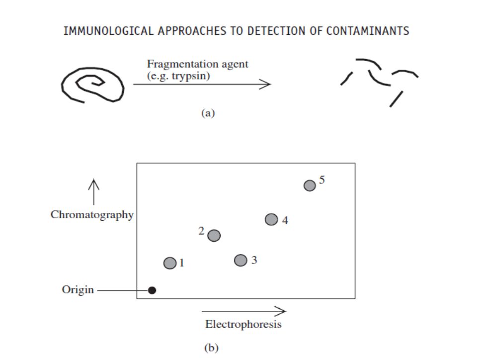
Peptide mapping:- A major concern relating to biopharmaceuticals produced in high-expression recombinant systems is the potential occurrence of point mutations in the product’s gene, leading to an altered primary structure (i.e. amino acid sequence). The approach most commonly used to detect alterations in amino acid sequence is peptide (fingerprint) mapping.
. The approach most commonly used to detect alterations in amino acid sequence is peptide (fingerprint) mapping.")
38
Peptide mapping entails exposure of the protein product to a reagent that promotes hydrolysis of peptide bonds at specific points along the protein backbone. This generates a series of peptide fragments. These fragments can be separated from each other by a variety of techniques, including one- or two-dimensional electrophoresis, and RP-HPLC in particular. A standardized sample of the protein product when subjected to this procedure will yield a characteristic peptide fingerprint, or map,

40
Two-dimensional separation of the peptides is far more likely to resolve each peptide completely from the others. In the case above, for example, chromatography (in the vertical dimension) alone would not have been sufficient to resolve peptides 1 and 3 fully. During biopharmaceutical production, each batch of the recombinant protein produced should yield identical peptide maps.
 alone would not have been sufficient to resolve peptides 1 and 3 fully. During biopharmaceutical production, each batch of the recombinant protein produced should yield identical peptide maps.")
41
N-terminal sequencing:-
N-terminal sequencing of the first 20–30 amino acid residues of the protein product has become a popular quality control test for finished biopharmaceutical products. The technique is useful, as it: Positively identifies the protein; Confirms (or otherwise) the accuracy of the amino acid sequence of at least the N-terminus of the protein; Readily identifies the presence of modified forms of the product in which one or more amino acids are missing from the N-terminus.
 the accuracy of the amino acid sequence of at least the N-terminus of the protein; Readily identifies the presence of modified forms of the product in which one or more amino acids are missing from the N-terminus.")
42
N-terminal sequencing is normally undertaken by Edman degradation.
Facilitate fast and automated determination of up to the first 100 amino acids from the N-terminus of most proteins, and usually requires a sample size of less than 1 μmol to do so.

43
Analysis of secondary and tertiary structure:-
Although a protein’s three-dimensional conformation may be studied in great detail by X-ray crystallography or NMR spectroscopy, routine application of such techniques to biopharmaceutical manufacture is impractical, both from a technical and an economic standpoint. More recently proton-NMR has also been applied to studying higher orders of protein structure.

44
Endotoxin and other pyrogenic contaminants:-
Pyrogens are substances that, when they enter the blood stream, influence hypothalamic regulation of body temperature, usually resulting in fever and in severe cases results in patient death. Pyrogens represent a diverse group of substances, including various chemicals, particulate matter and endotoxin (LPS), a molecule derived from the outer membrane of Gram-negative bacteria.
, a molecule derived from the outer membrane of Gram-negative bacteria.")
45
In many instances the influence of pyrogens on body temperature is indirect.
For example, entry of endotoxin into the bloodstream stimulates the production of IL-1 by macrophages. It is the IL-1 that directly initiates the fever response (hence its alternative name, ‘endogenous pyrogen’). Effective implementation of GMP (good manufacturing practice) minimizes the likelihood of product contamination by pyrogens. For example, GMP dictates that chemical reagents used in the manufacture of process buffers be extremely pure.
. Effective implementation of GMP (good manufacturing practice) minimizes the likelihood of product contamination by pyrogens. For example, GMP dictates that chemical reagents used in the manufacture of process buffers be extremely pure.")
46
Such raw materials, therefore, are unlikely to contain chemical contaminants displaying pyrogenic activity. Furthermore, GMP encourages filtration of virtually all parenteral products through a 0.45 or 0.22 μm filter at points during processing and prior to filling in final product containers (even if the product can subsequently be sterilized by autoclaving). As an additional safeguard, the final product will usually be subject to a particulate matter test by QC before final product release.
. As an additional safeguard, the final product will usually be subject to a particulate matter test by QC before final product release.")
47
Contamination of the final product with endotoxin is more difficult to control because:-
Many recombinant biopharmaceuticals are produced in Gram-negative bacterial systems; thus, the product source is also a source of endotoxin. Most biopharmaceutical preparations will be contaminated with low levels of Gram-negative bacteria at some stage of manufacture. This is one of many reasons why GMP dictates that the level of bioburden in the product stream should be minimized at all stages of manufacture.

48
The heat stability exhibited by endotoxin means that autoclaving of process equipment will not destroy endotoxin present on such equipment. Adverse medical reactions caused by endotoxin are witnessed in humans at dosage rates as low as 0.5 ng per kilogram body weight.

49
Endotoxin, the molecule:-
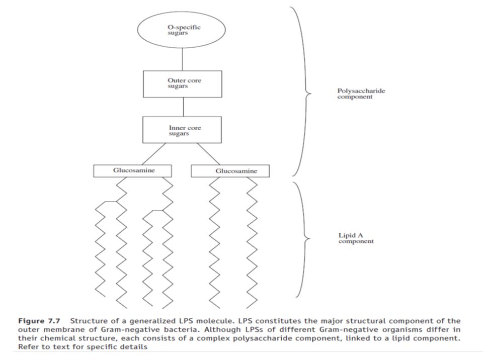
The structural detail of a generalized endotoxin (LPS) molecule is presented in Figure 7.7. As its name suggests, LPS consists of a complex polysaccharide component linked to a lipid (lipid A) moiety. Most of the LPS biological activity (pyrogenicity) is associated with its lipid A moiety. This usually consists of six or more fatty acids attached directly to sugars such as glucosamine.
 molecule is presented in Figure 7.7. As its name suggests, LPS consists of a complex polysaccharide component linked to a lipid (lipid A) moiety. Most of the LPS biological activity (pyrogenicity) is associated with its lipid A moiety. This usually consists of six or more fatty acids attached directly to sugars such as glucosamine.")
51
Pyrogen detection:- Pyrogens may be detected in parenteral preparations (or other substances) by a number of methods. Two such methods are widely employed in the pharmaceutical industry. 1. The rabbit pyrogen test: This entails parenteral administration of the product to a group of healthy rabbits, with subsequent monitoring of rabbit temperature using rectal probes. Increased rabbit temperature above a certain point suggests the presence of pyrogenic substances.

52
The product is considered to have passed the test if the total (summed) increase of the temperature of all three animals (rabbits) is less than 1.15 C. If the total increase recorded is greater than 2.65 C then the product has failed. However, it is also subject to a number of disadvantages, including: it is expensive (there is a requirement for animals, animal facilities and animal technicians); excitation/poor handling of the rabbits can affect the results obtained, usually prompting a false positive result; subclinical infection/poor overall animal health can also lead to false positive results.
; excitation/poor handling of the rabbits can affect the results obtained, usually prompting a false positive result; subclinical infection/poor overall animal health can also lead to false positive results.")
53
d. use of different rabbit colonies/breeds can yield variable results.
e. Another issue of relevance is that certain biopharmaceuticals (e.g. cytokines such as 1L-1 and TNF) themselves induce a natural pyrogenic response. 2. in vitro assay; the Limulus ameobocyte lysate (LAL) test. This is based upon endotoxin-stimulated coagulation of amoebocyte lysate obtained from horseshoe crabs. This test is now the most widely used assay for the detection of endotoxins in biopharmaceutical and other pharmaceutical preparations.
 themselves induce a natural pyrogenic response. 2. in vitro assay; the Limulus ameobocyte lysate (LAL) test. This is based upon endotoxin-stimulated coagulation of amoebocyte lysate obtained from horseshoe crabs. This test is now the most widely used assay for the detection of endotoxins in biopharmaceutical and other pharmaceutical preparations.")
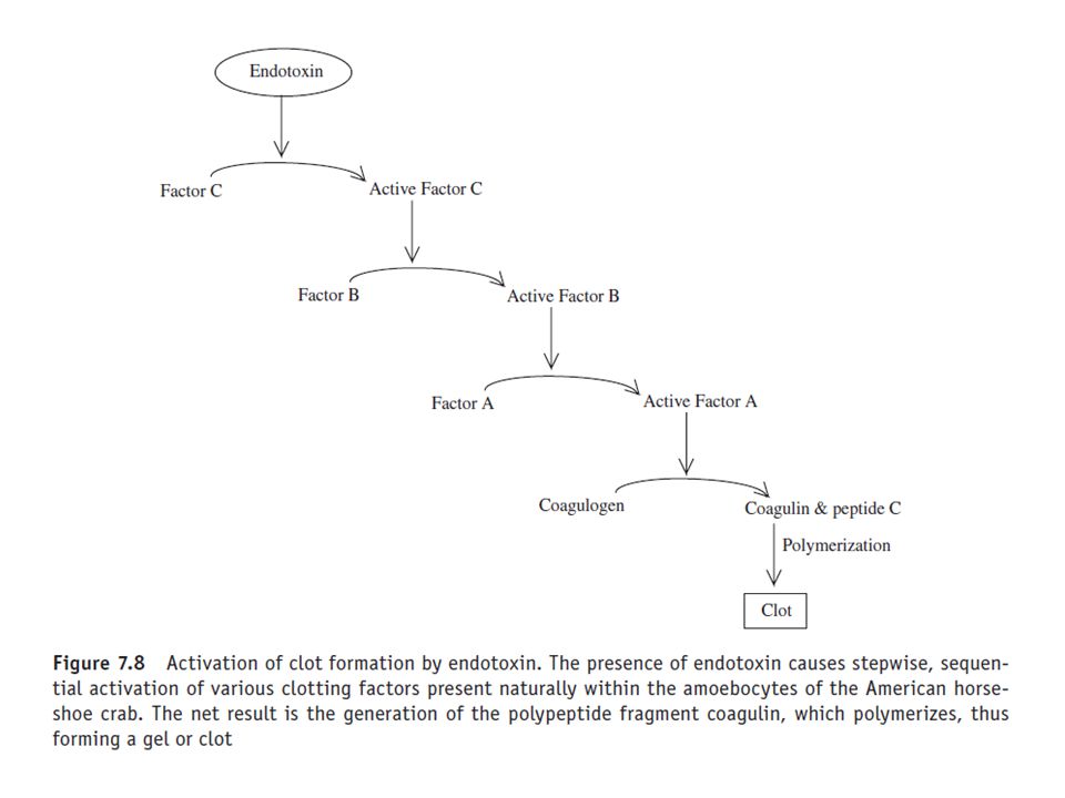
54
Development of the LAL assay was based upon the observation that the presence of Gram negative bacteria in the vascular system of the American horseshoe crab, Limulus polyphemus, resulted in the clotting of its blood. Tests on fractionated blood showed that the factor responsible for coagulation resided within the crab’s circulating blood cells, i.e. the amoebocytes. Further research revealed that the bacterial agent responsible of initiation of clot formation was endotoxin.

55
The endotoxin molecule activates a coagulation cascade quite similar in design to the mammalian blood coagulation cascade (Figure 7.8). Activation of the cascade also requires the presence of divalent cations such as calcium or magnesium. The LAL reagent is prepared by extraction of blood from the horseshoe crab, followed by isolation of its amoebocytes by centrifugation. After a washing step, the amoebocytes are lysed and the lysate dispensed into pyrogen-free vials. The assay is normally performed by making a series of 1:2 dilutions of the test sample using (pyrogen-free) WFI-water for injections-(and pyrogen-free test tubes).
 WFI-water for injections-(and pyrogen-free test tubes).")
57
A reference standard endotoxin preparation is treated similarly.
LAL reagent is added to all tubes, incubated for 1 h, and these tubes are then inverted to test for gel (i.e. clot) formation, which would indicate presence of endotoxin. More recently, a colorimetric-based LAL procedure has been devised. This allows spectrophotometric analysis of the test sample, facilitating more accurate end-point determination.
 formation, which would indicate presence of endotoxin. More recently, a colorimetric-based LAL procedure has been devised. This allows spectrophotometric analysis of the test sample, facilitating more accurate end-point determination.")
58
The LAL system displays several advantages when compared with the rabbit test, most notably:-
1. Sensitivity – endotoxin levels as low as a few picograms per millilitre of sample assayed will be detected. 2. Cost – the assay is far less expensive than the rabbit assay. 3. Speed – depending upon the format used, the LAL assay may be conducted within 15–60 min. Its major disadvantage is its selectivity: it only detects endotoxin-based pyrogens.

59
Removing of Endotoxin:
Endotoxin present in the earlier stages of production is often effectively removed from the product during chromatographic fractionation. The endotoxin molecule’s highly negative charge often facilitates its effective removal from the product stream by ion-exchange chromatography. Gel-filtration chromatography also serves to remove endotoxin from the product. Although individual LPS molecules exhibit an average molecular mass of less than 20 kDa, these molecules aggregate in aqueous environments and generate supramolecular structures of molecular mass 100–1000 kDa.

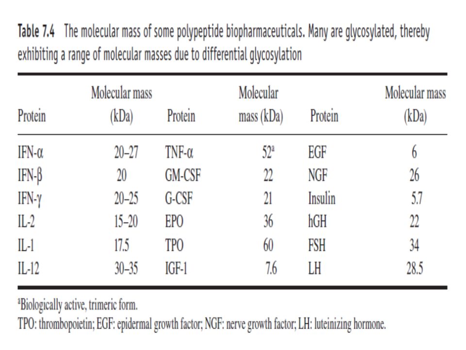
60
The molecular mass of most biopharmaceuticals is considerably less than 100 kDa (Table 7.4).
The proteins would thus elute from gel-filtration columns much later than contaminating endotoxin aggregates. Should the biopharmaceutical exhibit a molecular mass approaching or exceeding 100 kDa, then effective separation can still be achieved by inclusion of a chelating agent such as EDTA in the running buffer. This promotes depolymerization of the endotoxin aggregates into monomeric (20 kDa) form.
 form.")
62
DNA as a contaminant: The clinical significance of DNA-based contaminants in biopharmaceutical products remains unclear. The concerns relating to the presence of DNA in modern biopharmaceuticals focus primarily upon the presence of active oncogenes in the genome of several producer cell types (e.g. monoclonal antibody production in hybridoma cell lines). Parenteral administration of DNA contaminants containing active oncogenes to patients is considered undesirable.
. Parenteral administration of DNA contaminants containing active oncogenes to patients is considered undesirable.")
63
The concern is that uptake and expression of such DNA in human cells could occur.
There is some evidence to suggest that naked DNA can be assimilated by some cells at least, under certain conditions. Guidelines to date state that an acceptable level of residual DNA in recombinant products is of the order of 10 pg per therapeutic dose.

64
DNA Detection: DNA hybridization studies (e.g. the ‘dot blot’ assay) utilizing radiolabelled DNA probes allows detection of DNA contaminants in the product, to levels in the nanogram range. The process begins with isolation of the contaminating DNA from the product. This can be achieved, for example, by phenol and chloroform extraction and ethanol precipitation. The isolated DNA is then applied as a spot (i.e. a ‘dot’) onto nitrocellulose filter paper, with subsequent baking of the filter at 80°C under vacuum. This promotes (a) DNA denaturation, yielding single strands, and (b) binding of the DNA to the filter.
 utilizing radiolabelled DNA probes allows detection of DNA contaminants in the product, to levels in the nanogram range. The process begins with isolation of the contaminating DNA from the product. This can be achieved, for example, by phenol and chloroform extraction and ethanol precipitation. The isolated DNA is then applied as a spot (i.e. a ‘dot’) onto nitrocellulose filter paper, with subsequent baking of the filter at 80°C under vacuum. This promotes (a) DNA denaturation, yielding single strands, and (b) binding of the DNA to the filter.")
65
A sample of total DNA derived from the cells in which the product is produced is then radiolabelled with 32P using the process of nick translation. It is heated to 90°C (promotes denaturation, forming single strands) and incubated with the baked filter for several hours at 40°C. Lowering the temperature allows reannealing of single strands via complementary base-pairing to occur. Labelled DNA will reanneal with any complementary DNA strands immobilized on the filter. After the filter is washed (to remove non-specifically bound radiolabelled probe) it is subjected to autoradiography, which allows detection of any bound probe.
 and incubated with the baked filter for several hours at 40°C. Lowering the temperature allows reannealing of single strands via complementary base-pairing to occur. Labelled DNA will reanneal with any complementary DNA strands immobilized on the filter. After the filter is washed (to remove non-specifically bound radiolabelled probe) it is subjected to autoradiography, which allows detection of any bound probe.")
66
Quantification of the DNA:
Quantification of the DNA isolated from the product involves concurrent inclusion in the dot blot assay of a set of spots, containing known quantities of DNA, and being derived from the producer cell. After autoradiography, the intensity of the test spot is compared with the standards. DNA Removal: In many instances there is little need to incorporate specific DNA removal steps during downstream processing. Endogenous nucleases liberated upon cellular homogenization come into direct contact with cellular DNA, resulting in its degradation.

67
Commercial DNase’s are sometimes added to crude homogenate to reduce DNA-associated product viscosity. Most chromatographic steps are also effective in separating DNA from the product stream. Ion-exchange chromatography is particularly effective, as DNA exhibits a large overall negative charge.

68
Microbial and viral contaminants :-
Finished-product biopharmaceuticals, along with other pharmaceuticals intended for parenteral administration, must be sterile (the one exception being live bacterial vaccines). The presence of microorganisms in the final product is unacceptable for a number of reasons: Parenteral administration of contaminated product would likely lead to the establishment of a severe infection in the recipient patient.
. The presence of microorganisms in the final product is unacceptable for a number of reasons: Parenteral administration of contaminated product would likely lead to the establishment of a severe infection in the recipient patient.")
69
Microorganisms may be capable of metabolizing the product itself, thus reducing its potency.
Microbial-derived substances secreted into the product could adversely affect the recipient’s health. Examples include endotoxin secreted from Gram-negative bacteria.

70
Sterilization of biopharmaceuticals by filtration, followed by aseptic filling into a sterile final-product container, inherently carries a greater risk of product contamination. Biopharmaceutical products are also subjected to screening for the presence of viral particles prior to final product release. Although viruses could be introduced, for example, via infected personnel during downstream processing, proper implementation of GMP minimizes such risk. Any viral particles found in the finished product are most likely derived from raw material sources.

71
Examples could include HIV or hepatitis viruses present in blood used in the manufacture of blood products. Such raw materials must be screened before processing for the presence of likely viral contaminants. Producer cell lines are screened during product development studies to ensure freedom from a variety of pathogenic advantageous agents, including various species of bacteria, fungi, yeast, mycoplasma, protozoa, parasites, viruses and prions. Suitable microbiological precautions must subsequently be undertaken to prevent producer cell banks from becoming contaminated with such pathogens.

72
Removal of Viruses: Gel-filtration chromatography, for example, effectively separates viral particles from most proteins on the basis of differences in size. 2. Filtration through a 0.22 μm filter effectively removes microbial agents from the product stream, but fails to remove most viral types. Repeat filtration through a 0.1 μm filter is more effective in this regard. 3. Alternatively, incorporation of an ultrafiltration step (preferably at the terminal stages of downstream processing) also proves effective.
 also proves effective.")
73
4. Heating the product to between 40 and 60°C for several hours inactivates a broad range of viruses. Many biopharmaceuticals can be heated to such temperatures without being denatured themselves. Such an approach has been used extensively to inactivate blood-borne viruses in blood products. 5. Exposure of product to controlled levels of UV radiation can also be quite effective, while having no adverse effect on the product itself.

74
Viral assays: Viral assays currently available will detect only a specific virus, or at best a family of closely related viruses. Current viral assays fall into one of three categories: 1. immunoassays; 2. assays based on viral DNA probes; 3. bioassays.

75
1. Immunoassays capable of detecting a wide range of viruses are available commercially.
The sensitivity, ease, speed and relative inexpensiveness of these assays render them particularly attractive. 2. An alternative assay format entails the use of virus-specifi cDNA probes. These can be used to screen the biopharmaceutical product for the presence of viral DNA. The assay strategy is similar to the dot blot assays used to detect host-cell-derived DNA contaminants, as discussed earlier.

76
3. Viral bioassays: different formats have also been developed.
One format entails incubation of the final product with cell lines sensitive to a range of viruses. The cells are subsequently monitored for cytopathic effects or other obvious signs of viral infection. A range of mouse-, rabbit- or hamster-antibody production tests may also be undertaken. These bioassays entail administration of the product to a test animal. Any viral agents present will elicit production of antiviral antibodies in that animal.

77
Serum samples (withdrawn from the animal approximately 4 weeks after product administration) are screened for the presence of antibodies recognizing a range of viral antigens. This can be achieved by enzyme immunoassay, in which immobilized antigen is used to screen for the virus-specific antibodies. These assay systems are extremely sensitive, as minute quantities of viral antigen will elicit strong antibody production. A single serum sample can also be screened for antibodies specific to a wide range of viral particles. Time and expense factors, however, militate against this particular assay format.

78
Miscellaneous contaminants
Could include buffer components, precipitants (ethanol or other solvents, salts, etc.), proteolytic inhibitors, glycerol, anti-foam agents, etc. In addition to these, other contaminants may enter the product during downstream processing in a less controlled way. Examples could include metal ions leached from product-holding tanks/pipework, or breakdown products leaking from chromatographic media. For this reason, high-quality glass vials are often used.
, proteolytic inhibitors, glycerol, anti-foam agents, etc. In addition to these, other contaminants may enter the product during downstream processing in a less controlled way. Examples could include metal ions leached from product-holding tanks/pipework, or breakdown products leaking from chromatographic media. For this reason, high-quality glass vials are often used.")
79
In some instances it may be necessary to demonstrate that all traces of specific contaminants have been removed prior to final product filling. This would be true, for example, of many proteolytic inhibitors added during the initial stages of downstream processing to prevent proteolysis by endogenous proteases. Some such inhibitors may be inherently toxic, and many could (inappropriately) inhibit endogenous proteases of the recipient patient. Various chemical-coupling methods may be used to attach affinity ligands to the chromatographic support material.
 inhibit endogenous proteases of the recipient patient. Various chemical-coupling methods may be used to attach affinity ligands to the chromatographic support material.")
80
Some such procedures entail the use of toxic reagents, which, if not entirely removed after coupling, could leach into the product. Improvements in the chemical stability of modern chromatographic media, however, have reduced such difficulties, and most manufacturers have carried out extensive validation studies regarding the stability of their product. The possibility exists, however, that uncharacterized contaminants may persist, remaining undetected in the final product. As an additional safety measure, finished products are often subjected to ‘abnormal toxicity’ or ‘general safety’ tests.

81
Standardized protocols for such tests are outlined in various international pharmacopoeias.
These normally entail parenteral administration of the product to at least five healthy mice. The animals are placed under observation for 48 h and should exhibit no ill effects (other than expected symptoms). The death or illness of one or more animals signals a requirement for further investigation, usually using a larger number of animals.
. The death or illness of one or more animals signals a requirement for further investigation, usually using a larger number of animals.")
Similar presentations


 SDS PAGE Isoelectric Point Isoelectric focusing.>")

>")

