Download presentation
Presentation is loading. Please wait.

1
Evaluation of the novel nanocell (in vitro/in vivo) Dr.Ramraj Panthee Chulabhorn Research Institute
 Dr.Ramraj Panthee Chulabhorn Research Institute")
2
Outline Introduction In vitro studies In vivo studies Summary

3
Introduction Novel nanocellNovel nanocell Confocal microscopeConfocal microscope In vitro In vitro Cell culture studies Cell culture studies Co- culture studies Co- culture studies In vivoIn vivo Nude mice tumor studiesNude mice tumor studies

4
The novel nanocell structure FTY720 Doxorubisin Doxorubisin +5FU(D+5F) PLGAconjugation Both) PLGA conjugation (Both) 200 nm 100 nm (F) Nanocell
 PLGAconjugation Both) PLGA conjugation (Both) 200 nm 100 nm (F) Nanocell")
5
The novel nanocell Function 24 1 200 nm 100 nm 3 Nanocell Tumor cell BVs shutdown Nc into tumor cell

6
Confocal microscope Structure and function Laser ray Laser ray 3 dimensional picture 3 dimensional picture Computerized device Computerized device

7
In vitro studies controls In vitro studies controls Vehicle is phosphate buffer solution(PBS) FTY720 Doxo + 5FU Doxorubicin 5FU Dox+FTY Dox v
 FTY720 Doxo + 5FU Doxorubicin 5FU Dox+FTY Dox v")
8
Cell culture studies + Liver cancer cells Calf serum culture medium 24 hours 5%5% Cells + Drugs Viable rate Drugs action on liver cancer cells Survival

9
Cont. Cell culture studies Plated for 24 hrs,Plated for 24 hrs, Doxorubicin +5FU Conjugated mixed for 48hrs.Doxorubicin +5FU Conjugated mixed for 48hrs. Evaluation: Trypan blue exclusion method.Evaluation: Trypan blue exclusion method. Ec 50 calculation curve fitting Ec 50 calculation curve fitting Cellculture Trypan blue – cell viability Ec 50 - 50% test cells effective by drugs

10
Tumor-endothelium co- culture studies Tumor-endothelium co- culture studies Liver cancer cells with GFP Expre..Liver cancer cells with GFP Expre.. Endothelial cellsEndothelial cells MatrigelmatrixMatrigelmatrix Paraformaldehyde(4 %)Paraformaldehyde(4 %) Incubation- propidium IodideIncubation- propidium Iodide Exposed to different treatmentExposed to different treatment Liver cancer cell Endothelial cell
Paraformaldehyde(4 %) Incubation- propidium IodideIncubation- propidium Iodide Exposed to different treatmentExposed to different treatment Liver cancer cell Endothelial cell.")
11
Microscopic studies of cell Control 30 h FTY720 DOXO+5FU Modified picture from: Nature vol. 436 July 2005 S.sengupta et.al.. Control 12 h Nanocell 12h Nanocell 30h Nanocell in 12h action,BV Nanocell in 30h tumor

12
Microscopic studies (cont.) Yellow stained – Liver cancer cellsYellow stained – Liver cancer cells Red stained - endothelial cellsRed stained - endothelial cells Doxorubicin +5FU –Tumor reducedDoxorubicin +5FU –Tumor reduced FTY720 – Vascular shutdownFTY720 – Vascular shutdown Nanocell in 12hours little effectNanocell in 12hours little effect Nanocell in 30hours complete tumor reducedNanocell in 30hours complete tumor reduced
 Yellow stained – Liver cancer cellsYellow stained – Liver cancer cells Red stained - endothelial cellsRed stained - endothelial cells Doxorubicin +5FU –Tumor reducedDoxorubicin +5FU –Tumor reduced FTY720 – Vascular shutdownFTY720 – Vascular shutdown Nanocell in 12hours little effectNanocell in 12hours little effect Nanocell in 30hours complete tumor reducedNanocell in 30hours complete tumor reduced")
13
Stereological quantification of co-culture Vascular componentVascular component : VEHICLE 12hr-no change In 30hrs. Vascular changed Model picture from Nature vol.436 2005 ------ 12 h--- -------30 h--------- Vehicle Nanocell Vehicle FTY720 5Fluouracil +Doxo

14
Stereological quantification of co-culture Tumor componentTumor component : Vehicle 12hrs no change Doxo+5FU 30hrs Model picture from Nature vol.436 2005 Vehicle Nanocell vehicle FTY720 Doxo+5F Tumor component

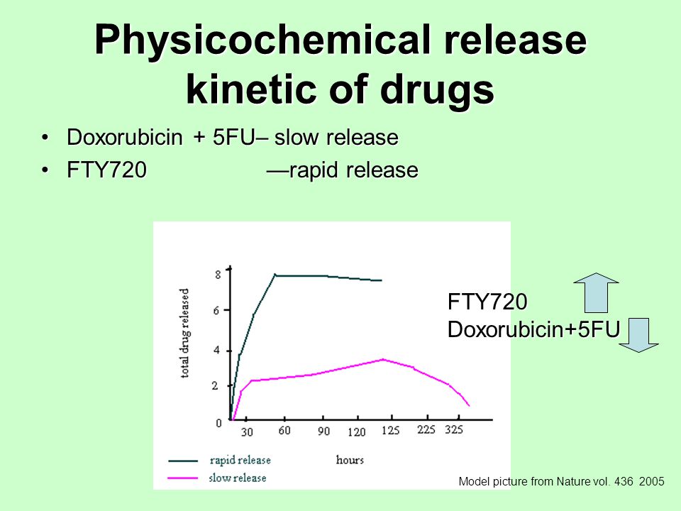
15
Physicochemical release kinetic of drugs Doxorubicin + 5FU– slow releaseDoxorubicin + 5FU– slow release FTY720 —rapid releaseFTY720 —rapid release FTY720Doxorubicin+5FU Model picture from Nature vol. 436 2005
16
In vivo nanocell studies Nude mouse Nude mouse 1.High sensitive to antigen 2.Well manifestation of tumor

17
In vivo nanocells (cont.) in vivo tumor studies GFP expressionGFP expression Liver cancer cellsLiver cancer cells Implanted male nude miceImplanted male nude mice Tumor vol. 50mm 3Tumor vol. 50mm 3 Treatment started with 100µ l ivTreatment started with 100µ l iv FTY720 andFTY720 and doxorbicin+5FUdoxorbicin+5FU Animals were killed in periodsAnimals were killed in periods Necropsies tumor - histopathologyNecropsies tumor - histopathology

18
In vivo Nanocell Nude mouse tumor Liver cancer cells injected into sub - cutaneous tissue Tumor 50mm 3 in size

19
Tumor studies Size of tumor with different controls Modified picture from Nature vol.436,july 2005 S. sengupta et.al. D+5F+F F 5F N D+F V

20
Effects of treatment SurvivalSurvival ToxicityToxicity Traditional chemotherapy 30days survive Nanocell combined 65 days survive Less toxic WBC count show - normal Without any treatment20days survive Source-Nature:436:2005 S.Sengupta et.al

21
Tissue distribution studies NC + Fluorescence dyeNC + Fluorescence dye Injected – miceInjected – mice Mice were killed – 5, 10, 24 hrs. after injectionMice were killed – 5, 10, 24 hrs. after injection Organs collection –liver, lungs, spleen, tumor and serum after necropsedOrgans collection –liver, lungs, spleen, tumor and serum after necropsed Wt. – organs and fluorescence extractedWt. – organs and fluorescence extracted Purpose - Distribution of drugs into the tissues Result – Nanocells were detected within 5 hours

22
Summary A Summary A novel nanocell therapy Slow release Target specific Less toxic Cytotoxic Lifespan

Similar presentations


>")

 Antibody Siltuximab in Mouse Xenograft Models of Lung Cancer Lanxi Song, MS, Matthew A. Smith, PhD,>")


