Download presentation
Presentation is loading. Please wait.

1
Blood Anatomy & Physiology

2
Functions of blood Transportation Transportation Heat regulation Heat regulation

3
Composition of blood Made of plasma & formed elements Made of plasma & formed elements Plasma: fluid portion of blood Plasma: fluid portion of blood Formed elements Formed elements Red blood cells (erythrocytes) Red blood cells (erythrocytes) White blood cells (leukocytes) White blood cells (leukocytes) Platelets (thrombocytes) Platelets (thrombocytes)
 Red blood cells (erythrocytes) White blood cells (leukocytes) White blood cells (leukocytes) Platelets (thrombocytes) Platelets (thrombocytes)")
4
Watch US blood cells clip Watch US blood cells clip

5
Hematocrit or Packed Cell Volume (PCV) Percent of red blood cells in whole blood Percent of red blood cells in whole blood Buffy coat (less than 1%): white blood cells & platelets Buffy coat (less than 1%): white blood cells & platelets
 Percent of red blood cells in whole blood Percent of red blood cells in whole blood Buffy coat (less than 1%): white blood cells & platelets Buffy coat (less than 1%): white blood cells & platelets")
6
Erythrocytes (RBCs) Mature RBC has no nucleus, ribosomes, mitochondria Mature RBC has no nucleus, ribosomes, mitochondria Small biconcave discs Small biconcave discs Primary component: hemoglobin (1/3 of cell volume) Primary component: hemoglobin (1/3 of cell volume) Flexible because of stretchable fibers called spectrin Flexible because of stretchable fibers called spectrin
 Mature RBC has no nucleus, ribosomes, mitochondria Mature RBC has no nucleus, ribosomes, mitochondria Small biconcave discs Small biconcave discs Primary component: hemoglobin (1/3 of cell volume) Primary component: hemoglobin (1/3 of cell volume) Flexible because of stretchable fibers called spectrin Flexible because of stretchable fibers called spectrin")
7
Function of RBCs Transportation of oxygen & carbon dioxide depends on hemoglobin & an enzyme carbonic anhydrase Transportation of oxygen & carbon dioxide depends on hemoglobin & an enzyme carbonic anhydrase

8
Hemoglobin 200-300 million molecules of hemoglobin in each RBC 200-300 million molecules of hemoglobin in each RBC Each hemoglobin molecule contains 4 protein chains called globin & each chain is bound to a red pigment, heme, which each heme contains an iron molecule Each hemoglobin molecule contains 4 protein chains called globin & each chain is bound to a red pigment, heme, which each heme contains an iron molecule One hemoglobin molecule can unite with 4 oxygen molecules One hemoglobin molecule can unite with 4 oxygen molecules

9
Erythropoiesis Formation of RBC Formation of RBC Begins in the bone marrow from hematopoietic stem cells which form all blood cells Begins in the bone marrow from hematopoietic stem cells which form all blood cells In series of steps lose nuclei to become reticulocyte which is released into circulation which become mature RBC which is smaller In series of steps lose nuclei to become reticulocyte which is released into circulation which become mature RBC which is smaller

10
Erythropoietin Hormone released by the kidney when blood oxygen levels decline which then stimulates the bone marrow to increase production of RBCs Hormone released by the kidney when blood oxygen levels decline which then stimulates the bone marrow to increase production of RBCs

11
Destruction of RBCs Life span of RBC is about 105-120 days Life span of RBC is about 105-120 days Macrophages in lining of blood vessels in spleen & liver phagocytose old or damaged RBC Macrophages in lining of blood vessels in spleen & liver phagocytose old or damaged RBC Hemoglobin broken down & amino acids, iron & pigment bilirubin released Hemoglobin broken down & amino acids, iron & pigment bilirubin released Iron used to form new hemoglobin & bilirubin transported to liver & excreted into the intestines in bile Iron used to form new hemoglobin & bilirubin transported to liver & excreted into the intestines in bile

12
Leukocytes (WBCs) 5 types 5 types All have nuclei All have nuclei Larger than RBCs Larger than RBCs Granulocytes (have large granules in cytoplasm) Granulocytes (have large granules in cytoplasm) Neutrophils Eosinophils Basophils Agranulocytes Agranulocytes Lymphocytes Monocytes
 5 types 5 types All have nuclei All have nuclei Larger than RBCs Larger than RBCs Granulocytes (have large granules in cytoplasm) Granulocytes (have large granules in cytoplasm) Neutrophils Eosinophils Basophils Agranulocytes Agranulocytes Lymphocytes Monocytes")
13
Neutrophils About 65% of the total WBC count About 65% of the total WBC count Multilobed nucleus Multilobed nucleus Small light purple granules in cytoplasm Small light purple granules in cytoplasm Function: cell defense by phagocytosis of microorganisms Function: cell defense by phagocytosis of microorganisms Life span: hours to 3 days Life span: hours to 3 days

14
Eosinophils Usually 2 lobed nucleus Usually 2 lobed nucleus Large orange-red staining granules Large orange-red staining granules Function: cellular defense usually against parasites & involved in allergic reactions Function: cellular defense usually against parasites & involved in allergic reactions Life span: 10-12 days Life span: 10-12 days

15
Basophils Usually 2 lobed nucleus Usually 2 lobed nucleus Sparse, large purple staining granules Sparse, large purple staining granules Least numerous WBC Least numerous WBC Function: secrete heparin & histamine Function: secrete heparin & histamine Life span: hours to 3 days Life span: hours to 3 days

16
Lymphocytes Smallest of WBC, about 25% of total WBC count Smallest of WBC, about 25% of total WBC count Large spherical nuclei with scant pale blue cytoplasm Large spherical nuclei with scant pale blue cytoplasm T-lymphocytes: directly attack infected or cancerous cell T-lymphocytes: directly attack infected or cancerous cell B-lymphocytes: produce antibodies against specific antigens B-lymphocytes: produce antibodies against specific antigens Life span: days to years Life span: days to years

17
Monocytes Largest of the WBC Largest of the WBC Kidney bean shaped nuclei with large quantities of blue-gray cytoplasm Kidney bean shaped nuclei with large quantities of blue-gray cytoplasm Phagocytic cell capable of ingesting bacteria, debris, cancerous cells Phagocytic cell capable of ingesting bacteria, debris, cancerous cells In tissue called macrophages In tissue called macrophages Life span: months Life span: months

18
WBC Formation Neutrophils, eosinophils, basophils originate in bone marrow Neutrophils, eosinophils, basophils originate in bone marrow Most lymphocytes & monocytes originate in lymphatic tissue Most lymphocytes & monocytes originate in lymphatic tissue

19
Watch US platelets clip Watch US platelets clip

20
Platelets Small, nearly colorless, irregular Small, nearly colorless, irregular 3 important physical properties 3 important physical properties Agglutination Agglutination Adhesiveness Adhesiveness Aggregation Aggregation

21
Functions of platelets Hemostasis: stoppage of blood flow Hemostasis: stoppage of blood flow Damage to blood vessels vascular spasm temporary platelet plug by sticky platelets secrete ADP, thromboxane & fatty acid (arachidonic acid) which are involved in coagulation Damage to blood vessels vascular spasm temporary platelet plug by sticky platelets secrete ADP, thromboxane & fatty acid (arachidonic acid) which are involved in coagulation Coagulation: blood clotting Coagulation: blood clotting
 which are involved in coagulation Damage to blood vessels vascular spasm temporary platelet plug by sticky platelets secrete ADP, thromboxane & fatty acid (arachidonic acid) which are involved in coagulation Coagulation: blood clotting Coagulation: blood clotting")
22
Formation & Life Span of Platelets Formed in bone marrow, lungs & spleen by fragmentation of very large cell, megakaryocyte Formed in bone marrow, lungs & spleen by fragmentation of very large cell, megakaryocyte Life span: 7 days Life span: 7 days

23
Watch US blood type clip Watch US blood type clip

24
Blood Types Refers to the type of antigens, called agglutinogens, present on RBC membrane Refers to the type of antigens, called agglutinogens, present on RBC membrane Important blood antigens: A, B, Rh Important blood antigens: A, B, Rh Agglutinins: antibodies dissolved in plasma that react with specific blood group antigens Agglutinins: antibodies dissolved in plasma that react with specific blood group antigens

25
ABO System Type A: Antigen A on RBCs Type A: Antigen A on RBCs Type B: Antigen B on RBCs Type B: Antigen B on RBCs Type AB: Antigen A & B on RBCs Type AB: Antigen A & B on RBCs Type O: Neither A nor B on RBCs Type O: Neither A nor B on RBCs Plasma never contains Ab against Ag present on it own RBCs Plasma never contains Ab against Ag present on it own RBCs Plasma does contain AB against those Ag not present on its RBCs Plasma does contain AB against those Ag not present on its RBCs

28
The Rh System Rh positive: Rh antigen is present on RBCs Rh positive: Rh antigen is present on RBCs Rh negative: RBCs have no Rh antigen Rh negative: RBCs have no Rh antigen Blood does not normally contain anti-Rh antibodies except thru previous transfusion or pregnancy Blood does not normally contain anti-Rh antibodies except thru previous transfusion or pregnancy

29
Blood Plasma 90% water, 10 % solutes 90% water, 10 % solutes Most of the solutes are proteins (formed by liver) Most of the solutes are proteins (formed by liver) Albumin: help maintain osmotic balance Albumin: help maintain osmotic balance Globulins: immune mechanism Globulins: immune mechanism Fibrinogen: blood clotting Fibrinogen: blood clotting Remaining solutes are food substances, metabolic products, respiratory gases, hormones, etc Remaining solutes are food substances, metabolic products, respiratory gases, hormones, etc
 Most of the solutes are proteins (formed by liver) Albumin: help maintain osmotic balance Albumin: help maintain osmotic balance Globulins: immune mechanism Globulins: immune mechanism Fibrinogen: blood clotting Fibrinogen: blood clotting Remaining solutes are food substances, metabolic products, respiratory gases, hormones, etc Remaining solutes are food substances, metabolic products, respiratory gases, hormones, etc")
30
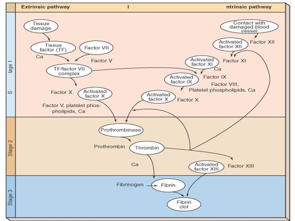
Coagulation Four components critical to coagulation Four components critical to coagulation Prothrombin Prothrombin Thrombin Thrombin Fibrinogen Fibrinogen Fibrin Fibrin Three stages Three stages Stage I Stage II Stage III

31
Stage I Production of thromboplastin activator by either: Production of thromboplastin activator by either: Extrinsic pathway: chemicals released from damaged tissues Extrinsic pathway: chemicals released from damaged tissues Intrinsic pathway: chemicals present in the blood Intrinsic pathway: chemicals present in the blood

32
Stage II Conversion of prothrombin to thrombin by the prothrombin activator produced in stage I Conversion of prothrombin to thrombin by the prothrombin activator produced in stage I

33
Stage III Conversion of fibrinogen to fibrin and production of fibrin clot by thrombin produced in Stage II Conversion of fibrinogen to fibrin and production of fibrin clot by thrombin produced in Stage II

35
Coagulation facts Many of clotting factors require calcium ion as a cofactor Many of clotting factors require calcium ion as a cofactor Liver synthesizes both prothrombin & fibrinogen. Vitamin K is necessary for production of prothrombin Liver synthesizes both prothrombin & fibrinogen. Vitamin K is necessary for production of prothrombin

36
Conditions that oppose clotting Smooth surface of lining of blood vessels does not allow platelets to stick Smooth surface of lining of blood vessels does not allow platelets to stick Antithrombins: substances in blood that oppose or inactivate thrombin Antithrombins: substances in blood that oppose or inactivate thrombin Ex: Heparin Ex: Heparin

37
Conditions that hasten clotting Rough spot in the blood vessel lining Rough spot in the blood vessel lining Abnormally slow blood flow Abnormally slow blood flow

38
Clot Dissolution Fibrinolysis: physiologic mechanism that dissolves clots Fibrinolysis: physiologic mechanism that dissolves clots

39
Image Citations Slide 5: Hematocrit, 12/20/06, http://www.drstandley.com/labvalues_hematology.shtml Slide 5: Hematocrit, 12/20/06, http://www.drstandley.com/labvalues_hematology.shtml http://www.drstandley.com/labvalues_hematology.shtml Slide 13: Neutrophil, 12/27/06, http://faculty.une.edu/com/abell/histo/histolab3a.htm Slide 13: Neutrophil, 12/27/06, http://faculty.une.edu/com/abell/histo/histolab3a.htm http://faculty.une.edu/com/abell/histo/histolab3a.htm Slide 14: Eosinophil, 12/27/06, http://faculty.une.edu/com/abell/histo/histolab3a.htm Slide 14: Eosinophil, 12/27/06, http://faculty.une.edu/com/abell/histo/histolab3a.htm http://faculty.une.edu/com/abell/histo/histolab3a.htm Slide 14: eosinophil1a, 12/27/06, http://cellbio.utmb.edu/microanatomy/blood/more_eosinophils.htm Slide 14: eosinophil1a, 12/27/06, http://cellbio.utmb.edu/microanatomy/blood/more_eosinophils.htm http://cellbio.utmb.edu/microanatomy/blood/more_eosinophils.htm Slide 15: Basophils, 12/27/06, http://faculty.une.edu/com/abell/histo/histolab3a.htm Slide 15: Basophils, 12/27/06, http://faculty.une.edu/com/abell/histo/histolab3a.htm http://faculty.une.edu/com/abell/histo/histolab3a.htm Slide 16: Medium lymphocyte, 12/27/06, http://www.anatomy.dal.ca/Human_Histology/Lab7/61LO4.html Slide 16: Medium lymphocyte, 12/27/06, http://www.anatomy.dal.ca/Human_Histology/Lab7/61LO4.html http://www.anatomy.dal.ca/Human_Histology/Lab7/61LO4.html Slide 17: Monocyte, 12/27/06, http://www.med- ed.virginia.edu/courses/path/innes/nh/wcbmaturation.cfm Slide 17: Monocyte, 12/27/06, http://www.med- ed.virginia.edu/courses/path/innes/nh/wcbmaturation.cfmhttp://www.med- ed.virginia.edu/courses/path/innes/nh/wcbmaturation.cfmhttp://www.med- ed.virginia.edu/courses/path/innes/nh/wcbmaturation.cfm Slide 21: Megakaryocyte, 12/30/06, http://www.med- ed.virginia.edu/courses/path/innes/nh/platelets.cfm Slide 21: Megakaryocyte, 12/30/06, http://www.med- ed.virginia.edu/courses/path/innes/nh/platelets.cfmhttp://www.med- ed.virginia.edu/courses/path/innes/nh/platelets.cfmhttp://www.med- ed.virginia.edu/courses/path/innes/nh/platelets.cfm

Similar presentations


 Leukocytes (WBC) Lymphocytes Monocytes Granulocytes neutrophils.>")


