Download presentation
Presentation is loading. Please wait.

1
K. Kaya Graduate School of Environmental Studies Tohoku University Chemistry and Toxicology of Cyanobacterial toxins

2
Causes of Water Quality Deterioration Industrial Waste water Waste Water from Home 1) Pollutants ( PCDD 、 Endocrine Disruptors 、 Herbicides etc ) 2) Eutrophication ( Roading of Nitrogen and phosphorus→ Occurrence of Toxic Cyanobacterial Waterblooms )
 Pollutants ( PCDD 、 Endocrine Disruptors 、 Herbicides etc ) 2) Eutrophication ( Roading of Nitrogen and phosphorus→ Occurrence of Toxic Cyanobacterial Waterblooms )")
3
Noctiluca bloom in California

4
Anabaenopsis sp. bloomAnabaenopsis sp. bloom in Bedetti Lake, Santo Tome, Santa Fe, Argentina

5
Toxic Scum of Anabaena sp. in a Drinking Water Reservoir in Finland

8
Estimation of Asian Water Resource in 21 Century by UNEP Increases in Population in Asia ( 1/3 of world population), Food Production and Industrial Activities Increase in Water Demand Scarcity of Freshwater Resource Localized Torrential Downpour by Global Warming and Reducing of Forest Scarcity of Freshwater and Eutrophication
, Food Production and Industrial Activities Increase in Water Demand Scarcity of Freshwater Resource Localized Torrential Downpour by Global Warming and Reducing of Forest Scarcity of Freshwater and Eutrophication")
9
N P Toxic waterblooms are occurred by enrichment of phosphorus and nitrogen in waterbodies (Eutrophication). Toxins

10
Toxic Cyanobacterial blooms of Dianchi Lake in Kunming City, Yunnan, PR China

12
(1990)
")
13
Bloom-forming cyanobacteria) O. agardhii A. spiroides M. viridis M. aeruginosa
 O. agardhii A. spiroides M. viridis M. aeruginosa")
14
Cyanobacteria are Prokaryotes Comparison of Physiological Functions between Cyanobactera and Higher Plants Cyanobacteria Higher Plants Respiration thylakoid mitochondria Photosynthesis thylakoid(HCO 3 - ) chloroplast(CO 2 ) oxygen production oxygen production Nitrogen source N 2, NO 3 - NO 3 - Genetic Double Strand DNA DNA-Histon Complex (Chromatin) movement slide non
 chloroplast(CO 2 ) oxygen production oxygen production Nitrogen source N 2, NO 3 - NO 3 - Genetic Double Strand DNA DNA-Histon Complex (Chromatin) movement slide non")
15
CO 2 HCO 3 - CO 3 2- 84610 12 pH 100% pH Dependency of Soluble Carbonate Ions and cyanobacteria Optimal pH of cyanobacteria

16
Toxins Produced by Cyanobacteria ・ Neurotoxins Anatoxin-a, Anatoxin-a(s), Saxitoxin ・ Hepatotoxins microcystin, Nodularin, Cylindrospermopsin ・ Cytotoxins Hapalindoles ・ Ichthyotoxins Thionsulfolipid
, Saxitoxin ・ Hepatotoxins microcystin, Nodularin, Cylindrospermopsin ・ Cytotoxins Hapalindoles ・ Ichthyotoxins Thionsulfolipid")
17
Hepatotoxins

18
Neurotoxins

19
Cytotoxins

20
Thionsulfolipid from Synechococcus sp. Ichthyotoxin Kaya, K., et al.(1993) Biochim. Biophys. Acta, 1169, 39-45. tautomerism

21
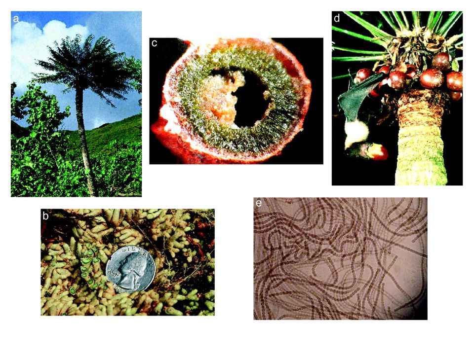
ALS/PDC (The amyotrophic lateral sclerosis / parkinsonism-dementia complex) The rate of ALS/PDC in Chamorro people in Guam is higher than those of other people. ALS/PDC is related with fruit-bat soup as a domestic food of the Chamorro. Neurotoxin (chronic)
.")
24
Biomagnification of cyanobacterial BMAA in Guam.

25
Satellite photograph of a Trichodesmium bloom by using SeaWiFS imagery for spectral imaging at 443, 490, and 550 nm off the eastern coast of Florida on October 30, 1998.

26
HPLC chromatograph of BMAA peak in Chroococcidiopsis indica GT-3-26 (solid line) and BMAA authenticated standard (dashed line) obtained by using fluorescence detection.
 and BMAA authenticated standard (dashed line) obtained by using fluorescence detection.")
27
Representative chromatogram depicting BMAA in the frontal superior gyrus tissue of a Canadian Alzheimer’s patient

28
Microcystins, Nodularins Bioactive Compounds Isolated from Cyanobacteria Adda unit Ahmf unit Ureido unit Oscillatoric acid unit Other Tricyclo Ahp unit Ampa unit Other Ahd unit Choi unit Saa unit Fatty acid unit Other Cyclic Pep. Cyclic Depsipep. Linear Pep. Cyclic Pep. Peptides Alkaloids Macrolides Lipids Other Puwainaphycins Oscillamides Oscillatorin Laxaphycins Microviridins Micropeptins Cryptophycins Majusculamides Microginens Aeruginosins Aeruginoguanidines Spiroidesin Radiosumin Anatoxins, Aphantoxin Cylindrospermopsin Tolytoxins Thionsulfolipid Cracin, Fischerellin A Bioactive Compounds

29
Cyanopeptolin A Oscillapeptin G 3-Amino-6-hydroxy-2-piperidone (Ahp) Ahp-Containing-Cyclic Depsipeptides H N N H NH N H N OH N H N H 3 C O H 3 C O O O O O OH O H 3 C O O CH 3 H 3 C CH 3 OH CH 3 NH 2 O OH O OH HO
 Ahp-Containing-Cyclic Depsipeptides H N N H NH N H N OH N H N H 3 C O H 3 C O O O O O OH O H 3 C O O CH 3 H 3 C CH 3 OH CH 3 NH 2 O OH O OH HO")
30
Micropeptin A, B M. a. T. Okino et al (1993) Micropeptin 90 M. a. NIES-90 K. Ishida et al (1995) Micropeptin T-20 M. a. NIES (T-20) T. Okano et al (1999) Oscillapeptin O. a. NIES-204 H. J. Shin et al (1995) Oscillapeptin A, B O. a. NIVA CYA 18 T. Sano & K. Kaya (1998) Oscillapeptin C O. a. CCAP 1459/16 T. Sano (1996) Oscillapeptin D O. a. (China) T. Sano et al (1998) Nostocyclin Nostoc sp. K. Kaya et al (1996) Aeruginopeptin 95A, 95-B M.a. TAC 95 K. Harada et al (1993) Aeruginopeptin 228-A M.a. 228A K. Harada et al (1993) Cyanopeptolin A-D M.a. PCC 7806 C. Marchin et al (1993) Microcystilode A M. a. NO-15-1840 S. Tsukamoto et al (1993) Cyanopeptolin S M. a. bloom C. Jakobi et al (1993) Cyanopeptolin SS M.a. bloom J. Weckesser et al (1996) Anabaenopeptilide 90-A, 90-B A. c. 90 K. Fujii et al (1995) Anabaenopeptilide 202-A, 202-B A. l. 202A2/41 K. Fujii et al (1995) A90720A Microchaete loktakensis A. Y. Lee et al (1994) Variants of Ahp-containing cyclic depsipeptide Variants Source Reference Oscillapeptin G O. a. NIVA CYA 18 T. Sano & K. Kaya (1996)
 Micropeptin T-20 M. a. NIES (T-20) T. Okano et al (1999) Oscillapeptin O. a. NIES-204 H. J. Shin et al (1995) Oscillapeptin A, B O. a. NIVA CYA 18 T. Sano & K. Kaya (1998) Oscillapeptin C O. a. CCAP 1459/16 T. Sano (1996) Oscillapeptin D O. a. (China) T. Sano et al (1998) Nostocyclin Nostoc sp. K. Kaya et al (1996) Aeruginopeptin 95A, 95-B M.a. TAC 95 K. Harada et al (1993) Aeruginopeptin 228-A M.a. 228A K. Harada et al (1993) Cyanopeptolin A-D M.a. PCC 7806 C. Marchin et al (1993) Microcystilode A M. a. NO S. Tsukamoto et al (1993) Cyanopeptolin S M. a. bloom C. Jakobi et al (1993) Cyanopeptolin SS M.a. bloom J. Weckesser et al (1996) Anabaenopeptilide 90-A, 90-B A. c. 90 K. Fujii et al (1995) Anabaenopeptilide 202-A, 202-B A. l. 202A2/41 K. Fujii et al (1995) A90720A Microchaete loktakensis A. Y. Lee et al (1994) Variants of Ahp-containing cyclic depsipeptide Variants Source Reference Oscillapeptin G O. a. NIVA CYA 18 T. Sano & K. Kaya (1996).")
31
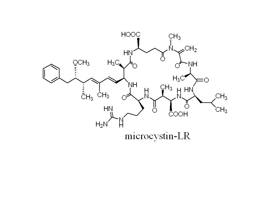
Comparison of Acute toxicity between Cyanotoxins and Artificial Toxic Chemicals. Cyanotoxins and Toxic chemicals LD 50 ( g/kg) Remarks Palytoxin* 1 0.15 mouse 2,3,7,8-TCDD * 2 0.6 guinea pig (oral adm.) mouse(274 g/kg) Tetrodotoxin* 3 8 mouse Sarin 17 rabbit mouse (170 g/kg) Anatoxin-a(s) 20 mouse (IP) Microcystin-LR 100 mouse (IP) Anatoxin-a 200 mouse (IP) Sodium cyanate 2200 rabbit (IP) The underlines express artificial toxic chemicals * 1 : sea anemone toxin 、 * 2 : the most toxic in PCDD * 3 : globefish toxin
 Remarks Palytoxin* mouse 2,3,7,8-TCDD * guinea pig (oral adm.) mouse(274 g/kg) Tetrodotoxin* 3 8 mouse Sarin 17 rabbit mouse (170 g/kg) Anatoxin-a(s) 20 mouse (IP) Microcystin-LR 100 mouse (IP) Anatoxin-a 200 mouse (IP) Sodium cyanate 2200 rabbit (IP) The underlines express artificial toxic chemicals * 1 : sea anemone toxin 、 * 2 : the most toxic in PCDD * 3 : globefish toxin.")
34
Structure of Biosynthesis Gene of Microcystin

35
mcyE mcyD mcyG mcy J mcy I mcy H mcy F PCR-primer for detection of toxin gene mcyA About 550 bases

36
NIES 88 mcyD mcyG mcyJ mcyD mcyG mcyJ NIES 99 Toxic Non-toxic Toxin gene of toxic strain 940bp 420bp PCR Detection of Microcystin Gene

37
Mouse Liver enlarged by microcystin-RR

39
Microcystin Shock Microcystin Bile Acid Tranport System Receptor Hyperphophorylatesd Protein Cytoskeletal changes Membrane Structure Changes PAF Inhibition of Protein Phosphatase Arachidonic acid Cyclooxygenase PLA 2 TXA 2 IL-1 TNF- PAF Arachidonic acid Cyclooxygenase TXA 2 Hepatocytes Macrophages Ca 2+ ion PGI 2 Enclosure A Enclosure B Enclosure C Dianch Lake Side

40
Cancer Promotion by Microcystin (MC) Phosphorylated Protein (Activation) Function ( Proliferation, Differentiation ) Protein KinasesProtein Phosphatases Protein (Inactivation) Inhibition of Protein Phosphatase by MC Hyper- Phospho- rylation Deformation of Cells Cancer Promotion TNF-
 Phosphorylated Protein (Activation) Function ( Proliferation, Differentiation ) Protein KinasesProtein Phosphatases Protein (Inactivation) Inhibition of Protein Phosphatase by MC Hyper- Phospho- rylation Deformation of Cells Cancer Promotion TNF- ")
41
WHO Guideline for microcystin-LR is 1 g/litre (drinking water), 1997 (Falconer, I. R. etal.(1994), Toxicity of the blue-green alga (cyanobacterium) Microcystis aeruginosa in drinking water to growing pigs, as an animal model for human injury and risk assessment. Environ. Toxicol. Water Qual. Intern. J., 9, 131-139. ) Outline of the Experiment 1) Animal : Pig ( Body weight 60 – 65 kg 、 5 heads/group ) 2) Administration : Oral (microcystin containing water) 3) Dosage : 1312, 796, 280 and 0 g/kg/day 4) experimental Period : 8 weeks
, Toxicity of the blue-green alga (cyanobacterium) Microcystis aeruginosa in drinking water to growing pigs, as an animal model for human injury and risk assessment. Environ. Toxicol. Water Qual. Intern. J., 9, ) Outline of the Experiment 1) Animal : Pig ( Body weight 60 – 65 kg 、 5 heads/group ) 2) Administration : Oral (microcystin containing water) 3) Dosage : 1312, 796, 280 and 0 g/kg/day 4) experimental Period : 8 weeks.")
42
Outline of Risk Assessment Minimum Dose ( 280 g/kg/day ) Liver tissue damage. Safety Factor 1) Only 1% of lifetime exposure------------------A safety factor of 10 is applied 2) Use of pig data as an animal model for human injury -------------------------------------A safety factor of 10 is applied 3) Difference of health condition due to age, other causes of liver damage, and other-----A safety factor of 10 is applied Thus a safety factor 1000 is applied to the lowest doserate. This provide a guideline safe intake for humans of 0.28 g/kg/day, Which should result in no adverse effect as seen by direct liver injury To apply this to a 60kg adults drinking 2L water/day, a consumption, Of water containing 8.4 g microcystin/L should be safe.
 Only 1% of lifetime exposure A safety factor of 10 is applied 2) Use of pig data as an animal model for human injury A safety factor of 10 is applied 3) Difference of health condition due to age, other causes of liver damage, and other-----A safety factor of 10 is applied Thus a safety factor 1000 is applied to the lowest doserate. This provide a guideline safe intake for humans of 0.28 g/kg/day, Which should result in no adverse effect as seen by direct liver injury To apply this to a 60kg adults drinking 2L water/day, a consumption, Of water containing 8.4 g microcystin/L should be safe..")
43
4) For tumor prmortion, additional safety factor of 5 or 10 is required Thus a conservative estimate for water safety is 0.84 g microcystin/ L or approximately 1 g/L.
 For tumor prmortion, additional safety factor of 5 or 10 is required Thus a conservative estimate for water safety is 0.84 g microcystin/ L or approximately 1 g/L.")
44
Determination Methods for Total Microcystin 1)Molecular biological method i) PCR of Toxin gene 2) Biochemical methods i) Protein Phosphatase Inhibition ii) ELISA 3) Physical methods i) HPLC/UV or MS 4) Chemical i) MMPB metho ii) GSH method
Molecular biological method i) PCR of Toxin gene 2) Biochemical methods i) Protein Phosphatase Inhibition ii) ELISA 3) Physical methods i) HPLC/UV or MS 4) Chemical i) MMPB metho ii) GSH method")
45
1) Inhibition of Protein Phosphatase 2A 2) Enzyme-Linked Immunosorbent Assay (ELISA) Biochemical Determination
 Inhibition of Protein Phosphatase 2A 2) Enzyme-Linked Immunosorbent Assay (ELISA) Biochemical Determination")
46
Microcystin Bile Acid Tranport System Receptor Hyperphophorylatesd Protein Cytoskeletal changes Membrane Structure Changes PAF Inhibition of Protein Phosphatase Arachidonic acid Cyclooxygenase PLA 2 TXA 2 IL-1 TNF- PAF Arachidonic acid Cyclooxygenase TXA 2 Hepatocytes Macrophages Caイオン PGI 2 Inhibitory Activity Not only microcystin but also other compounds inhibit

47
ELISA Secondary antibody HR P Substrate Color MCLR-BSA Perimary antibody microcystin

48
HPLC Analysis of Unknown Microcystins - Kaya’s Lab. Method - Check points 1)Absorption ratio at 239 nm / 280 nm 2)Division of Peak Shape 3)UV Spectrum
Absorption ratio at 239 nm / 280 nm 2)Division of Peak Shape 3)UV Spectrum.")
49
55% MeOH pH 3.0, 1ml/min, Mightysil 4.6x150 mmRt of LR was 25 min Unknown microcystins LR

51
Rt, 15.4 minRt, 28 min Rt, 30 min Rt, 31 min (shoulder)
")
52
MMPB method for total microcystin determination Chemical Determination R:CH 2 ( normal microcystin ) →quantitative addition of GSH (C=CH 2 +GSH→CH-CH 2 -SG) R:C=CH-CH 3 (Dhb-microcystin ) →non-reaction with GSH Selective Determination KMnO 4 +NaIO 4 GSH method
 →quantitative addition of GSH (C=CH 2 +GSH→CH-CH 2 -SG) R:C=CH-CH 3 (Dhb-microcystin ) →non-reaction with GSH Selective Determination KMnO 4 +NaIO 4 GSH method")
53
MMPB法 Kaya,K. and Sano, T, Anal. Chim. Acta 386 (1999) 107-112
")
54
SIM Profile of MMPB by LC/MS m/z 207(MMPB) m/z 210 (MMPB-d 3 ) MMPB-d 3 MMPB Method
 m/z 210 (MMPB-d 3 ) MMPB-d 3 MMPB Method")
55
H CH 3 1 2 3 4 5 [L-Ala 7 ] microcystin 6 L-Ala 7 7 CH 3 CH 3 Microcystin groups according to the amino acid at unit 7
![H CH [L-Ala 7 ] microcystin 6 L-Ala 7 7 CH 3 CH 3 Microcystin groups according to the amino acid at unit 7](http://images.slideplayer.com/12/3514455/slides/slide_55.jpg "H CH [L-Ala 7 ] microcystin 6 L-Ala 7 7 CH 3 CH 3 Microcystin groups according to the amino acid at unit 7")
57
Detection of N -TNB-dimethyl glutamate by LC/MS

58
Microcystin Fr. TLC (Identification of individual variants) (Total microcystin) Colorimetry OVERALL PROCEDURE GSH Kaya, K. et al Anal. Chim. Acta 450 (2001) 73-80
 (Total microcystin) Colorimetry OVERALL PROCEDURE GSH Kaya, K. et al Anal. Chim. Acta 450 (2001)")
59
RRSampleLRAC-Sample

60
OCH 3 CH 3 CH 3 HN NCH 2 O HOOC NH H 3 C O NH HN O O H 3 C H N O CH 3 CH 3 H N COOH CH 3 CH 3 O O N H H 2 N HN microcystin CH CH 3 Dhb- H 3 C CH 3 1 2 34 5 6 7 L-amino acid L-amino acid D-Ala D -Leu D-Glu ( Z ) and ( E ) Adda
 and ( E ) Adda")
61
OCH 3 CH 3 CH 3 HN NCH 2 O HOOC NH H 3 C O NH HN O O H 3 C H N O CH 3 CH 3 H N COOH CH 3 CH 3 O O N H H 2 N HN Dhb-microcystin C CH 3 1 2 34 5 6 7 L-amino acid L-amino acid D-Ala D-Glu Mdh H N H NHO a 5.6-5.7 ppm ( E )-Dhb- C CH 3 H N H NH O b 6.4-6.5 ppm ( Z )-Dhb- Sano, T.Beattie, K., Codd, G. A., and Kaya, K. J. Nat. Prod. 61, 851-853 (1998) Sano, T. a nd Kaya, K. Tetrahedron 54, 463-470 (1998)
 Sano, T. a nd Kaya, K. Tetrahedron 54, (1998).")
63
[Asp 3, (E)-Dhb 7 ]microcystin RR O. agardhii [Asp 3, (E)-Dhb 7 ]microcystin HtyR O. agardhii [Asp 3, (E)-Dhb 7 ]microcystin HilR P. rubescens [Asp 3, ADMAdda 5, (E)-Dhb 7 ]microcystin RR Nostoc sp. [Asp 3, ADMAdda 5, (E)-Dhb 7 ]microcystin HtyR Nostoc sp. [Asp 3, ADMAdda 5, (E)-Dhb 7 ]microcystin LR Nostoc sp. [Asp 3, (Z)-Dhb 7 ]microcystin HtyR O. agardhii [Asp 3, (Z)-Dhb 7 ]microcystin LR O. agardhii Dhb-microcystin Dhb-microcystin has not been found from Microcystis.
![[Asp 3, (E)-Dhb 7 ]microcystin RR O. agardhii [Asp 3, (E)-Dhb 7 ]microcystin HtyR O.](http://images.slideplayer.com/12/3514455/slides/slide_63.jpg "agardhii [Asp 3, (E)-Dhb 7 ]microcystin HilR P. rubescens [Asp 3, ADMAdda 5, (E)-Dhb 7 ]microcystin RR Nostoc sp. [Asp 3, ADMAdda 5, (E)-Dhb 7 ]microcystin HtyR Nostoc sp. [Asp 3, ADMAdda 5, (E)-Dhb 7 ]microcystin LR Nostoc sp. [Asp 3, (Z)-Dhb 7 ]microcystin HtyR O. agardhii [Asp 3, (Z)-Dhb 7 ]microcystin LR O. agardhii Dhb-microcystin Dhb-microcystin has not been found from Microcystis..")
64
世界地図: http://www.sekaichizu.jp/ Geographical Distribution of Dhb-MC

65
OCH 3 CH 3 CH 3 HN NCH 2 O HOOC NH H 3 C O NH HN O O H 3 C H N O CH 3 CH 3 H N COOH CH 3 CH 3 O O N H H 2 N HN microcystin CH CH 3 Dhb- H 3 C CH 3 1 2 34 5 6 7 L-amino acid L-amino acid D-Ala D -Leu D-Glu ( Z ) and ( E ) Adda D- [D-Leu 1 ] microcystin LR
![OCH 3 CH 3 CH 3 HN NCH 2 O HOOC NH H 3 C O NH HN O O H 3 C H N O CH 3 CH 3 H N COOH CH 3 CH 3 O O N H H 2 N HN microcystin CH CH 3 Dhb- H 3 C CH L-amino acid L-amino acid D-Ala D -Leu D-Glu ( Z ) and ( E ) Adda D- [D-Leu 1 ] microcystin LR](http://images.slideplayer.com/12/3514455/slides/slide_65.jpg "OCH 3 CH 3 CH 3 HN NCH 2 O HOOC NH H 3 C O NH HN O O H 3 C H N O CH 3 CH 3 H N COOH CH 3 CH 3 O O N H H 2 N HN microcystin CH CH 3 Dhb- H 3 C CH L-amino acid L-amino acid D-Ala D -Leu D-Glu ( Z ) and ( E ) Adda D- [D-Leu 1 ] microcystin LR")
66
[D-Leu 1 ]microcystin LR found from Microcystis aeruginosa isolated from Brazil and Canada.
![[D-Leu 1 ]microcystin LR found from Microcystis aeruginosa isolated from Brazil and Canada.](http://images.slideplayer.com/12/3514455/slides/slide_66.jpg "[D-Leu 1 ]microcystin LR found from Microcystis aeruginosa isolated from Brazil and Canada.")
67
Summary 1)Dhb-microcystins were found from cells of O. agardhii, P. rubescens,and Nostoc sp. isolated from North European countries. 2)[D-Leu 1 ]microcystin was isolated from cells of M. aeruginosa collected from Brazil and Canada, but has not been found any other area. Problems Are toxin genes in cyanobacteria localized ? Do migratory birds carry cyanobacteria ?
[D-Leu 1 ]microcystin was isolated from cells of M. aeruginosa collected from Brazil and Canada, but has not been found any other area. Problems Are toxin genes in cyanobacteria localized . Do migratory birds carry cyanobacteria .")
68
SELECTIVE CONTROL OF TOXIC MICROCYSTIS WATERBLOOMS USING LYSINE AND MALONIC ACID

69
Why do we need selective control of toxic cyanobacterial waterblooms? In Europe, they do not need selective control of toxic cyanobacteria, since they use only drinking. Therefore, They remove phosphate completely in eutrophicated lake water for control of toxic cyanobacteria. As the result, there is no phytoplankton in the lake, also Zooplankton and fish.

70
In Asia, inland residents have utilized freshwater fish for a major protein source. Therefore, aquaculture is important, and eutrophication is necessary for growth of phytoplankton, zooplankton and fish, but exclusion of toxic cyanobacteria is necessary for human health and aquaculture. As the result, we need to develop a method of selective control of toxic cyanobacteria. As the opposite situation of the European,

71
Kaya, K. and Sano, T.(1996) Algicidal compounds in yeast extract as a component of microbial culture media. Phycologia, 35(6 Supp.), 117-119 Two algicidal compounds, lysine and malonic acid, were identified from Yeast extract. Lysine was toxic to only Microcystis (cyano- Bacteria, blue-green algae). Cells of Microcystis viridis NIES-102 were completely killed within 48 hr by lysine at the concentration of 1.0 ppm, whereas lysine was non-toxic to Anabaena and Chlorella species. Also, cells of M. viridis were killed by malonic acid at the concentration of 40 ppm. Why did we select lysine and malonic acid for the control?
 Algicidal compounds in yeast extract as a component of microbial culture media. Phycologia, 35(6 Supp.), Two algicidal compounds, lysine and malonic acid, were identified from Yeast extract. Lysine was toxic to only Microcystis (cyano- Bacteria, blue-green algae). Cells of Microcystis viridis NIES-102 were completely killed within 48 hr by lysine at the concentration of 1.0 ppm, whereas lysine was non-toxic to Anabaena and Chlorella species. Also, cells of M. viridis were killed by malonic acid at the concentration of 40 ppm. Why did we select lysine and malonic acid for the control .")
72
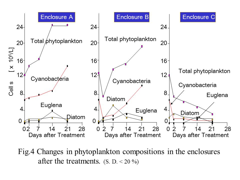
We examined effects of lysine and malonic acid on Microcystis Blooms using enclosures. Enclosure 10 m 1.3-1.5 m 10 – 20 cm ABC Lake Sediments Enclosure A: Control Enclosure B: Lysine treatment Enclosure C: Lysine plus malonic acid treatment 1 m 0.5 m Sampling Point

73
Enclosure A Enclosure B Enclosure C Dianchi Lake Side

74
Macrophytes: Seeds of macrophytes (Myriophllum spicatum and Potamogeton crispus L) and water chestnuts (Trapa sp.) were contained in the lake sediment. Monitoring: Water pH, DO, Chlorophill-a, Lysine, Malonic acid, Microcystin, Cell numbers of phytoplankton (cyanobacteria, dyatom,eugllena) and zooplankton (cradoceran ) Results were expressed as average of three sampling points with S. D. Lysine and malonic acid treatments: Lysine was dissolved with water at the concentration of 100g/L, and sprayed with an insecticide sprayer (lysine 10 g/m 2 ). Malonoc acid was sprayed as the same manner as the lysine treatment (malonic acid 10g/m 2 ). Methods:
 and zooplankton (cradoceran ) Results were expressed as average of three sampling points with S. D. Lysine and malonic acid treatments: Lysine was dissolved with water at the concentration of 100g/L, and sprayed with an insecticide sprayer (lysine 10 g/m 2 ). Malonoc acid was sprayed as the same manner as the lysine treatment (malonic acid 10g/m 2 ). Methods:.")
75
1 B 3 The enclosure surfaces on Day 3 after the treatments A: Control B: lysine C: lysine + malonic acid A C Microcystis aeruginosa

76
6 8 10 Lysine [ mg/L] 1 0 7 14 0 Days after Treatment 2 4 35 Enclosure C Enclosure B Enclosure A Fig.1 Decrease in lysine concentration in the enclosures after the treatments. ( The zero day means immediately after the treatments, S. D. < 5 %)
![Lysine [ mg/L] Days after Treatment Enclosure C Enclosure B Enclosure A Fig.1 Decrease in lysine concentration in the enclosures after the treatments.](http://images.slideplayer.com/12/3514455/slides/slide_76.jpg "( The zero day means immediately after the treatments, S. D. < 5 %).")
77
8.0 8.5 9.0 9.5 pH 7.5 207142128 0 Days after Treatment Enclosure A Enclosure B Enclosure C Fig.2 Changes in pH in the enclosures after the treatments ( S. D. < 5%; *p < 0.05 ) * *
 * *.")
78
60 80 100 120 Chlorophyll a [ g/L] 2071421 28 0 20 40 Days after Treatment Enclosure A Enclosure B Enclosure C Fig.3 Changes in biomass in the enclosures after the treatment of lysine and malonic acid. (S. D. < 20 %)
![Chlorophyll a [ g/L] Days after Treatment Enclosure A Enclosure B Enclosure C Fig.3 Changes in biomass in the enclosures after the treatment of lysine and malonic acid.](http://images.slideplayer.com/12/3514455/slides/slide_78.jpg "(S. D. < 20 %).")
80
Cladoceran [individuals/L] 207142128 0 Days after Treatment Enclosure A Enclosure B Enclosure C 100 200 300 400 500 600 700 800 Fig.5 changes in individual number of cladoceran in the enclosures after the treatments. (S. D. < 20 %)
![Cladoceran [individuals/L] Days after Treatment Enclosure A Enclosure B Enclosure C Fig.5 changes in individual number of cladoceran in the enclosures after the treatments.](http://images.slideplayer.com/12/3514455/slides/slide_80.jpg "(S. D. < 20 %).")
81
9 12 15 Total microcystin [mg/L] 207142128 0 Days after Treatment 3 6 Enclosure A Enclosure B Enclosure C Fig.6 Changes in total microcystyin contents in the enclosures after the treatments. (S. D. < 10 %)
![9 Total microcystin [mg/L] Days after Treatment 3 6 Enclosure A Enclosure B Enclosure C Fig.6 Changes in total microcystyin contents in the enclosures after the treatments.](http://images.slideplayer.com/12/3514455/slides/slide_81.jpg "(S. D. < 10 %).")
82
The enclosures surfaces on day 28 after the treatments A: Control B: lysine C: lysine + malonic acid A B C Myriophllum spicatum water chestnut (Trapa sp.) Microcystis aeruginosa
 Microcystis aeruginosa")
83
Conclusion: The treatment with lysine plus malonic acid is an effective method for the control of toxic Microcystis blooms. The ecological and water qualitative changes derived from the treatment suggested that the incorporation cycles of nitrogen and phosphorus in eutrophicated water were switched from toxic cyanobacteria (Microcystis) to non- toxic macrophytes.
 to non- toxic macrophytes..")
84
Another Methods for Cyanobacterial Control

86
Dry up ( of Dam Sediments ) Cyanobacterial (Microcystis) cells are resting on dam sediment at low water temperature (below 10 ºC) in winter season. When the dam sediment were dried up, the germination rates of the cells on the sediment were dependent on the water content in the sediment. Water level under normal conditions S1(31m from WL) S2(16m from WL) Water level in the experiment (36m from WL) (WL)
 S2(16m from WL) Water level in the experiment (36m from WL) (WL).")
87
Water content in the sediment (WCS) and germination rate (GR) 100 0 7142130 Day after dry up % of WCS (bars) 100 75 50 25 0 0 50 75 100 % of GR (closed circles) S1 S2 S1 S2
 and germination rate (GR) Day after dry up % of WCS (bars) % of GR (closed circles) S1 S2 S1 S2")
88
In Europe: Prof. G. A. Codd (UK) and Dr. H.C. Utkilen (Norway) In North America,and parts of Central America and the Caribbean: Prof. Wayne Carmichael (USA) In South America, and parts of the Central America and the Caribbean: Prof. Sandra Azevedo (Brazil) In Africa: Dr. William R. Harding (South Africa) In Asia (western sector): Dr. Suvendra Bagchi (India) In Asia (eastern sector): Prof. Kunimitsu Kaya (Japan) In Australasia and parts of Oceania: M.D. Burch (Australia) UNESCO-CYANONET Committee Member
 In North America,and parts of Central America and the Caribbean: Prof. Wayne Carmichael (USA) In South America, and parts of the Central America and the Caribbean: Prof. Sandra Azevedo (Brazil) In Africa: Dr. William R. Harding (South Africa) In Asia (western sector): Dr. Suvendra Bagchi (India) In Asia (eastern sector): Prof. Kunimitsu Kaya (Japan) In Australasia and parts of Oceania: M.D. Burch (Australia) UNESCO-CYANONET Committee Member.")
89
WWC KK GAC&HCU MDB SB WRH SA

90
T hankyou foryourattention !

Similar presentations


 as a measure of oxygen- demanding wastes in water. Distinguish between aerobic and anaerobic.>")


? All are secondary metabolites of cyanobacteria. Cyanotoxins grouped into 2 categories: –Cytoxins –Biotoxins.>")
 Microcystin- LR Toxin in Callander Bay Presented by: Jamie Lavigne Supervised by: Dr. Reehan Mirza Department.>")
