Download presentation
Presentation is loading. Please wait.

1
Chapter 10: The Knee

2
Anatomy The bones that comprise the knee joint: Tibia Fibula Femur
Patella There are two joints in the knee: Tibiofemoral joint Patellafemoral joint

3
Anatomy The medial and lateral meniscus rest between the femur and tibia. They are responsible for shock absorption, improved bony correlation, joint lubrication, improved weight distribution, and decreased friction. The patella guides the quadriceps, decreases friction during movement, and protects the femoral condyles.

4
Anatomy of the Knee Knee is a hinge joint
Articulation (point of contact) Consists of 3 bones Stabilized by Four major ligaments Cartilage Strong musculature Knee is able to rotate
 Consists of 3 bones. Stabilized by. Four major ligaments. Cartilage. Strong musculature. Knee is able to rotate.")
5
Cartilage Ends of the tibia and femur Top of tibia End of femur
Covered/cushioned by Pieces of tough cartilage tissue Called menisci Help to stabilize the knee joint Without bones would rub & wear down quickly Top of tibia Flat like a tabletop End of femur Rounded (called condyles) Without stabilization, femur would move a lot on the tibia
 Without stabilization, femur would move a lot on the tibia.")
6
Cartilage-Menisci Lateral & Medial Thicker on sides
Thinner in the middle Form a dish-shaped hollow Attached to the top of the tibia Provide a seat for the femoral condyles to sit in Femur moves but will not roll off

7
Ligaments 4 primary knee ligaments ACL & PCL Medial collateral (MCL)
Helps provide stability to inside of knee Lateral collateral (LCL) Helps provide stability to outside of knee Anterior cruciate ligament (ACL) Keeps tibia from moving forward on the femur Posterior cruciate ligament (PCL) Prevents tibia from moving backward on the femur ACL & PCL pass through the middle of the knee joint Cross each other (i.e cruciate means “cross-shaped”)
 Helps provide stability to outside of knee. Anterior cruciate ligament (ACL) Keeps tibia from moving forward on the femur. Posterior cruciate ligament (PCL) Prevents tibia from moving backward on the femur. ACL & PCL. pass through the middle of the knee joint. Cross each other (i.e cruciate means cross-shaped )")
8
Ligaments of the Knee Anterior cruciate ligament (ACL)
Posterior cruciate ligament (PCL) Medial collateral ligament (MCL) Lateral collateral ligament (LCL)
 Medial collateral ligament (MCL) Lateral collateral ligament (LCL)")
9
Anatomy Muscles of the Knee Quadriceps muscles
Responsible for knee extension Hamstring muscles Responsible for knee flexion Calf muscles Assist in knee flexion Other muscles that act at the knee Sartorius Popliteus Plantaris Gracilis

10
Muscles of the Knee Provide Primary muscles spanning the knee Movement
Stability Primary muscles spanning the knee Quadriceps group (perform knee extension) Vastus medialis, vastus lateralis, vastus intermedius & rectus femoris Hamstring group (perform knee flexion) Biceps femoris, semimembranosus & semitendinosus Help prevent forward movement of the tibia on the femur By the location of their attachments
 Vastus medialis, vastus lateralis, vastus intermedius & rectus femoris. Hamstring group (perform knee flexion) Biceps femoris, semimembranosus & semitendinosus. Help prevent forward movement of the tibia on the femur. By the location of their attachments.")
11
Primary Muscles Spanning the Knee
Quadriceps group Vastus medialis, vastus lateralis, vastus intermedius & rectus femoris Perform knee extension

12
Primary Muscles Spanning the Knee
Hamstring group Biceps femoris, semimembranosus & semitendinosus Perform knee flexion Help prevent forward movement of the tibia on the femur By the location of their attachments

13
Knee Alignment Concerns
Genu valgum (knock-knees) Genu varum (bow legs) Genu recurvatum (hyperextension) Q-angle Greater than 20 increases risk for injury Leg length discrepancy
 Genu varum (bow legs) Genu recurvatum (hyperextension) Q-angle. Greater than 20 increases risk for injury. Leg length discrepancy.")
14
Preventing Knee Injuries
Ligament sprains Most common injuries seen at the knee Muscles provide stability to the knee Help resist abnormal bony movement Athletes should develop strength in the muscles (quads, hams, gastrocnemius/calf, hip abductors & hip adductors) Gastrocnemius-heel raises Some trainers & athletes use preventative knee braces Designed to protect medial collateral ligament Tearing can result from a blow to the lateral side
 Gastrocnemius-heel raises. Some trainers & athletes use preventative knee braces. Designed to protect medial collateral ligament. Tearing can result from a blow to the lateral side.")
15
Treating Knee Injuries & Conditions
Knee is exposed to many forces Makes it vulnerable to injuries Especially the ligaments Tendon & bone injuries also occur Patella & menisci are subject to unique types of athletics-related injuries

16
Ligament Injuries Ligament sprains of the knee Can be Mild Moderate
Severe

18
Injuries of the Knee

19
Patellar Fracture Signs & Symptoms: Pain directly over bone
Slight to moderate swelling Pain, especially in the first 30º of movement Treatment: Immobilize and refer to a physician for x-rays Requires lengthy immobilization during recovery

20
Patellar Dislocation Signs & Symptoms: Moderate to extreme pain
Moderate swelling Complete loss of ROM in knee Obvious deformity laterally Treatment: Refer to physician for reduction RICE therapy Progressive strengthening program

21
Patella Dislocation Patella forced to lateral aspect of the knee
Often occurs when the knee is bent and forced to twist inward Signs & symptoms Obvious deformity Athlete is often in distress EMS must be called Unless team physician is present only a physician should reduce a dislocated patella Complications may result Posterior aspect of patella may be injured further

22
OUCH!!!!!!!!!

23
Patella-Femoral Stress Syndrome
Signs & Symptoms: Pain and tenderness in lateral aspect of patella Slight swelling Crepitus or popping with extension Treatment: RICE therapy Closed kinetic chain exercises 0-40

24
Patellar Femoral Syndrome
Fancy name for a set of symptoms that include pain/discomfort around patella Often caused by patellar tracking problems Knee bends Patella is grated across the femur Causing cartilage on back of patella to soften or wear away Known as chondromalacia

25
Chondromalacia Signs & Symptoms: Pain underneath the patella
Grinding or popping during motion Slight chronic swelling Special Tests: Clarke’s Sign Treatment: RICE Therapy Quadriceps strengthening

26
Patellar Femoral Syndrome Chondromalacia
Characterized by achiness around the patella Especially with prolonged sitting in the same position Athlete reports a grinding sensation with flexion/extension Grinding can be felt by placing hand over knee

27
Patella Injuries (Chondromalacia)
")
28
Osgood-Schlatter’s Disease
Signs & Symptoms: Pain at insertion of patella tendon Tenderness to palpation Enlarged tibial tuberosity Pain with jumping or running Treatment: RICE therapy Decrease activity or cross-train

29
Osgood-Schlatter Disorder
Irritation at the site of the patellar tendon attachment To front of the tibia Called tibial turberosity Repeated stress causes the patellar tendon to partially pull away from the bone Called Osgood-Schlatter’s disorder

30
Osgood-Schlatter Disorder
Signs & symptoms Discomfort of the knee Swelling Tenderness Pain during activity Possible bump below knee cap (bony growth at the top of the tibia) Can remain even after symptoms have disappeared Care Restrict activity until resolved Stationary bicycling Use pain as a guide Modify activities based on pain level reported by athlete Ice before & after activity Special pad made to fit over front of tibia Often improves by age 16 or 17 (but known to last into early 20’s)
 Can remain even after symptoms have disappeared. Care. Restrict activity until resolved. Stationary bicycling. Use pain as a guide. Modify activities based on pain level reported by athlete. Ice before & after activity. Special pad made to fit over front of tibia. Often improves by age 16 or 17 (but known to last into early 20’s)")
31
Patellar Tendinitis Signs & Symptoms:
Pain in patella tendon or at inferior pole of patella Pain increases with activity Squeaking noise with motion Slight swelling Treatment: Modality treatment Ice or ice massage Ultrasound

32
Muscle & Tendon Injuries
Patellar tendinitis Overuse disorder Characterized by quadriceps weakness Tenderness over the patellar tendon Minimal swelling Condition is also called jumper's knee Athletes that do lots of jumping often get this condition (basketball, volleyball) Early stages Athlete typically has pain after activity Treatment Trainer attempts to control inflammation Apply ice Modify athlete’s activity level Restricting running & jumping Rehabilitation program Address any flexibility problems or weakness of the leg
 Early stages. Athlete typically has pain after activity. Treatment. Trainer attempts to control inflammation. Apply ice. Modify athlete’s activity level. Restricting running & jumping. Rehabilitation program. Address any flexibility problems or weakness of the leg.")
33
Patella Tendon Rupture
Signs & Symptoms: Extreme pain with an immediate drop in pain Significant swelling Window shade effect Complete loss of knee extension Previous history of chronic tendinitis Treatment: Surgical repair is the only treatment option 6-8 months minimum recovery

34
Ruptured Patella Tendon

35
Knee Dislocation Signs & Symptoms:
Immediate pain that may decrease dramatically Obvious deformity (usually anteriorly) Significant swelling Decreased blood flow and neural sensation Treatment: Splinted and transported to hospital immediately Surgical intervention is often required for neurovascular and ligament repair
 Significant swelling. Decreased blood flow and neural sensation. Treatment: Splinted and transported to hospital immediately. Surgical intervention is often required for neurovascular and ligament repair.")
36
Knee Contusion Signs & Symptoms: Pain at affected site
Moderate swelling and discoloration Loss of ROM Decreased weight bearing Treatment: RICE therapy

37
Patella Injuries (Bursitis)
Prepatellar bursitis is the inflammation of a small sac of fluid located in front of the kneecap. This inflammation can cause many problems in the knee.

38
Causes Bursitis is the inflammation of a bursa. The prepatellar bursa can become irritated and inflamed in a number of ways. A direct blow or a fall onto the knee can damage the bursa.

39
Meniscus Contusion Signs & Symptoms:
Pain, especially at full extension Loss of ROM in extension Slight swelling Treatment: RICE therapy

40
Meniscus Tears Signs & Symptoms:
Pain, especially when moved similarly to the mechanism of injury Pain with full extension or flexion Diffuse swelling in the joint (effusion) Pain along the line of the joint Sensation of locking or giving out Clicking or popping sound with movement
 Pain along the line of the joint. Sensation of locking or giving out. Clicking or popping sound with movement.")
41
Meniscal Injuries Meniscal injuries damage the cushioning tissue between the tibia and the femur, inside the knee joint, on both sides (medial and lateral) of the knee.
 of the knee.")
42
Causes They are highly vulnerable to injury from abrupt rotations of the knee while it is bearing weight, for example, when you turn to hit a tennis ball, rotating your thigh (femur) while leaving your foot stationary.
 while leaving your foot stationary.")
43
Types of Meniscal Tears

44
MRI Torn Medial Meniscus

46
Arthroscopic Repair

47
Posterior horn tear with multiple flaps

48
Meniscus Tears Special Tests: McMurray’s Test Apley’s Compression Test
Bounce Home Test Treatment: Referral to a physician Surgery is often required for full recovery. RTP depends on surgical option selected.

49
ACL Sprain Signs & Symptoms: Pain in the joint
Athlete hears ‘pop’ at time of injury Sense of looseness in joint, giving away, or shifting Swelling that increases rapidly post-injury

50
Anterior Cruciate Ligament Injuries
Keeps tibia from moving forward on the femur If ligament is injured Athlete is often disabled Complaining of the knee “giving away”, collapsing & popping Most serious of all knee ligament injuries Most frequently surgically reconstructed

51
Anterior Cruciate Ligament Injuries
Often injured as the athlete is attempting to change directions quickly Twists the lower leg May hear a popping sound during the twisting Also injured during excessive hyperextension

52
Anterior Cruciate Ligament Injuries
Signs & symptoms Rapid swelling Loss of knee function Immediate treatment PRICE Knee immobilizer Crutches Follow up with an orthopedist is necessary Athlete rarely can continue a high level of function with a torn ACL

53
Anterior Cruciate Ligament Injuries
Often needs to be surgically reconstructed Determination that must be made by the athlete, surgeon & athlete’s family Depends on the amount of instability that exists Level of function desired by the athlete Age of the athlete

54
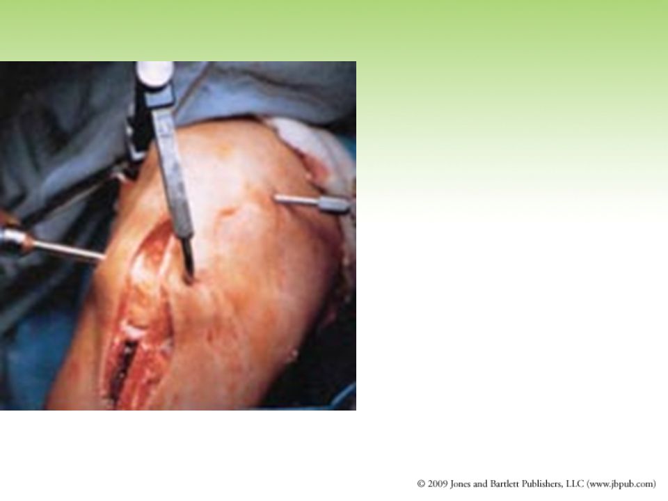
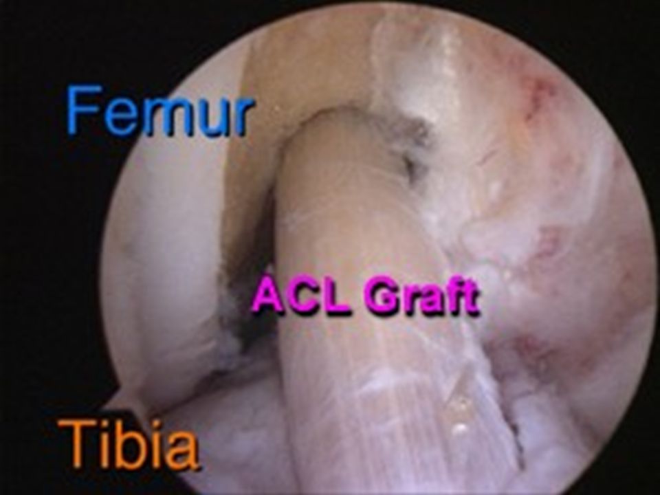
Anterior Cruciate Ligament Injuries Knee Arthroscopy

55
Anterior Cruciate Ligament Injuries Knee Arthroscopy

56
ACL has poor healing potential
PCL has intermediate healing potential MCL heals on its own

61
Allograft Bone Tendon Bone

63
Patella Tendon Graft

64
Anterior Cruciate Ligament Injuries Rehabilitation
Focuses on strengthening the hamstrings Helps stabilize the tibia Helps regain full function Even with aggressive ACL rehab May be six months before athlete can return to participation

65
ACL Sprain, cont. Special Tests: Anterior Drawer Test Lachman’s Test
Pivot Shift Test Treatment: Grade 1 or 2 may be treated conservatively. Grade 3 tear will require surgery.

66
PCL Sprain Signs & Symptoms: Pain in posterior aspect of knee
Slight swelling Joint laxity Loose feeling with walking Special Tests: Posterior Sag Test Posterior Drawer Test Treatment: Grade 1 or 2 may be treated conservatively Grade 3 tear will require surgery

67
Posterior Cruciate Ligament Injuries
Prevents posterior tibial movement on the femur Frequently injured when athlete falls and a bent knee bears full weight Knee is forcefully hyperflexed Blow delivered to the front of the tibia

70
Posterior Cruciate Ligament Injuries
Assessment Trainer determines mechanism of injury Athlete reports having heard a pop Often little swelling with PCL injury Initial treatment PRICE Referral to a physician

71
Posterior Cruciate Ligament Injuries
Physicians disagree about whether or not surgery should be performed on a severe PCL injury Even complete PCL tears can be rehabilitated without surgery Rehabilitation programs for mild/moderate PCL sprains Focus on strengthening the quadriceps Regaining full function Many athletes can become functional again After initial pain & swelling are controlled After knee is strengthened

72
MCL Sprain Signs & Symptoms: Pain increasing with severity
Joint stiffness Slight to moderate swelling Decreased ROM Joint laxity medially

73
Medial Collateral Ligament Sprains
Frequently injured when an athlete receives a blow to the outside of the knee Causes knee to bend inward (valgus stress) Stresses the MCL Mild sprain Medial joint line pain Little if any swelling No joint laxity when stressed by trainer during assessment Full knee flexion & extension
 Stresses the MCL. Mild sprain. Medial joint line pain. Little if any swelling. No joint laxity when stressed by trainer during assessment. Full knee flexion & extension.")
74
Medial Collateral Ligament Sprains
Moderate MCL sprain Mild swelling Discomfort Some joint laxity when stressed by the trainer during assessment Severe MCL injury Moderate or severe amount of swelling Loss of function Great deal of joint laxity when stressed by the trainer during assessment

75
Medial Collateral Ligament Sprains
Treated with PRICE Mild injury Elastic wrap for compression/support Moderate/severe injury Knee put in an immobilizer Trainer should consider possibility of damage to the menisci or an ACL injury Rehabilitation Focuses on strengthening the muscles that cross the medial aspect of the knee

76
MCL Sprain, Cont. Special Tests: Valgus Stress Test
Apley’s Distraction Test Treatment: RICE Therapy Immobilization Progressive strengthening program

77
LCL Sprain Signs & Symptoms: Pain over lateral aspect of knee
Slight to moderate swelling Joint laxity laterally Joint stiffness Decreased ROM

78
Lateral Collateral Ligament Injuries
Occur less frequently than MCL injuries Signs & symptoms are similar to MCL Except discomfort is at the lateral aspect of the knee Treatment Same as MCL Rehabilitation (regaining joint stability) Strengthening exercises focus on the lateral thigh muscles & hamstrings
 Strengthening exercises focus on the lateral thigh muscles & hamstrings.")
79
LCL Sprain, cont. Special Tests: Varus Stress Test
Apley’s Distraction Test Treatment: RICE Therapy Immobilization Progressive strengthening program

80
Discussion Questions What would be your response to an athlete who wants to play with an ACL tear? How would you react on the field if an athlete dislocated their knee?

Similar presentations


>")


