Download presentation
Presentation is loading. Please wait.

1
Serum Electrophoresis AND IMMUNOFIXATION june 2013 Dr. Nitin A Inamdar Department of Biochemistry Tata Memorial Center 9869515089

4
Serum/protein electrophoresis: – plasma cell dyscrasia, nephrotic syndrome, etc. Hemoglobin electrophoresis: – thalassemia, hemoglobinopathies like sickle cell anemia, HbC, HbD, etc. Urine electrophoresis: – BJP in plasma cell dyscrasia

5
Electrophoresis Electro: Under influence of electric field Phoresis: Separation or migration or movement

6
Serum Mixture of proteins Amino acids: – Amino group (NH2) – Carboxyl group (COOH) Two amino acids join each other with polypeptide bond to form polypeptide chain & many polypeptide chains make a protein. Electrophoresis Ph 8.6/0.075M Barbitone buffer 1%Agarose Constant current 5mAM/Slide

7
Immunity Immune cell types – T lymphocytes Mature in thymus – B lymphocytes Mature in marrow – Antigen- presenting cells (APCs)
")
8
Immunity Humoral response – Secondary response Memory cells start response to same antigen

10
Immunity Antibody classes = Immunoglobulins – IgD, IgM, IgG In blood &/or attached to B lymphocytes – IgA In saliva, sweat, tears, etc. – IgE Bound to basophils & mast cells Causes release of histamine; allergy; parasitic infection

11
Immunity Antibodies – Large complex proteins Y-shaped 4 polypeptide chains – 2 “heavy” – 2 “light” Constant region Variable region with antigen binding sites

12
Programmed cell death is part of normal development Falling of leaves: Apoptosis

15
In Multiple Myeloma Normal Plasma cell become Abnormal Plasmacell

16
Indication of serum electrophoresis Diagnostic: – Diagnosis of plasma cell dyscrasia – Diagnosis of Waldenstrom’ macroglbulinemia Monitoring of disease: – Monitoring of plasma cell dyscrasia

17
WHO Classification of Plasma Cell Neoplasms Plasma cell myeloma (MM) Plasma cell myeloma variants Non secretory myeloma Indolent Myeloma Smoldering Myeloma Plasma cell leukemia Plasmacytomas Solitary plasmacytoma of bone Extramedullary plasmacytoma Immunoglobulin deposition diseases Primary Amyloidosis Systemic light and heavy chain deposition disease Osteosclerotic myeloma (POEMS syndrome) Heavy chain disease Gamma /Mu / Alpha
 Plasma cell myeloma variants Non secretory myeloma Indolent Myeloma Smoldering Myeloma Plasma cell leukemia Plasmacytomas Solitary plasmacytoma of bone Extramedullary plasmacytoma Immunoglobulin deposition diseases Primary Amyloidosis Systemic light and heavy chain deposition disease Osteosclerotic myeloma (POEMS syndrome) Heavy chain disease Gamma /Mu / Alpha")
18
Electrophoresis separates proteins based on their physical properties. Serum is placed on a specific medium, and a charge is applied. The net charge (positive or negative) and the size and shape of the subsets of these proteins are used in interpreting the results.
 and the size and shape of the subsets of these proteins are used in interpreting the results..")
19
Method of detection 1.Serum protein electrophoresis 2.Quantitative immunoglobulins (nephelometry) 3.Immunofixation, immunoelectrophoresis and immunodiffusion 4.Urine studies 5.Free light chain assay
 3.Immunofixation, immunoelectrophoresis and immunodiffusion 4.Urine studies 5.Free light chain assay")
20
Equipment used for the gel electrophoresis power supply (direct current) electrophoresis chamber containers for staining and destaining gel applicator
 electrophoresis chamber containers for staining and destaining gel applicator")
21
Serum protein electrophoresis on agarose gel is a type of horizontal gel electrophoresis

22
Albumin Globulins: – α1, – α2, – β1, – β2, – γ

23
Normal serum electrophoresis pH=8.6, 1% agarose gel, sodium barbitone buffer Anode +veCathode -ve

25
Abnormal serum electrophoresis showing M band Anode +veCathode -ve

26
M bandNormal study

27
Diagnostic criteria for plasma cell dyscrasia Symptomatic plasma cell myeloma: BM clonal plasma cells or Plasmacytoma Marrow plasmacytosis (>30%) M-component in serum or urine Serum: Ig G >3 g/dL, Ig A >2.5 g/dL or Urine: >/= 1g/ day of λ or κ [BJP] Asymptomatic plasma cell myeloma: M band in serum at myeloma levels > 3 gm/dL and /or 10% or more clonal plasma cell in BM No related organ or tissue impairment (end organ damage or bony lesion) or myeloma-related symptoms MUGS: M-Component, present but less than 3 gm/dL Marrow Plasmacytosis < 10% No Lytic bone lesions or related organ impairment etc
![ Diagnostic criteria for plasma cell dyscrasia Symptomatic plasma cell myeloma: BM clonal plasma cells or Plasmacytoma Marrow plasmacytosis (>30%) M-component in serum or urine Serum: Ig G >3 g/dL, Ig A >2.5 g/dL or Urine: >/= 1g/ day of λ or κ [BJP] Asymptomatic plasma cell myeloma: M band in serum at myeloma levels > 3 gm/dL and /or 10% or more clonal plasma cell in BM No related organ or tissue impairment (end organ damage or bony lesion) or myeloma-related symptoms MUGS: M-Component, present but less than 3 gm/dL Marrow Plasmacytosis < 10% No Lytic bone lesions or related organ impairment etc](http://images.slideplayer.com/12/3502306/slides/slide_27.jpg " Diagnostic criteria for plasma cell dyscrasia Symptomatic plasma cell myeloma: BM clonal plasma cells or Plasmacytoma Marrow plasmacytosis (>30%) M-component in serum or urine Serum: Ig G >3 g/dL, Ig A >2.5 g/dL or Urine: >/= 1g/ day of λ or κ [BJP] Asymptomatic plasma cell myeloma: M band in serum at myeloma levels > 3 gm/dL and /or 10% or more clonal plasma cell in BM No related organ or tissue impairment (end organ damage or bony lesion) or myeloma-related symptoms MUGS: M-Component, present but less than 3 gm/dL Marrow Plasmacytosis < 10% No Lytic bone lesions or related organ impairment etc")
30
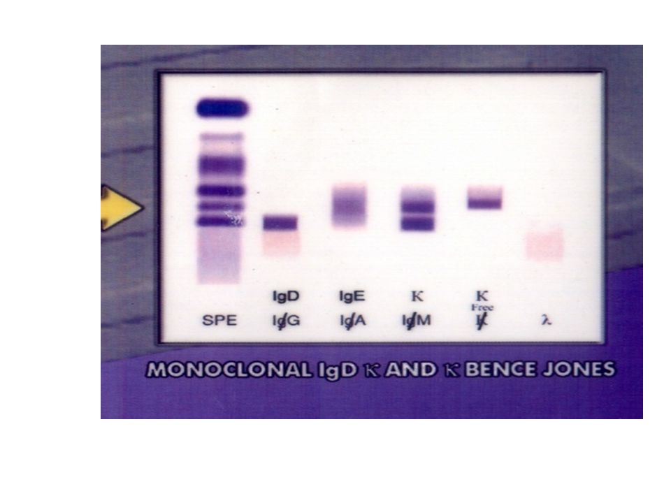
Immuno-fixation electrophoresis After separation of serum proteins by electrophoresis fix the separated protein with antiserum of immunoglobulin heavy chain and light chains.

42
Bis-albuminemia

45
ABNORMAL?

46
Intravascular hemolysis

47
ABNORMAL

49
Nephrotic syndrome

50
Hypergammaglobulinemia

51
M band in β region

52
M band in γ region

Similar presentations


 that react specifically with the antigen that stimulated.>")


