Download presentation
Presentation is loading. Please wait.

1
Neural Integration The sensory pathways Chapter 15

2
Afferent Division of the Nervous System
Receptors Sensory neurons Sensory pathways

3
Afferent Division – location in CNS
Somatic Sensory info Sensory cortex of cerebrum Cerebellum Visceral Sensory info Reflex centers in brainstem Reflex centers in diencephalon

4
The somatic sensory system
Sensory stimuli that reach the conscious level of perception Specialized cells that monitor specific conditions in the body or external environment General Senses: Temp, pain, touch, pressure, vibration, proprioception Simple receptors located anywhere on body Special Senses: Are located in sense organs such as the eye or ear Olfaction, vision, gustation, hearing, equilibrium Complex receptors located in specialized sense organs

5
General Properties: Sensory Division
Table 10-1 (1 of 2)
")
6
From Sensation to Perception

7
Sensory Pathways – from sensation to perception
Stimulus as physical energy sensory receptor Receptor acts as a transducer Intracellular signal usually change in membrane potential Stimulus threshold action potential to CNS Integration in CNS cerebral cortex or acted on subconsciously

8
Sensory Receptors Transduction – conversion of environmental stimulus into action potential by sensory receptor Receptors specific for particular type of stimulus Specificity is due to structure of receptor

9
From Sensation to Perception
A stimulus is a change in the environment that is detected by a receptor Sensation: the awareness of changes in the internal and external environment Perception: the conscious interpretation of those stimuli

10
Classification by Location
Exteroceptors Respond to stimuli arising outside the body Receptors in the skin for touch, pressure, pain, and temperature Most special sense organs Interoceptors (visceroceptors) Respond to stimuli arising in internal viscera and blood vessels Sensitive to chemical changes, tissue stretch, and temperature changes
 Respond to stimuli arising in internal viscera and blood vessels. Sensitive to chemical changes, tissue stretch, and temperature changes.")
11
Classification by Location
Proprioceptors Respond to stretch in skeletal muscles, tendons, joints, ligaments, and connective tissue coverings of bones and muscles Inform the brain of one’s movements

12
Four types of General Sensory Receptors
Pain: nociceptor Temperature: thermoreceptor Physical: mechanoreceptor Chemicals: chemoreceptors All can be found in both somatic (exteroceptors) and visceral (interoceptors) locations except: Proprioceptors (a mechanoreceptor) are somatic only report the positions of skeletal muscles and joints
 and visceral (interoceptors) locations except: Proprioceptors (a mechanoreceptor) are somatic only. report the positions of skeletal muscles and joints.")
13
Pain Receptors: Nociceptors
(noci = harm) sensitive to pain-causing stimuli (e.g. extreme heat or cold, excessive pressure, inflammatory chemicals) Free nerve ending Mode of Action: Injured cells release arachidonic acid Arachidonic acid is converted into prostaglandins by the interstitial enzyme cyclo-oxygenase Prostaglandins activate nociceptors Many pain medications like aspirin function to inhibit cyclo-oxygenase Pain levels are modulated by endorphins which inhibit CNS function
 sensitive to pain-causing stimuli (e.g. extreme heat or cold, excessive pressure, inflammatory chemicals) Free nerve ending. Mode of Action: Injured cells release arachidonic acid. Arachidonic acid is converted into prostaglandins by the interstitial enzyme cyclo-oxygenase. Prostaglandins activate nociceptors. Many pain medications like aspirin function to inhibit cyclo-oxygenase. Pain levels are modulated by endorphins which inhibit CNS function.")
14
Thermoreceptors Detect temperature
Found in skin, skeletal muscle, liver, and hypothalamus Consist of free nerve endings Phasic receptors that adapt easily Cold response are more superficial and receptors that respond to heat – deeper Temperature out of the range of the thermoreceptors will activate nociceptors

15
Mechanoreceptors Detect membrane distortion Three receptor types:
Tactile Receptors Proprioceptors Baroreceptors

16
Mechanoreceptors - Tactile Receptors
Detect touch, pressure and vibration on skin Detect hair movement Detect fine touch Detect deep pressure respond to itch (respond among other to histamine) and light touch (detect changes in shape like bending)
 and light touch (detect changes in shape like bending)")
17
Receptor type Structure Location Function Meissner’s corpuscle/tactile corpuscle Few spiral terminals surrounded by CT capsule Between dermal papillae in hairless skin Touch, pressure Pacinian corpuscle/lamellated corpuscle Single dendrite surrounded by capsule with up to 60 layers of collagen fibers Skin, interosseous membrane, viscera Deep pressure. Respond only when the pressure is first applied (on/off pressure stimulation) Ruffini’s corpuscle Receptor endings enclosed by flatten capsule All skin, joint capsule Stretching of skin – continuous pressure
 Ruffini’s corpuscle. Receptor endings enclosed by flatten capsule. All skin, joint capsule. Stretching of skin – continuous pressure.")
18
Mechanoreceptors - Proprioceptors
Detect positions of joints and muscles Muscle spindles Modified skeletal muscle cell Monitor skeletal muscle length Golgi tendon organs Dendrites around collagen fibers at the muscle-tendon junction Monitor skeletal muscle tension Joint capsule receptors - Monitor pressure, tension and movement in the joint

19
Receptor type Structure Location Function Muscle spindles Spindle-shape proprioceptors. Modified skeletal muscle fibers enclosed in CT capsule Perimysium of skeletal muscles Detect muscle stretch and initiate reflex that resist stretch Golgi tendon organs Proprioceptors. Consist of bundle of collagen fibers enclosed in CT capsule with sensory endings coiling between and around the fibers In tendons close to skeletal muscle insertion When tendon fibers are stretched by muscle contraction the nerve endings are activated by compression. When activated, the contraction of the muscle is inhibited which causes relaxation Joint receptors Proprioceptors (combination of several receptors types – Pacinian, Raffini, free ending and Golgi tendon) Joints’ CT capsule Monitor stretch in in the articular capsule and provide information on the position and motion of the joint (conscious)
 Joints’ CT capsule. Monitor stretch in in the articular capsule and provide information on the position and motion of the joint (conscious)")
20
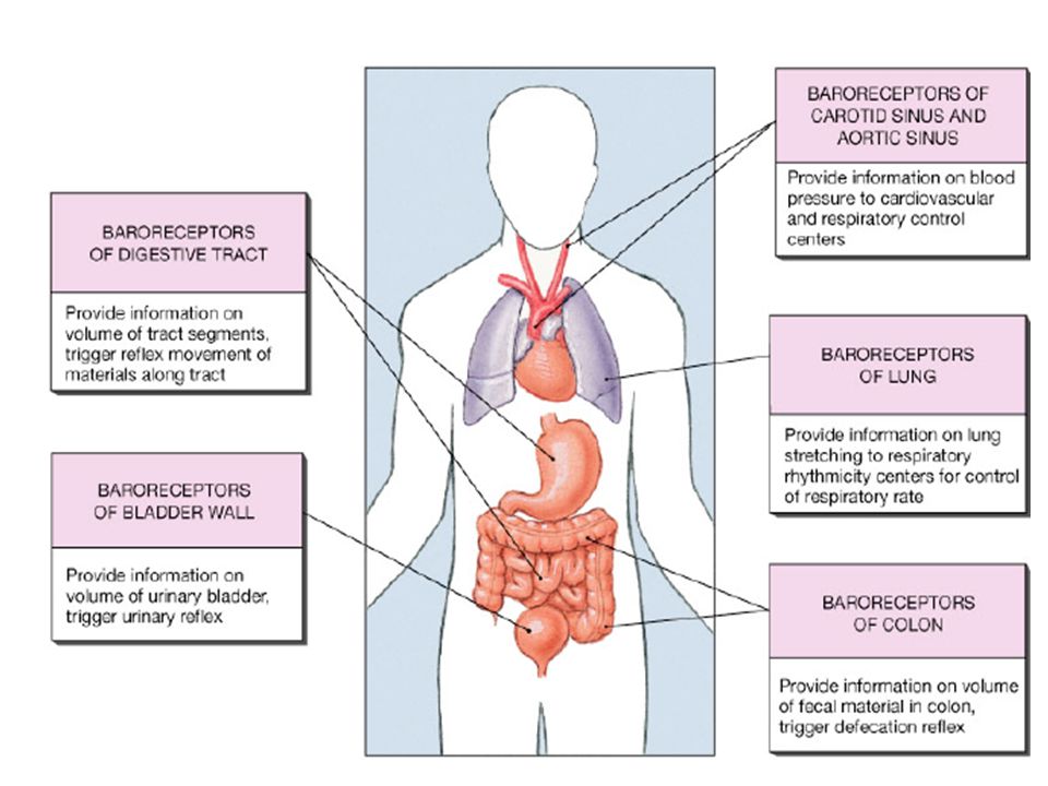
Mechanoreceptors - Baroreceptors
Detect pressure changes Found in elastic tissue of blood vessels and organs of digestive, reproductive and urinary tracts

22
Chemoreceptors Detect change in concentration of specific chemicals or compounds pH, CO2, sodium etc. Found in respiratory centers of the brain and in large arteries

23
Sensory Receptors Table 10-2

24
Processing of the sensory information
Levels of neural integration in sensory systems: Receptor level — the sensor receptors Circuit level — ascending pathways in the CNS Perceptual level — neuronal circuits in the cerebral cortex

25
Processing at the Receptor Level
3 Perceptual level (processing in cortical sensory centers) Motor cortex Somatosensory cortex Thalamus Reticular formation Cerebellum Pons Medulla 2 Circuit level (processing in ascending pathways) Spinal cord Free nerve endings (pain, cold, warmth) Muscle spindle 1 Receptor level (sensory reception and transmission to CNS) Joint kinesthetic receptor Figure 13.2
 Motor. cortex. Somatosensory. cortex. Thalamus. Reticular. formation. Cerebellum. Pons. Medulla. 2. Circuit level. (processing in. ascending pathways) Spinal. cord. Free nerve. endings (pain, cold, warmth) Muscle. spindle. 1. Receptor level. (sensory reception. and transmission. to CNS) Joint. kinesthetic. receptor. Figure")
26
Processing at the Receptor Level
The receptor must have specificity for the stimulus energy (as previously discussed) The receptor’s receptive field must be stimulated The stimulus need to be converted to a nerve impulse Receptors have different levels of adaptation Information is encoded in the frequency of the stimuli – the greater the frequency, the stronger is the stimulus.
 The receptor’s receptive field must be stimulated. The stimulus need to be converted to a nerve impulse. Receptors have different levels of adaptation. Information is encoded in the frequency of the stimuli – the greater the frequency, the stronger is the stimulus.")
27
The stimulation of the receptive field affects the discharge of the sensory neurons
The receptive field is the a specific physical area that, when stimulated, affect the discharge of the stimulus. Most receptive fields activation will result in message sending – excitatory receptive field Sensory receptors in the CNS can have inhibitory receptive field (example: vision fields to determine borders). Sensory neurons of neighboring receptive field may exhibit Convergence many sub-threshold stimuli to sum in the postsynaptic neuron Overlapping with another receptor’s receptive field – sending 2 sensations from the same area (pressure and pain) The smaller the receptive field the greater the ability of the brain to localize the site
. Sensory neurons of neighboring receptive field may exhibit. Convergence many sub-threshold stimuli to sum in the postsynaptic neuron. Overlapping with another receptor’s receptive field – sending 2 sensations from the same area (pressure and pain) The smaller the receptive field the greater the ability of the brain to localize the site.")
28
Sensory Neurons: Two-Point Discrimination
convergence Two-point discrimination (a) Compass with points separated by 20 mm Skin surface Primary sensory neurons Secondary sensory neurons One signal goes to the brain. Figure 10-3a
 Compass with points separated by 20 mm. Skin surface. Primary sensory neurons. Secondary sensory neurons. One signal goes to the brain. Figure 10-3a.")
29
Sensory Neurons: Two-Point Discrimination - overlapping
Two signals go to the brain. Compass with points separated by 20 mm Primary sensory neurons Skin surface Secondary sensory neurons (b) Figure 10-3b
 Figure 10-3b.")
30
Receptive Fields of Sensory Neurons - overlapping
Primary sensory neurons The primary sensory neurons converge on one secondary sensory neuron. Information from the secondary receptive field goes to the brain. Secondary sensory neuron The receptive fields of three primary sensory neurons overlap to form one large secondary receptive field. SECTION THROUGH SPINAL CORD Figure 10-2

31
Properties of Stimulus: Location
Lateral inhibition enhances contrast and makes a stimulus easier to perceive Stimulus Stimulus Pin Skin A B C Frequency of action potentials Tonic level Primary neuron response is proportional to stimulus strength. Primary sensory neurons Pathway closest to the stimulus inhibits neighbors. Secondary neurons A B C Frequency of action potentials Inhibition of lateral neurons enhances perception of stimulus. Tertiary neurons Tonic level A B C Figure 10-6

32
Transduction allows sensory receptors to respond to stimuli – converting sensation into a nerve impulse Sensory transduction – the process that enables a sensory receptor to respond to a stimulus. The sensory transduction induces a receptor potential in the peripheral terminal of the sensory neuron A receptor potential is a depolarization event that if brings the membrane to a threshold, will become a nerve impulse (AP) The conversion from receptor potential to AP happens in the trigger zone that can be in the first node of Ranvier. In some cases, the peripheral terminal is a separate sensory cell (ex. Photo receptors). In this case there is an involvement of a synapse and NT
 The conversion from receptor potential to AP happens in the trigger zone that can be in the first node of Ranvier. In some cases, the peripheral terminal is a separate sensory cell (ex. Photo receptors). In this case there is an involvement of a synapse and NT.")
33
Receptors adaptation The duration of a stimulus is coded by duration of action potentials. A longer stimulus generates longer series of APs. If a stimulus persists, some receptors adapt or stop responding There are 2 classes of receptors according to how they adapt: Tonic receptors – slowly adapting – they fire rapidly when first activated, than they slow and maintain firing as long as the stimulus is present (baroreceptors, proprioceptors) Phasic receptors – rapidly adapting receptors – rapidly firing when first activated but stop firing if the strength of stimulus remains constant This type of reaction allows the body to ignore information that was evaluated and found not to be a threat to homeostasis (smell)
 Phasic receptors – rapidly adapting receptors – rapidly firing when first activated but stop firing if the strength of stimulus remains constant. This type of reaction allows the body to ignore information that was evaluated and found not to be a threat to homeostasis (smell)")
34
Tonic Receptors Always active Signal at different rate when stimulated
Monitor background levels Figure 10-8a

35
Phasic Receptors Activated by stimulus
Become active for a short time whenever a change occurs Monitor intensity and rate of change of stimulus Figure 10-8b

36
Receptors adaptation The mechanisms for receptors’ adaptation depends on the receptors: Potassium channels in the receptor’s membrane open causing the membrane repolarization Sodium channels inactivated stopping depolarization Accessory structure may contribute to decrease sensitivity (muscle in the ear contract and limit the movement of the auditory oscicles)
")
37
Processing at the circuit Level
3 Perceptual level (processing in cortical sensory centers) Motor cortex Somatosensory cortex Thalamus Reticular formation Cerebellum Pons Medulla 2 Circuit level (processing in ascending pathways) Spinal cord Free nerve endings (pain, cold, warmth) Muscle spindle 1 Receptor level (sensory reception and transmission to CNS) Joint kinesthetic receptor Figure 13.2
 Motor. cortex. Somatosensory. cortex. Thalamus. Reticular. formation. Cerebellum. Pons. Medulla. 2. Circuit level. (processing in. ascending pathways) Spinal. cord. Free nerve. endings (pain, cold, warmth) Muscle. spindle. 1. Receptor level. (sensory reception. and transmission. to CNS) Joint. kinesthetic. receptor. Figure")
38
Processing at the circuit Level
A sensory pathway is a set of neurons arranged in series. The circuit level role is to deliver the impulses to the appropriate region in the cerebral cortex. The ascending tract typically consists of 3 neurons First order neurons cell bodies in a ganglion (dorsal or cranial) Impulses from skin and proprioceptors to spinal cord or brain stem to a 2nd order neuron Second order neuron In the dorsal horn of the spinal cord or in the medulary nuclei Transmit impulses to thalamus or cerebellum Third order neurons Cell bodies in the thalamus (no 3rd-order neurons in the cerebellum) Transmit signals to the somatosensory cortex of the cerebrum
 Impulses from skin and proprioceptors to spinal cord or brain stem to a 2nd order neuron. Second order neuron. In the dorsal horn of the spinal cord or in the medulary nuclei. Transmit impulses to thalamus or cerebellum. Third order neurons. Cell bodies in the thalamus (no 3rd-order neurons in the cerebellum) Transmit signals to the somatosensory cortex of the cerebrum.")
39
Pathways for somatic perception
Receptors for the somatic sensations are found both in the skin and viscera Receptor activation triggers AP in the 1st order neuron In the spinal cord, sensory neurons synapse with interneurons – 2nd order neurons All 2nd order neurons cross over at some point (sensations are being integrated in the opposite side) The synapse between the 2nd and the 3rd happens in the thalamus The axons of the 3rd order neurons project to the appropriate somatosensory area in the cerebral cortex
 The synapse between the 2nd and the 3rd happens in the thalamus. The axons of the 3rd order neurons project to the appropriate somatosensory area in the cerebral cortex.")
40
Processing at the circuit Level
Impulses ascend in : Non specific pathway that in general transmit pain, temperature and touch Give branches to reticular formation and thalamus on the way up Sends general information that is also involved in emotional aspects of perception Specific ascending pathways involve in more precise aspect of sensation

41
Thalamic Function The thalamus is the “gateway to the cerebral cortex”
Major relay station for most sensory impulses that arrive to the primary sensory areas in the cerebral cortex: taste, smell, hearing, equilibrium, vision, touch, pain, pressure, temperature Contributes to motor functions by transmitting information from the cerebellum and basal ganglia to the cerebral primary motor area Connects areas of the cerebrum Impulses of similar function are sorted out, edited, and relayed as a group

42
3 major somatosensory pathways –1) spinothalamic pathway
Conscious sensation of poorly localized sensations Anterior spinothalamic tracts – crude touch and pressure Lateral spinothalamic tracts – pain and temperature 1st order neurons synapse with the 2nd in the posterior gray horn at the level of entrance The 2nd cross before ascending to the thalamus 3rd order synapse at the level of the primary somatosensory cortex

44
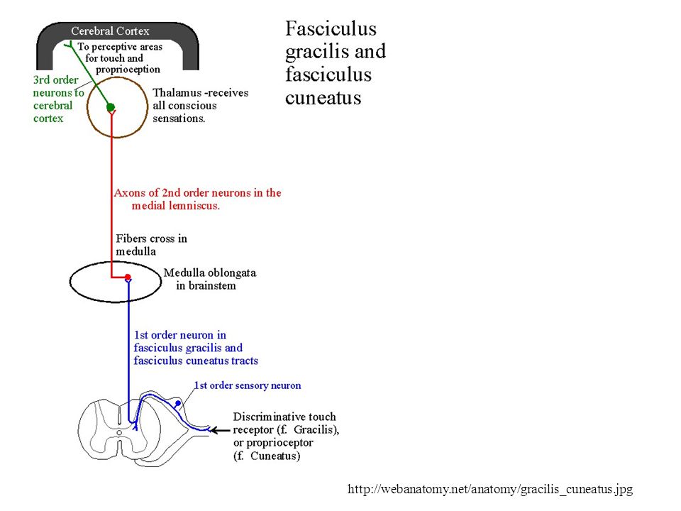
3 major somatosensory pathways - 2) Posterior column pathway
Sensation of precise touch, vibration and proprioception Includes Left and right fasciculus gracilis (inferior part of the body) Left and right fasciculus cuneatus (superior part of the body) First order neurons enter the CNS at the dorsal roots and the sensory roots of cranial nerves. Synapse with 2nd order in the medulla 2nd order neurons cross over in the brain stem 3rd order in the thalamus where the stimuli are sorted by the nature of stimulus and the region of body involved
 Left and right fasciculus cuneatus (superior part of the body) First order neurons enter the CNS at the dorsal roots and the sensory roots of cranial nerves. Synapse with 2nd order in the medulla. 2nd order neurons cross over in the brain stem. 3rd order in the thalamus where the stimuli are sorted by the nature of stimulus and the region of body involved.")
46
3 major somatosensory pathways – 3) The spinocerebellar pathway
Information about muscle, tendon and joint position from the spine to the cerebellum This information is subconscious 1st order neurons synapse in the dorsal horn 2nd order neurons ascend via anterior and posterior spinocerebellar tracts to the cerebellar cortex Used to coordinate movements In this pathway there is no 3rd order neuron

48
Pathway Sensation 1st order 2nd order 3rd order Final destination Spinothalamic pathway Lateral spinothalamic Pain and temperature Dorsal root ganglion Posterior horn Thalamus Primary sensory cortex (opposite side) Anterior spinothalamic Crude touch and pressure Posterior column pathway Fasciculus gracilis Proprioception, fine touch and pressure from inferior half of the body Medulla oblongata Fasciculus cuneatus Proprioception, fine touch and pressure from superior half of the body Spinocerebellar pathway Anterior and posterior Proprioception Not present Cerebellar cortex
 Anterior spinothalamic. Crude touch and pressure. Posterior column pathway. Fasciculus gracilis. Proprioception, fine touch and pressure from inferior half of the body. Medulla oblongata. Fasciculus cuneatus. Proprioception, fine touch and pressure from superior half of the body. Spinocerebellar pathway. Anterior and posterior. Proprioception. Not present. Cerebellar cortex.")
49
Somatic Senses Pathways
4 4 Sensations are perceived in the primary somatic sensory cortex. 3 3 Sensory pathways synapse in the thalamus. THALAMUS MEDULLA 2 2 Fine touch, vibration, and proprioception pathways cross the midline in the medulla. Fine touch, proprioception, vibration KEY 1 1 Pain, temperature, and coarse touch cross the midline in the spinal cord. Nociception, temperature, coarse touch Primary sensory neuron Secondary sensory neuron Tertiary neuron SPINAL CORD Figure 10-9, steps 1–4

50
Processing at the Perceptual Level
3 Perceptual level (processing in cortical sensory centers) Motor cortex Somatosensory cortex Thalamus Reticular formation Cerebellum Pons Medulla 2 Circuit level (processing in ascending pathways) Spinal cord Free nerve endings (pain, cold, warmth) Muscle spindle 1 Receptor level (sensory reception and transmission to CNS) Joint kinesthetic receptor Figure 13.2
 Motor. cortex. Somatosensory. cortex. Thalamus. Reticular. formation. Cerebellum. Pons. Medulla. 2. Circuit level. (processing in. ascending pathways) Spinal. cord. Free nerve. endings (pain, cold, warmth) Muscle. spindle. 1. Receptor level. (sensory reception. and transmission. to CNS) Joint. kinesthetic. receptor. Figure")
51
Processing at the Perceptual Level
Interpretation of sensory input occurs in the cerebral cortex The ability to identify the sensation depends on the specific location of the target neurons in the sensory cortex not on the nature of the message (all messages are action potentials)
")
52
The CNS integrate sensory information
Most of the somatic sensory information enters the spinal cord and travels via ascending pathways to the brain Some information goes directly to the brain through the cranial nerves Autonomic sensory information does not arrive conscious perception

53
Main Aspects of Sensory Perception
Perceptual detection – detecting that a stimulus has occurred and requires summation Magnitude estimation – the ability to detect how intense the stimulus is Spatial discrimination – identifying the site or pattern of the stimulus Feature abstraction – used to identify a substance that has specific texture or shape Quality discrimination – the ability to identify submodalities of a sensation (e.g., sweet or sour tastes) Pattern recognition – ability to recognize patterns in stimuli (e.g., melody, familiar face)
 Pattern recognition – ability to recognize patterns in stimuli (e.g., melody, familiar face)")
54
Somatosensation perception
The specific sensation depends on the 2nd and 3rd neurons The ability to localize the specific location of a stimulus depends on the stimulation of a specific area in the primary somatosensory cortex A sensory “homunculus” (little human) is a functional map of the primary somatosensory cortex
 is a functional map of the primary somatosensory cortex.")
55
Somatosensory Association Cortex
Located posterior to the primary somatosensory cortex and has connection with it Integrates sensory information like temperature and pressure coming from the primary somatosensory cortex. Forms understanding of the stimulus like size, texture, and relationship of parts Ex.: putting the hand in the pocket and feeling something. The center integrate previous information to identify objects without seeing them

56
The main Sensory Areas in the cerebral cortex
Figure 12.8a

57
Properties of the sensory system - summary
Stimulus – works on a receptor The receptor is a transducer that converts the stimulus into a change of membrane potential The message from the receptor will be sent in the form of action potential to the CNS Stimuli that will reach the cerebral cortex will be come conscious Somatosensory information ascends the spinal column along several pathways, which synapse at the midbrain &/or thalamus before reaching the cortex Sensory processes have different sub-modalities of somatosensory information Later stages of processing combine information across the sub-modalities, & with information from other senses

58
Pain pathways Pain is a protective mechanism
Pain is a subjective perception It is individual and can vary depending on emotional state Types of pain sensations: Fast pain – sharp and localized – in superficial parts of the body (cut, burn) Rapidly transferred to CNS by small myelinated fibers (within 0.1 seconds after stimulus applied) Slow pain – more diffused pain (associated with tissue destruction) Carried by small unmyelinated fibers Often fast pain will follow a slow one
 Rapidly transferred to CNS by small myelinated fibers (within 0.1 seconds after stimulus applied) Slow pain – more diffused pain (associated with tissue destruction) Carried by small unmyelinated fibers. Often fast pain will follow a slow one.")
59
Pain pathways Pain from the body – via spinal cord
Pain from face – via trigeminal (V) that enters the pons, descend to the medulla where they cross over and ascend to the thalamus The ascending pathway sends branches not only to thalamus and the cerebral cortex but also to the limbic system (emotions) and hypothalamus (autonomic reaction) The result is that pain may be accompanied by emotional distress and autonomic reactions such as nausea, vomiting or sweating
 that enters the pons, descend to the medulla where they cross over and ascend to the thalamus. The ascending pathway sends branches not only to thalamus and the cerebral cortex but also to the limbic system (emotions) and hypothalamus (autonomic reaction) The result is that pain may be accompanied by emotional distress and autonomic reactions such as nausea, vomiting or sweating.")
60
Pain perception Pain can be felt in skeletal muscle when anaerobic metabolism In cardiac muscle, pain is a result of ischemia (lack of oxygen due to reduced blood flow) during myocardial infraction (heart attack) Visceral pain is poorly localized and called referred pain
 during myocardial infraction (heart attack) Visceral pain is poorly localized and called referred pain.")
61
Pain perception – the gate control theory
Pain perception is subjected to modulation that can happen in several levels of the nervous system Pain can be magnified by past experiences Pain can be suppressed when in emergencies when surviving depends on ignoring the injury (minute 13.41)
")
62
The Gate-Control Theory of Pain
Pain can be suppressed in the dorsal horn level. Normally, tonically active inhibitory interneuron inhibit ascending pathways for pain Figure 10-12a

63
The Gate Control Theory of Pain Modulation
Fibers from nociceptors synapse on the inhibition interneuron When activated, the fibers send message to block the interneurons and pain travels to the brain Figure 10-12b

64
The Gate Control Theory of Pain Modulation
In the gate control theory of pain modulation fibers carrying sensory information about mechanical stimuli help block pain transmission Those fibers synapse on the interneuron and increase its inhibitory activity If both pain stimulus and nonpainful stimulus arrive at the same time, there will be partial inhibition of pain The sensation of pain will be perceived by the brain as lower Explains why rubbing a bumped elbow lessens the pain feeling Figure 10-12c

65
Visceral sensory pain pathways
Collected by interoceptors within the closed ventral body cavities The interoceptors include nociceptors, thermoreceptors, tactile receptors, baroreceptors and chemoreceptors The axons of the 1st order neuron usually travel with the autonomic motor fibers innervating the same visceral structures 2nd order neurons within the spinal cord use the spinothalamic pathway and arrive to the medulla oblongata Cranial nerves V, VII, IX and X carry visceral sensory information also to the medulla (all parasympathetic will be discussed with the ANS)
")
66
Referred Pain Skin (usual stimulus) Primary sensory neurons
Kidney (uncommon stimulus) Secondary sensory neuron Ascending sensory path to somatosensory cortex of brain (b) Figure 10-13b
 Secondary sensory neuron. Ascending sensory path to somatosensory cortex of brain. (b) Figure 10-13b.")
67
Sensory Pathways Figure 10-4 Primary somatic sensory cortex
Gustatory cortex Olfactory cortex Olfactory bulb Auditory cortex Visual cortex 1 Olfactory pathways from the nose project through the olfactory bulb to the olfactory cortex. Eye 2 Cerebellum 2 Most sensory pathways project to the thalamus. The thalamus modifies and relays information to cortical centers. 1 Nose Thalamus Sound Brain stem Equilibrium 3 3 Equilibrium pathways project primarily to the cerebellum. Tongue Somatic senses Figure 10-4

Similar presentations


 Introduction –Adrian’s work on sensory coding –Spinal cord and dorsal root ganglia.>")


