Download presentation
Presentation is loading. Please wait.

1
WINDSOR UNIVERSITY SCHOOL OF MEDICINE
DEPARTMENT OF ANATOMY Head and Neck Dr. SREEKANTH THOTA

3
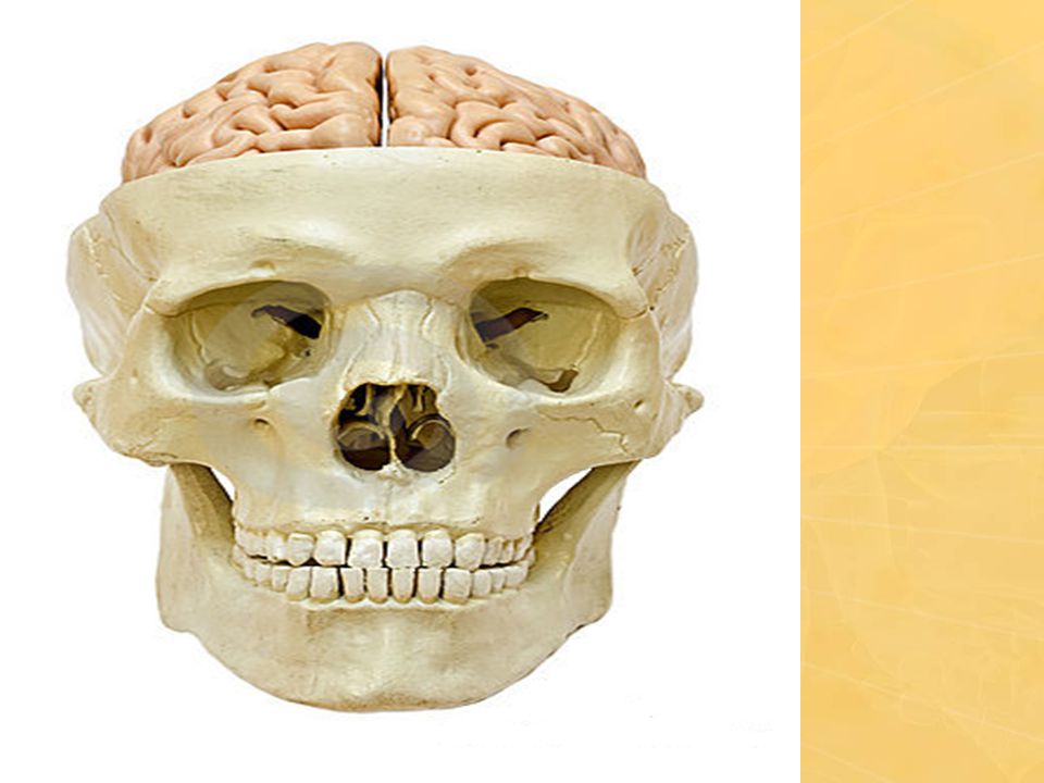
Bones of the Skull The skull bones are made up of external and internal tables of compact bone separated by a layer of spongy bone called the diploe.

4
Foramen In anatomy, a foramen is any opening.
Human skull, have numerous foramina through which nerves, arteries, veins and other structures pass.

6
The skull has 22 bones, excluding the ossicles of the ear.
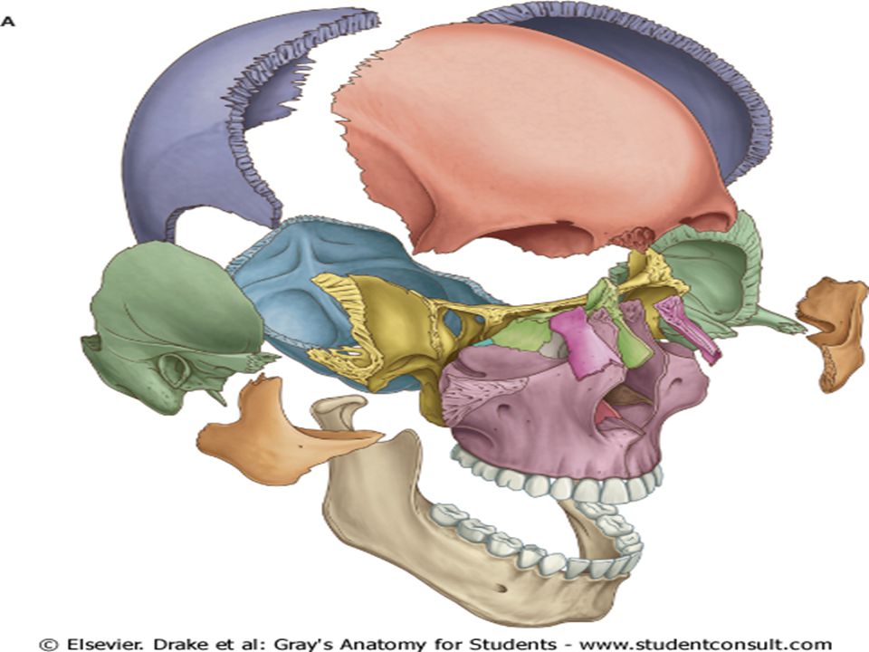
COMPONENT PARTS The skull has 22 bones, excluding the ossicles of the ear. Except for the mandible, which forms the lower jaw, the bones of the skull are attached to each other by sutures, are immobile, and form the cranium.

8
2.Lower anterior part-the facial skeleton (viscerocranium).
Subdivisons 1.The cranium can be subdivided into: an upper part the calvaria, which surrounds the cranial cavity containing the brain(Neurocranium). 2.Lower anterior part-the facial skeleton (viscerocranium).
. 2.Lower anterior part-the facial skeleton (viscerocranium).")
9
The cranium consists of the following bones, two of which are paired
Frontal bone: 1 Parietal bones: 2 Occipital bone: 1 Temporal bones: 2 Sphenoid bone: 1 Ethmoid bone: 1

10
The facial bones consist of the following, two of which are single:
Zygomatic bones: 2 Maxillae: 2 Nasal bones: 2 Lacrimal bones: 2 Vomer: 1 Palatine bones: 2 Inferior conchae: 2 Mandible: 1

11
External Views of the Skull
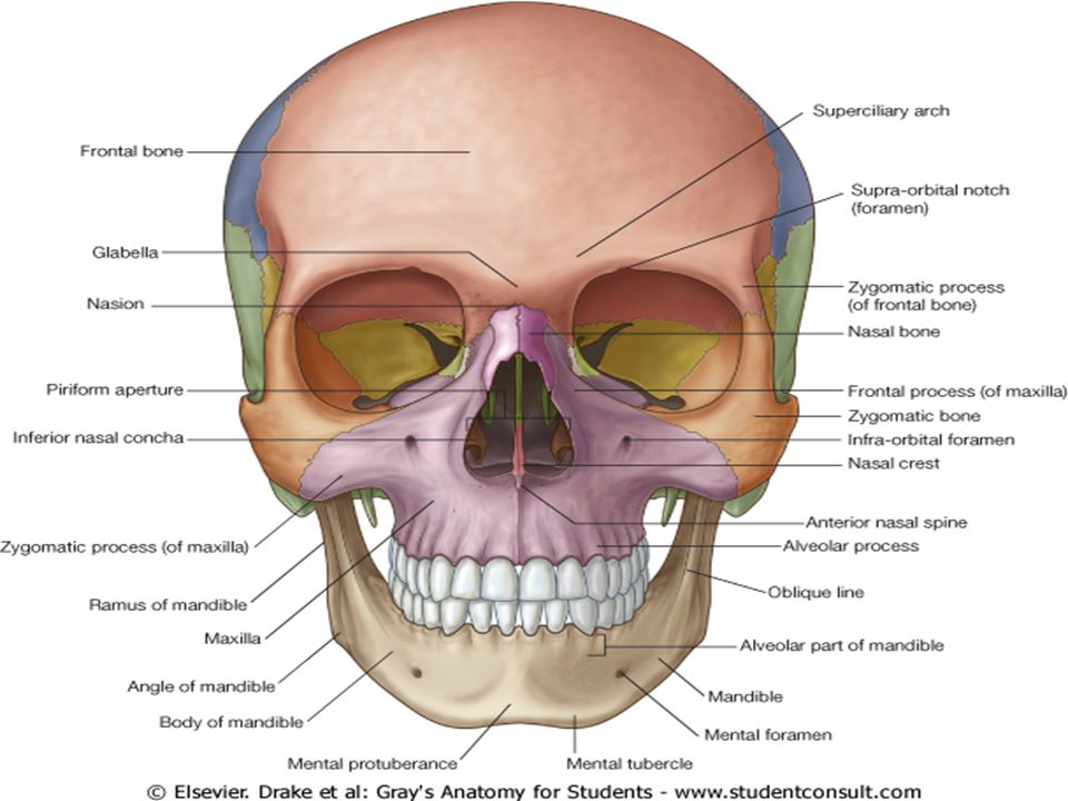
Anterior View of the Skull(Norma frontalis) The anterior view of the skull includes the forehead superiorly, and, inferiorly, the orbits, the nasal region, the part of the face between the orbit and the lower jaw
 The anterior view of the skull includes the forehead superiorly, and, inferiorly, the orbits, the nasal region, the part of the face between the orbit and the lower jaw.")
13
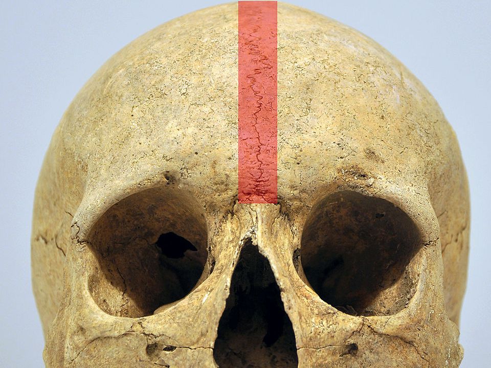
Metopic suture Suture that divides the two halves of the frontal bone of the skull in infants and children. It usually disappears by the age of six. In some individuals the suture can persist (totally or partly) into adulthood.
 into adulthood.")
15
Foramen of Anterior view
1. Supra-orbital foramen: Supra-orbital nerve and vessels. 2. Infra-orbital foramen: Infra-orbital nerve and vessels. 3. Mental foramen: Mental nerve and vessels

16
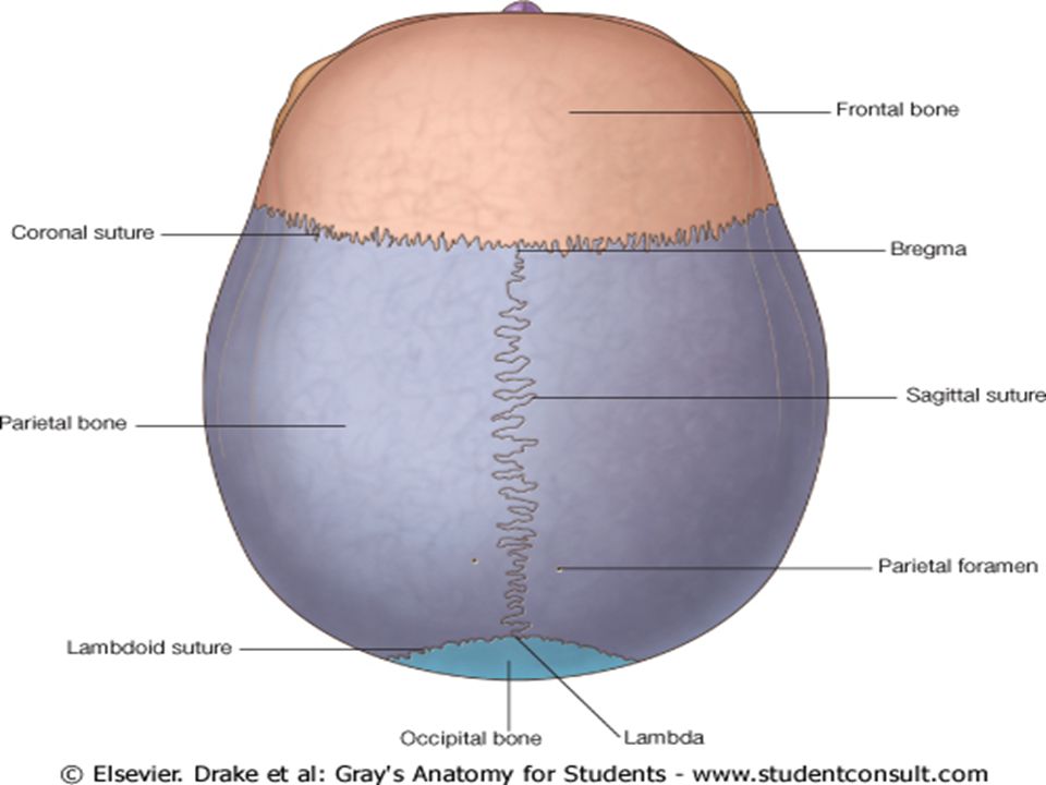
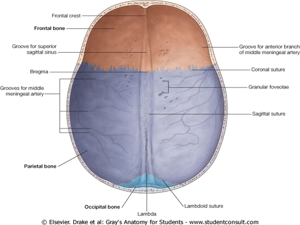
Superior view of the skull(Norma verticalis)
Parietal foramen: Emissary veins

18
Bregma and Lambda

19
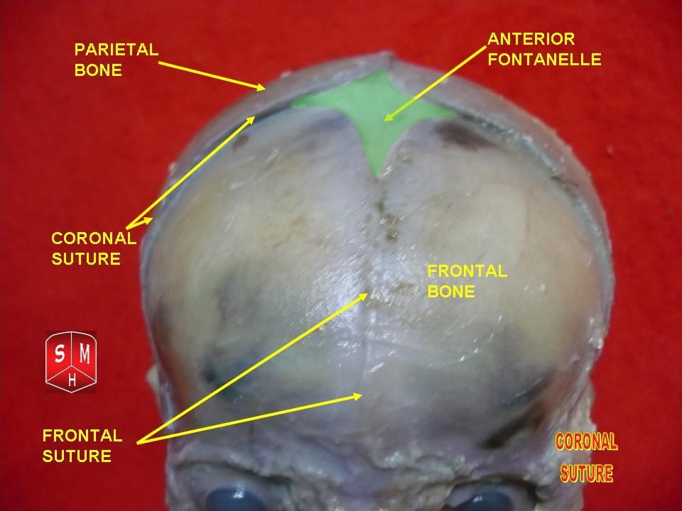
Anterior fontanelle Largest fontanelle, and is placed at the junction of the sagittal suture, coronal suture, and frontal suture; it is lozenge-shaped, and measures about 4 cm in its antero-posterior and 2.5 cm in its transverse diameter. Not completely closed until about the middle of the second year.

21
Clinical significance
Examination of an infant includes palpating the anterior fontanelle. A sunken fontanelle indicates dehydration, whereas a very tense or bulging anterior fontanelle indicates raised intracranial pressure.

22
Posterior view of the skull(Norma occipitalis)
")
23
Downloaded from: StudentConsult (on 10 December 2006 10:40 AM)
© 2005 Elsevier

24
Lateral view of skull(norma lateralis)
Zygomaticofacial foramen: Zygomaticofacial nerve

25
Downloaded from: StudentConsult (on 10 December 2006 10:40 AM)
© 2005 Elsevier

26
Fracture of the Pterion
A hard blow to the side of the head may fracture the thin bones forming the pterion, producing a rupture of the anterior branch of the middle meningeal artery crossing the pterion

27
Temporal bone

28
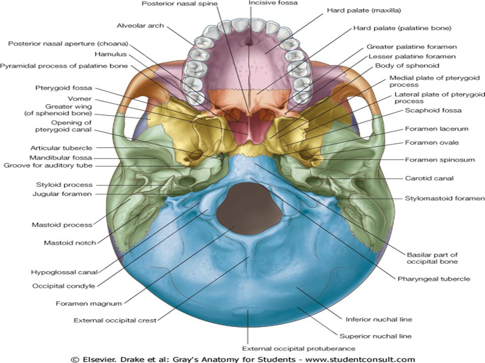
Inferior view of the skull.

30
Foramen 1. Incisive fossa 2. Greater and lesser palatine foramina
3.Jugular foramen 4.Carotid canal 5.Stylomastoid foramen 6.Foramen magnum

31
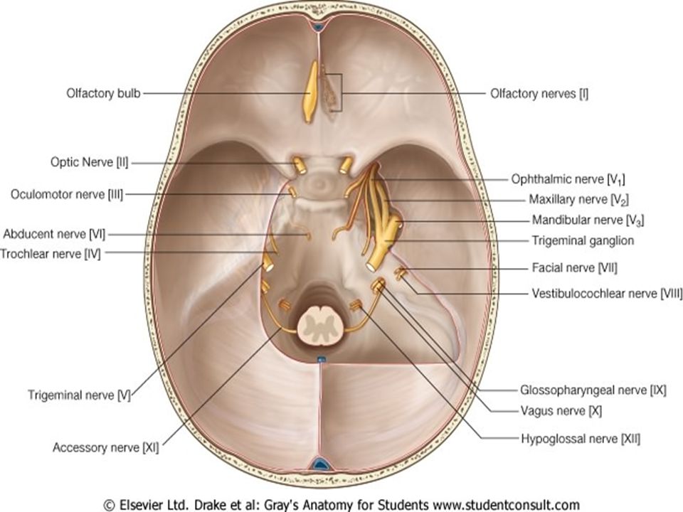
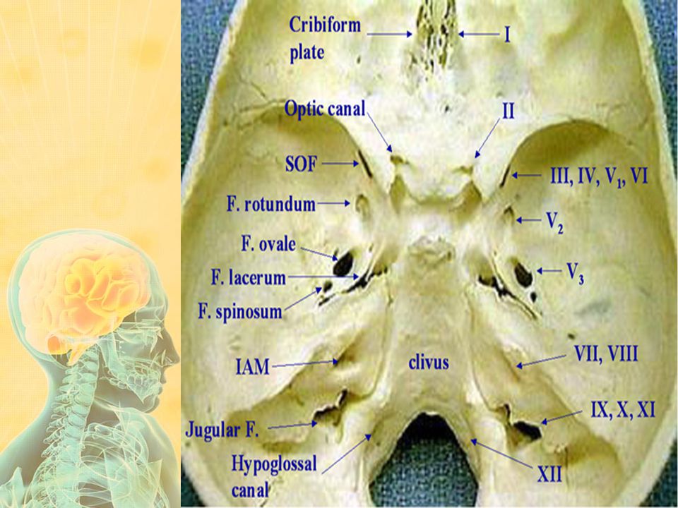
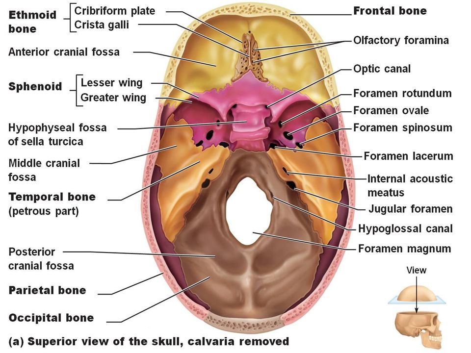
Skull Cranial fossa

33
Internal Surface of the Cranial Base
The internal surface of the cranial base has three large depressions that lie at different levels: the anterior, middle, and posterior cranial fossae .

35
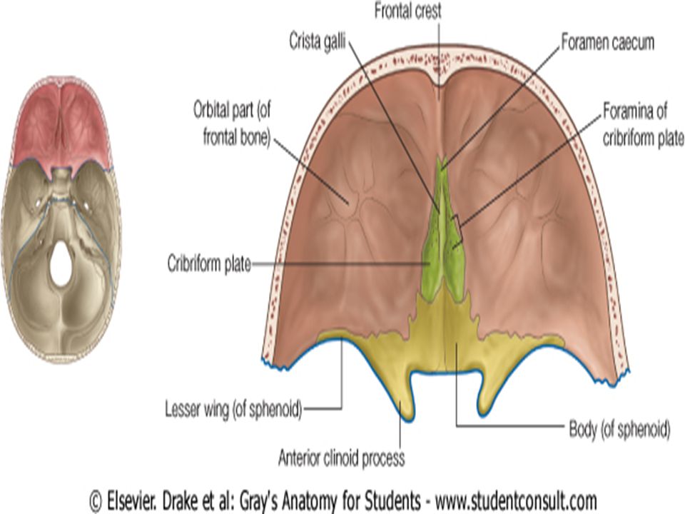
Anterior cranial fossa

38
Foramen of Anterior cranial fossa
1. Cribriform plate:olfactory nerves (CN-I) 2. Foramen cecum: occasional small emissary vein from nasal mucosa to superior sagittal sinus. 3. Anterior and posterior ethmoidal foramina: anterior and posterior ethmoidal nerves, arteries, and veins.
 2. Foramen cecum: occasional small emissary vein from nasal mucosa to superior sagittal sinus. 3. Anterior and posterior ethmoidal foramina: anterior and posterior ethmoidal nerves, arteries, and veins.")
39
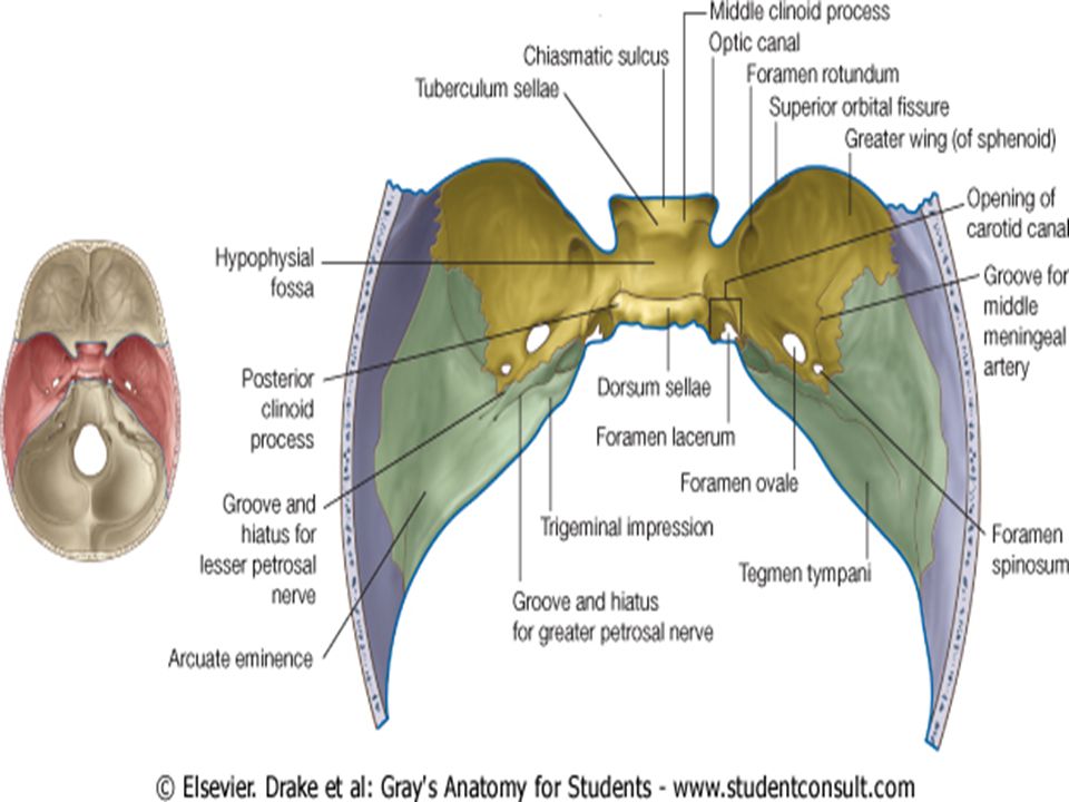
Middle cranial fossa-

41
Sphenoid bone

44
Foramen of Middle cranial fossa
Optic canal: optic nerve (CN II), ophthalmic artery. 2.Superior orbital fissure: oculomotor (CN III), trochlear (CN IV), and abducens (CN VI) nerves; ophthalmic division of trigeminal nerve (CN V1) and ophthalmic veins. 3. Foramen rotundum: Maxillary nerve
, ophthalmic artery. 2.Superior orbital fissure: oculomotor (CN III), trochlear (CN IV), and abducens (CN VI) nerves; ophthalmic division of trigeminal nerve (CN V1) and ophthalmic veins. 3. Foramen rotundum: Maxillary nerve.")
45
4. Foramen ovale: Mandibular nerve, the accessory meningeal artery, and the lesser petrosal nerve.
5. Foramen spinosum: middle meningeal artery.

46
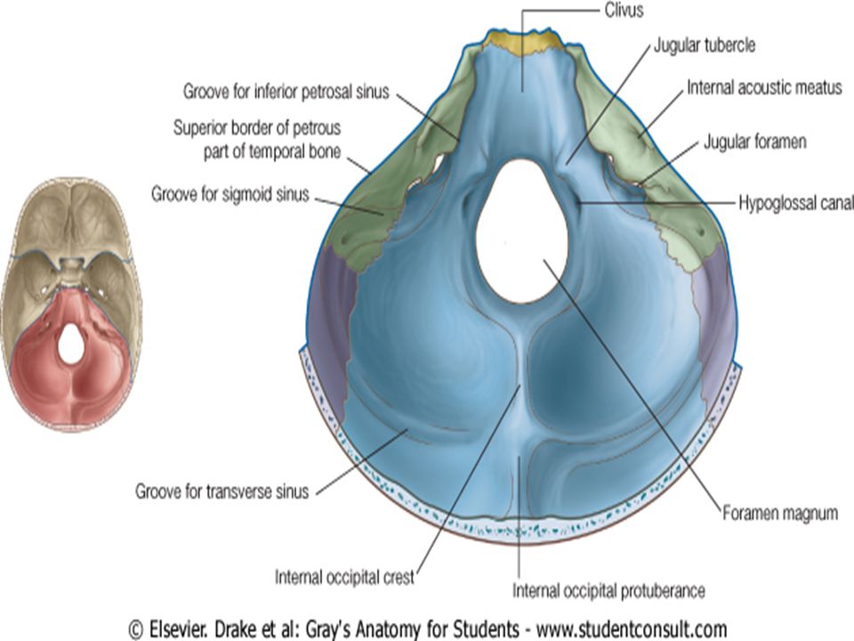
Posterior cranial fossa

47
Foramen of posterior cranial fossa
Foramen magnum: medulla, the ascending portions of the spinal accessory nerve (XI), and the vertebral arteries. Internal acoustic meatus:facial (VII) and vestibulocochlear (VIII) cranial nerves Jugular foramen: internal jugular vein (actually begins here), the glossopharyngeal (IX), the vagus (X) and the accessory (XI) nerves. Anterior condylar (hypoglossal) canal: hypoglossal (XII) nerve.
, and the vertebral arteries. Internal acoustic meatus:facial (VII) and vestibulocochlear (VIII) cranial nerves. Jugular foramen: internal jugular vein (actually begins here), the glossopharyngeal (IX), the vagus (X) and the accessory (XI) nerves. Anterior condylar (hypoglossal) canal: hypoglossal (XII) nerve.")
49
Fractures of the Calvaria
A depressed skull fracture is a break in a cranial bone (or "crushed" portion of skull) with depression of the bone in toward the brain. A compound fracture involves a break in, or loss of, skin and splintering of the bone.
 with depression of the bone in toward the brain. A compound fracture involves a break in, or loss of, skin and splintering of the bone.")
50
Contrecoup (counterblow) fracture
No fracture occurs at the point of impact, but one occurs on the opposite side of the cranium. When a moving object impacts the stationary head, coup injuries are typical, while contrecoup injuries are produced when the moving head strikes a stationary object

51
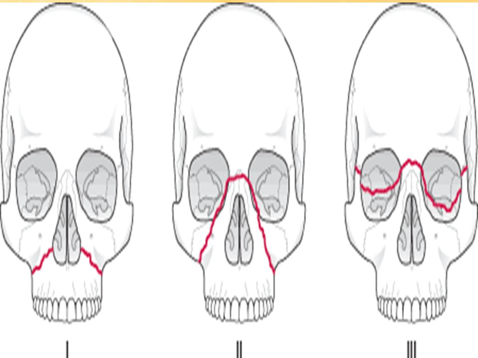
Fractures to the Maxilla and associated bones
Le Fort I Fracture = Horizontal Fracture Le Fort II Fracture = Pyramidal Fracture Le Fort III Fractures = Craniofacial Separation

53
Raccoon eyes (periorbital ecchymosis )
blood from skull fracture seeps into the soft tissue around the eyes Periorbital ecchymosis If bilateral, it is highly suggestive of basilar skull fracture They are most often associated with fractures of the anterior cranial fossa

Similar presentations