Download presentation
Presentation is loading. Please wait.

1
Pediatric Skull Xray Heather Patterson August 2, 2007

2
Objectives Brief review of anatomy Approach to pediatric skull xray Examples

3
Skull fractures Common in non-accidental trauma –80% in first year –Rare after 2y of age

4
Anatomy

6
Skull Xray Full series 3-4 views –AP –Towne’s view (AP with neck flexed) –Lateral x 2
 –Lateral x 2")
7
Skull Xray

11
Approach Follow cortex Identify suture lines Identify abnormal lines

12
What is the big deal? Risk of “growing fracture” –Leptomeningeal cysts –Long term sequelae

13
Growing fracture/Leptomeningeal Cyst Rare –<1% of skull fractures Pathophys –Dural deal with herniation of pia and arachnoid through tear –CSF pulsations lead to erosion of bone –Diastasis of fracture over time

14
Growing fracture/Leptomeningeal Cyst Imaging –Angular, linear lytic lesion –Scalloped margins Management –f/u with neurosurgery –Early intervention as needed

15
Case 1

17
Linear fracture R posterior parietal and occipital bones Extends through lambdoid suture

18
Case 2

19
R parietal skull fracture

20
Case 3

22
Linear fracture R occiput

23
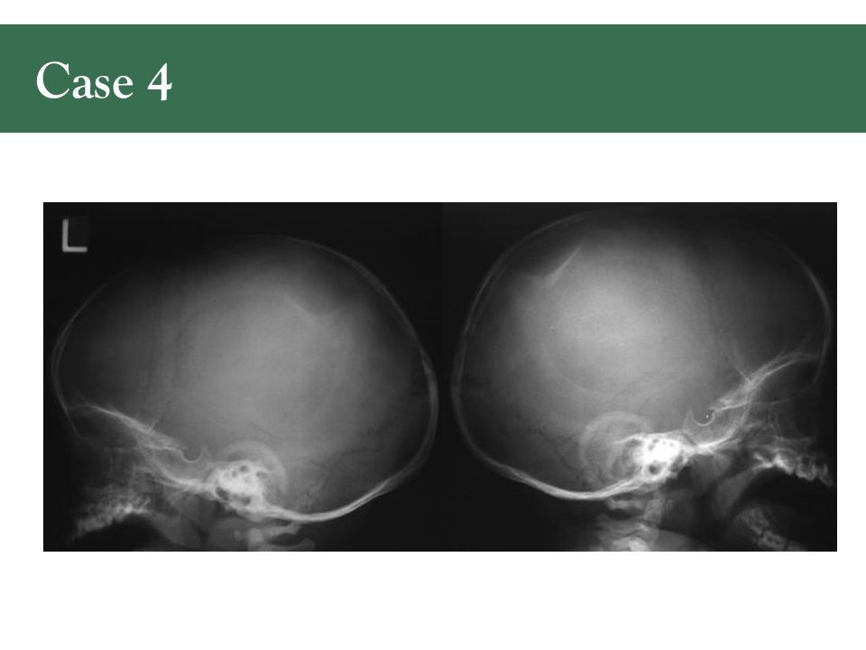
Case 4

25
Depressed skull fracture posterior right parietal bone

26
Case 5

27
R parietal fracture Communicates with lamboidal suture

28
Case 6

30
R parietal fracture

31
Case 7

33
L parietal fracture

34
Case 8

35
Persistent skull defect Encephalomalacic cystic defect –Consistent with leptomeningeal cyst

36
Uganda

Similar presentations


 Ultrasound Magnetic Resonance Imaging (MRI) Radioisotopes Studies.>")
 & hand RTEC 123 # 1 A LECTURE Contributions by: MOSBY – MERRILLS & BONTAGER XRAY2000.CO.UK rev 10/10/11 1.>")


