Download presentation
Presentation is loading. Please wait.

2
? Microsurgery Couching (extra capsular) (intracapsular)
(extracap. + IOL) PMMA Phaco Soft Bi/Mf Ls Mics Acc Aq ? Ph-E
 PMMA. Phaco. Soft. Bi/Mf. Ls. Mics. Acc. Aq. Ph-E.")
3
Complications of phacoemulsification
ZDMU , ALZHRA (S) EYE CENTRE , ZAHEDAN . Validad MD iranFCRS
 EYE CENTRE , ZAHEDAN . Validad MD iranFCRS.")
4
phaco. complication Preo. Intraop. Postop.

5
Preoperatory complication

6
Complications of anesthesia
GA LA TOPICAL

7
Transient, complete loss of vision
secondary to posterior diffusion of an ophthalmic viscosurgical device-lidocaine solution during complicated phacoemulsification. Falzon K - J Cataract Refract Surg - 01-AUG-2009; 35(8):
:")
8
The main drawback of local anesthesia is to enable the patient to perform movements during operation. Patient movements

9
Intra operatory complications

13
Conjunctival hydration
All cases where conjunctival hydration occurred were clear corneal incisions.

14
Capsulorhexis There was no need to convert the capsulorhexis
into an can-opener in # any situation. In intumescences cataracts we performed a smaller rexis to avoid its failure . After closing the rexis and cortex aspiration , broadening the rexis, bringing it to the normal diameter. In white cataracts (mature, hyper mature) used a colored substance to reveal the anterior capsule, under air protection. Rexis problems were not likely to influence the conduct the surgery. Thus, in 8 cases I have met the tendency of rexis disruption. After reintroduction of viscoelastic substances, the situations were resolved in a favorable way. There was no need to convert the capsulorexis into an can-opener in any situation. In intumescences cataracts we performed a smaller rexis to avoid its failure. After closing the rexis and cortex aspiration I started broadening the rexis, bringing it to the normal diameter. In 8 cases this maneuver was imposed. In white cataracts (mature, hyper mature) I used a colored substance to reveal the anterior capsule, under air protection. There were 2 cases in which, due to the rigidity of the capsule and the solidified cortex, rexis could not be conducted requiring the completing of it with scissors. In 5 cases the rexis edge was injured during the subsequent surgical maneuvers. In all cases the operation continued without incidents.
 used a colored substance to reveal the anterior capsule, under air protection. Rexis problems were not likely to influence the conduct the surgery. Thus, in 8 cases I have met the tendency of rexis disruption. After reintroduction of. viscoelastic substances, the situations were resolved in a favorable way. There was no. need to convert the capsulorexis into an can-opener in any situation. In intumescences cataracts we performed a smaller rexis to avoid its failure. After. closing the rexis and cortex aspiration I started broadening the rexis, bringing it to the. normal diameter. In 8 cases this maneuver was imposed. In white cataracts (mature, hyper mature) I used a colored substance to reveal the. anterior capsule, under air protection. There were 2 cases in which, due to the rigidity of the capsule and the solidified. cortex, rexis could not be conducted requiring the completing of it with scissors. In 5 cases the rexis edge was injured during the subsequent surgical maneuvers. In all. cases the operation continued without incidents.")
17
Preventing radial tears in the anterior capsule
The anterior chamber should be reinflated with an OVD. The vector forces of the tear should be changed to redirect the tear in a more central direction. If the tear is lost beneath the iris, the capsulorrhexis should be restarted from its origin, proceeding in the opposite direction (if possible, this new capsulorrhexis should finish by incorporating the original tear in an outside-in direction; however, the original tear is often too peripheral to permit this, and a single radial tear is created). . An alternative approach to a “lost” capsulorrhexis is to convert to a can-opener capsulectomy.
. . An alternative approach to a lost capsulorrhexis is to convert to a can-opener capsulectomy.")
18
Minimizing complications when radial tears are present
Hydro dissection or hydro delineation is performed gently The IOL should be placed with the haptics 90° away from the tear. Cracks during emulsification are made gently away from the area(s) with radial tears. the chamber is deepened each time the phacoemulsification or irrigation-aspiration tip is removed from the eye Minimizing complications when radial tears are present
 with radial tears. the chamber is deepened each time the phacoemulsification or irrigation-aspiration tip is removed from the eye. Minimizing complications when radial tears are present.")
19
Endocapsular phacoemulsification without hydrodissection: an effective technique for cataract surgery following anterior capsular tear An anterior capsular tear can result in a dropped nucleus with potentially sight-threatening complications. Hydrodissection is particularly likely to cause peripheral and posterior extension of such a tear. We offer a simple and effective approach to endocapsular phacoemulsification in this situation that avoids hydrodissection and which, in our experience, minimizes the risk of posterior tear propagation thereby improving surgical outcomes. The key points of this technique are: Fastidious attention to chamber stability and tamponade throughout the surgery. Use of continuous irrigation and OVD-BSS exchange when removing the irrigating instrument No primary hydro dissection Avoid cracking the lens Bowl out the lens using gentle sculpting The residual lens bowl often 'auto hydro dissects' due to irrigating fluid flow beneath the anterior capsule Aspirate the remaining cortex Position the IOL within the capsular bag or sulcus Avoid aspirating residual viscoelastic from behind the intraocular lens

21
Excessively small capsulorrhexis

22
Two major complications of hydro dissection are:
- inadequate hydro dissection - over inflation of the capsular bag. Hydro dissection Hydro dissection was developed to permit easy rotation of the nucleus in the capsular bag and to facilitate removal of various layers of the lens by eliminating their adhesion to surrounding tissues. Two major complications of hydro dissection are inadequate hydro dissection and over inflation of the capsular bag. The former results in a nucleus that does not rotate, which predisposes to zonular dehiscence if excessive force is exerted on the nucleus. This can be avoided by making an additional hydro dissection, particularly in quadrants that have not been hydro dissected before. U-shaped cannulas are useful to hydro dissect subincisional regions of the lens not accessible with straight or angulated cannulas. Over inflation of the capsular bag can predispose to nuclear prolapsed into the anterior chamber, which might compromise the ease or safety of nucleus emulsification. A serious complication of over inflation is posterior capsular rupture with loss of the nucleus into the vitreous. This is more likely to occur in eyes with long axial lengths, (hyper)mature cataracts, or with fragile posterior capsules, such as are found in patients who have posterior polar cataracts. The former results in a nucleus that does not rotate, which predisposes to zonular dehiscence if excessive force is exerted on the nucleus. This can be avoided by making an additional hydro dissection, particularly in quadrants that have not been hydro dissected before. U-shaped cannulas are useful to hydro dissect subincisional regions of the lens not accessible with straight or angulated cannulas. Overinflation of the capsular bag can predispose to nuclear prolapsed into the anterior chamber, which might compromise the ease or safety of nucleus emulsification. A serious complication of over inflation is posterior capsular rupture with loss of the nucleus into the vitreous. This is more likely to occur in eyes with long axial lengths, (hyper)mature cataracts, or with fragile posterior capsules, such as are found in patients who have posterior polar cataracts.
mature cataracts, or with fragile posterior capsules, such as are found in patients who have posterior polar cataracts. The former results in a nucleus that does not rotate, which predisposes to zonular dehiscence if excessive force is exerted on the nucleus. This can be avoided by making an additional hydro dissection, particularly in quadrants that have not been hydro dissected before. U-shaped cannulas are useful to hydro dissect subincisional regions of the lens not accessible with straight or angulated cannulas. Overinflation of the capsular bag can predispose to nuclear prolapsed into the anterior chamber, which might compromise the ease or safety of nucleus emulsification. A serious complication of over inflation is posterior capsular rupture with loss of the nucleus into the vitreous. This is more likely to occur in eyes with long axial lengths, (hyper)mature cataracts, or with fragile posterior capsules, such as are found in patients who have posterior polar cataracts.")
23
Phacoemulsification with viscodissection in posterior polar cataract: minimizing risk of posterior capsule tear Taskapili M - Ann Ophthalmol (Skokie) - 01-JUN-2007; 39(2): 145-9 the viscodissection technique was safer and more efficient than the non-dissection technique.

24
Descemet’s Detachment
Detachment of Descemet’s membrane can be a major postoperative complication; it results in persistent corneal edema and decreased visual acuity. To prevent Descemet’s detachment, the surgeon should carefully observe the inner lip at each phase of the procedure . To avoid blunt stripping of Descemet’s membrane during enlargement of the wound, a sharp metal or diamond blade is recommended.

26
Iris prolapse or damage
usually is caused when the anterior chamber is entered too posteriorly, such as near the iris root. If this is noted early in the case and interferes with the easy introduction of instruments into the eye, it is advisable to suture the incision & move to another location. A second and more ominous cause of iris prolapse is an acute increase of intraocular pressure (IOP) accompanied by choroidal effusion or hemorrhage. Iris prolapse or damage Iris prolapse usually is caused when the anterior chamber is entered too posteriorly, such as near the iris root. If this is noted early in the case and interferes with the easy introduction of instruments into the eye, it is advisable to suture the incision and move to another location. A second and more ominous cause of iris prolapse is an acute increase of intraocular pressure (IOP) accompanied by choroidal effusion or hemorrhage. In this instance, the surgeon should attempt to identify the cause and lower the IOP. Sometimes digital massage on the eye, pressing directly on the incision, can successfully lower the pressure. It is useful to examine the fundus to ascertain whether a choroidal effusion or hemorrhage exists. With choroidal effusion, aspiration of vitreous can be helpful, as can the administration of intravenous mannitol. If a choroidal hemorrhage occurs or if the increased IOP from an effusion is resistant to treatment, it usually is best to terminate surgery. The wound is sutured carefully; intraocular miotics are administered, and a peripheral iridectomy may be performed to help reposition the iris. For effusions, surgery can be deferred until later in the day or the next day, when the fluid dynamics of the eye have returned to a more normal state. If a limited choroidal hemorrhage has occurred, it is best to wait 2–3 weeks before attempting further surgery.
 accompanied by choroidal effusion or hemorrhage. Iris prolapse or damage. Iris prolapse usually is caused when the anterior chamber is entered too posteriorly, such as near the iris root. If this is noted early in the case and interferes with the easy introduction of instruments into the eye, it is advisable to suture the incision and move to another location. A second and more ominous cause of iris prolapse is an acute increase of intraocular pressure (IOP) accompanied by choroidal effusion or hemorrhage. In this instance, the surgeon should attempt to identify the cause and lower the IOP. Sometimes digital massage on the eye, pressing directly on the incision, can successfully lower the pressure. It is useful to examine the fundus to ascertain whether a choroidal effusion or hemorrhage exists. With choroidal effusion, aspiration of vitreous can be helpful, as can the administration of intravenous mannitol. If a choroidal hemorrhage occurs or if the increased IOP from an effusion is resistant to treatment, it usually is best to terminate surgery. The wound is sutured carefully; intraocular miotics are administered, and a peripheral iridectomy may be performed to help reposition the iris. For effusions, surgery can be deferred until later in the day or the next day, when the fluid dynamics of the eye have returned to a more normal state. If a limited choroidal hemorrhage has occurred, it is best to wait 2–3 weeks before attempting further surgery.")
27
Bleeding in the anterior chamber
Bleeding in the anterior chamber can come from intra operatory damage of iris. This occurs most frequently in temporal incisions located more posterior and deeper than normal. In these cases there are reached the blood vessels with higher risk of bleeding. it appears that there is no significant statistical correlation with anticoagulant therapy or chronic anti agregant Bleeding in the anterior chamber can come from intra operatory damage of iris. This occurs most frequently in temporal incisions located more posterior and deeper than normal. In these cases there are reached the blood vessels with higher risk of bleeding. In 11 cases (1.93%) I met bleeding in the anterior chamber during surgery. From our analysis it appears that there is no significant statistical correlation with anticoagulant therapy or chronic antiagregant (p> 0.05).
 I met bleeding in the anterior chamber during surgery. From our. analysis it appears that there is no significant statistical correlation with anticoagulant. therapy or chronic antiagregant (p> 0.05).")
28
Temporarily elevating the IOP with a balanced salt solution or an OVD.
Iris bleeding is caused by iris trauma. Intraocular bleeding can be stopped by: Temporarily elevating the IOP with a balanced salt solution or an OVD. Injecting a dilute solution of preservative-free epinephrine 1:5000 (or a weaker solution). Direct cautery (if the bleeding vessel can be identified) with a needle-tipped cautery probe.
. Direct cautery (if the bleeding vessel can be identified) with a needle-tipped cautery probe.")
29
Problems during phacoemulsification

30
shallow anterior chamber
Temporary loss of the chamber After adjusting the parameters of aspiration and irrigation and the introduction of viscoelastic substances with high molecular weight the situation was resolved favorably.

31
Hypertonic eye (2.81%)? All cases of hypertonic eye have been associated with temporary shallow anterior chamber earlier, the correlation being statistically significant. hypertonic eye & posterior capsule rupture has been reported ( was needed vitrectomy) Correlation was not statistically significant.
 Correlation was not statistically significant.")
32
Thermal injuries If for any reason the flow is blocked, a corneal burn can occur within 1–3 s

33
Corneal burn following phacoemulsification.
Corneal burn following phacoemulsification. In this patient who had an apparent filtering bleb, phacoemulsification was performed through a temporal, clear corneal incision. Posterior capsular rupture was suspected; the surgeon injected a highly retentive ophthalmic viscosurgical device beneath and in front of the nucleus to minimize the risk of posterior dislocation of the nucleus. Phacoemulsification was instituted with low flow and vacuum settings, and a severe corneal burn was immediately produced because of obstruction of the phacoemulsification tip by the viscoelastic material. The incision was closed with several interrupted sutures. Many of these pulled through the injured tissue, and as a result, additional suturing was required several days later. Postoperatively, the patient has 5 D of surgically induced astigmatism that has persisted for more than 5 years. additional suturing was required several days later. Postoperatively, the patient has 5 D of surgically induced astigmatism that has persisted for more than 5 years. Yanoff & Duker: Ophthalmology, 3rd ed.

34
acute increase of intraocular pressure (IOP)
the surgeon should attempt to identify the cause and lower the IOP. Sometimes digital massage on the eye, pressing directly on the incision, can successfully lower the pressure. It is useful to examine the fundus to ascertain whether a choroidal effusion or hemorrhage exists. With choroidal effusion, aspiration of vitreous can be helpful, as can the administration of intravenous mannitol. If a choroidal hemorrhage occurs or if the increased IOP from an effusion is resistant to treatment, it usually is best to terminate surgery. The wound is sutured carefully; intraocular miotics are administered, and a peripheral iridectomy may be performed to help reposition the iris. For effusions, surgery can be deferred until later in the day or the next day, when the fluid dynamics of the eye have returned to a more normal state. If a limited choroidal hemorrhage has occurred, it is best to wait 2–3 weeks before attempting further surgery.

37
Choroidal effusion also may be a precursor to suprachoroidal hemorrhage
presumably occurs from the rupture of a blood vessel that is placed under stretch. Risk factors include : hypertension, glaucoma, nanophthalmos, high myopia, and chronic intraocular inflammation.[26]

38
Broken capsules still occur at a rate between
0.45% for very experienced surgeons [1] & up to 14.7% for residents in training [2]. The frequency of retained lens fragments is estimated at 0.3% to 1.1% [3,4]. The challenge of cataract surgery is to minimize the risk of complications and to manage optimally complications that do occur.

39
Dropped nucleus B-scan ultrasonography 1 day after dislocation
of a lens nucleus into the vitreous cavity in a patient who has high myopia.

41
Loss of the nucleus into the vitreous
the early signs of posterior capsular rupture include: unusual deepening of the anterior chamber, decentration of the nucleus, or loss of efficiency of aspiration, which suggests occlusion of the tip with vitreous. If capsular rupture is noted, the steps outlined earlier should be taken to prevent nucleus loss. Loss of the nucleus into the vitreous cavity can sometimes be avoided by recognizing the early signs of posterior capsular rupture. These include unusual deepening of the anterior chamber, decentration of the nucleus, or loss of efficiency of aspiration, which suggests occlusion of the tip with vitreous. If capsular rupture is noted, the steps outlined earlier should be taken to prevent nucleus loss. Some controversy exists with regard to the appropriate management of loss of the nucleus into the vitreous. Most surgeons recommend completing the procedure with careful anterior vitrectomy and removal of remaining accessible lens material. In general, IOL implantation is permissible; one exception might be loss of an extremely hard, dense nucleus that would require removal through a limbal incision. If a significant amount of nuclear material has been retained, vitreoretinal surgery needs to be performed 1–2 days postoperatively. Patients whose eyes have small residual nuclear fragments may be observed and referred if increased IOP or uveitis refractory to medical treatment develops. Some surgeons advocate irrigating the vitreous with fluid in an attempt to float the nucleus back into position. An obvious concern is that this additional turbulence could increase vitreous traction on the retina and cause retinal tears.

42
Corneal edema on first postoperative day was significant problem
Usually Posterior capsular rupture is the most common intraoperative complication in initial cases. Corneal edema on first postoperative day was significant problem

44
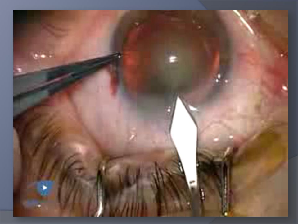
Zonular dialysis

46
Recent reports suggest that the visual prognosis of patients who have broken posterior capsules is excellent. The key factors are to minimize ocular trauma, meticulously clean prolapsed vitreous from the anterior segment, if present, and ensure secure fixation of the IOL.

47
Postoperative complications

48
Complications of phacoemulsification on the first postoperative day
The review yielded 392 patients. Six (1.53%) had intraocular pressure (> or = 30 mm Hg) requiring treatment, 1 (0.26%) had painless iris prolapse, 11 (2.81%) had corneal abrasions, and 7 (1.78%) were given a more intensive steroid regime (UVEITIS) Corneal edema No cases of fibrinous uveitis were recorded. J Cataract Refract Surg Jul;25(7):985-8.
 had intraocular pressure (> or = 30 mm Hg) requiring treatment, 1 (0.26%) had painless iris prolapse, 11 (2.81%) had corneal abrasions, and 7 (1.78%) were given a more intensive steroid regime (UVEITIS) Corneal edema. No cases of fibrinous uveitis were recorded. J Cataract Refract Surg Jul;25(7):")
49
Corneal edema Corneal edema is categorized according to severity in reversible and irreversible. • reversible corneal edema: - in 7 days: 8.42%; in 30 days: 1.03%; • irreversible corneal edema : – edemato bullous keratopathy (Corneal endothelial damage) a significant statistical correlation of the corneal edema post phaco with the phaco-time and the type of cataract, In 18 of the 48 cases of edema with remission in 7 days, they were associated with an increase in intraocular pressure (less than 35 mmHg on the first day post). The combination was significantly statistic with a precision of 99%.
 a significant statistical correlation of the corneal edema post phaco with the phaco-time and the type of cataract, In 18 of the 48 cases of edema with remission in 7 days, they were associated with an. increase in intraocular pressure (less than 35 mmHg on the first day post). The. combination was significantly statistic with a precision of 99%.")
50
Stromal and Epithelial Edema
Most common in pre-existing endothelial disorder Edema is most often caused by : Mechanical trauma, Prolonged intraocular irrigation, Inflammation, And elevated IOP. Toxic Solutions Vitreocorneal Adherence and Persistent Corneal Edema

51
Stromal and Epithelial Edema
Points in Management: If epithelial edema is present in the face of a compact stroma immediately after surgery, it is likely due to elevated lOP with intact endothelium. Corneal edema generally resolves completely within 4-6 weeks. As a rule, if the corneal periphery is clear, the corneal edema will usually resolve with time. Corneal edema persisting after 3 months usually does not clear and may require penetrating keratoplasty.

52
Wound dehiscence The sealing of the wound depends :
Small wounds, under 3.2 mm, are much less prone to this complication The sealing of the wound depends : the quality of the corneo-scleral tissue. the quality of incision This in turn depends on certain intraoperatory complications certain chronic diseases thermal injury of the wound If needed case can use a therapeutic contact lens.

53
Asymmetric pupil Intraoperatory injury with the phaco tip or instruments. (complicate cases) & some local or general associated conditions may cause pupil asymmetries. This will translate clinically by decreased visual acuity, lack of adaptation to strong light.

54
Intraocular inflammation
Post operatory intraocular inflammation can be acute and chronic (endophthalmitis). Chronic uveal inflammation may occur in weeks, months or years after cataract chronic inflammation significant statistical correlations between post-intraocular inflammation on the one hand and rupture of thecapsule.
. Chronic uveal inflammation may occur in weeks, months or years after cataract. chronic inflammation. significant statistical correlations. between post-intraocular inflammation on the one hand and rupture of thecapsule.")
55
Complications of vitreous loss at cataract surgery are as follows:
Cystoid macular edema Retinal detachment Persistent increase in intraocular pressure Intraocular lens dislocation or subluxation Choroidal detachment Endophthalmitis Corneal edema

56
Phacoemulsification Training
There were more cases of posterior capsule tears and vitreous loss in the first 80 cases performed by the residents, the posterior capsule tear rate peaked at more than 10% after 40 cases.

57
in-the-Bag Intraocular Lens Luxation
Spontaneous in-the-Bag Intraocular Lens Luxation into the Vitreous Cavity: Last- Stage Complication of Pseudoexfoliative Syndrome after Phacoemulsification The high prevalence of pseudoexfoliation (PEX) in the patients who had been operated for cataract phacoemulsification in our department could explain the occurrence of 8 posterior luxations of in-the-bag IOLs in only 1 year. Our study suggests that for the next years we will expect an increase in occurrence of spontaneous in-the- bag IOL luxations in the vitreous cavity. This condition could represent the last stage of PEX syndrome. Copyright © 2009 S. Karger AG, Basel [PUBLICATION ABSTRACT] A Balestrazzi, G M Tosi, M Alegente, C Mazzotta, et al. Ophthalmologica. Basel: Aug Vol. 223, Iss. 5; pg. 339, 4 pgs
 in the patients who had been operated for cataract phacoemulsification in our department could explain the occurrence of 8 posterior luxations of in-the-bag IOLs in only 1 year. Our study suggests that for the next years we will expect an increase in occurrence of spontaneous in-the- bag IOL luxations in the vitreous cavity. This condition could represent the last stage of PEX syndrome. Copyright © 2009 S. Karger AG, Basel [PUBLICATION ABSTRACT] A Balestrazzi, G M Tosi, M Alegente, C Mazzotta, et al. Ophthalmologica. Basel: Aug Vol. 223, Iss. 5; pg. 339, 4 pgs.")
58
National Cataract Register (NCR)
Recommendations offered for reducing capsule complications during cataract surgery National Cataract Register (NCR) type of anesthesia, history of trauma, ocular comorbidity, axial length, miosis, cornea pathology, and poor visibility , previous intraocular operation, iris synechias, small pupil, white cataract, brunescent/hard cataract, phacodonesis, presence of pseudoexfoliation, surgeon experience 3 years or less (operating under senior supervision) or more than 3 years of independent phacoemulsification practice, The investigators reviewed medical records of patients in the National Cataract Register (NCR) with (study group) and without (control group) capsule complications for the following preoperative parameters: patient age and gender, history of trauma, ocular co morbidity, ongoing topical medication treatment, previous intraocular operation, presence of pseudo exfoliation, cornea pathology, miosis, iris synechias, white cataract, brunescent/hard cataract, phacodonesis, axial length, planned postoperative refraction, and planned intraocular lens power. They also evaluated the following intraoperative variables: surgeon experience 3 years or less (operating under senior supervision) or more than 3 years of independent phacoemulsification practice, type of anesthesia, small pupil, mechanical dilation of the pupil, loose lens, use of trypan blue, and poor visibility because of corneal pathology.
 type of anesthesia, history of trauma, ocular comorbidity, axial length, miosis, cornea pathology, and poor visibility , previous intraocular operation, iris synechias, small pupil, white cataract, brunescent/hard cataract, phacodonesis, presence of pseudoexfoliation, surgeon experience 3 years or less (operating under senior supervision) or more than 3 years of independent phacoemulsification practice, The investigators reviewed medical records of patients in the National Cataract Register (NCR) with (study group) and without (control group) capsule complications for the following preoperative parameters: patient age and gender, history of trauma, ocular co morbidity, ongoing topical medication treatment, previous intraocular operation, presence of pseudo exfoliation, cornea pathology, miosis, iris synechias, white cataract, brunescent/hard cataract, phacodonesis, axial length, planned postoperative refraction, and planned intraocular lens power. They also evaluated the following intraoperative variables: surgeon experience 3 years or less (operating under senior supervision) or more than 3 years of independent phacoemulsification practice, type of anesthesia, small pupil, mechanical dilation of the pupil, loose lens, use of trypan blue, and poor visibility because of corneal pathology.")
62
Resident-performed phacoemulsification surgery generally safe March 27, 2009
Major complications occurred in 15 of 320 cases (4.7%) and involved 10 cases of vitreous loss; the other 5 cases involved malpositioned intraocular lens (IOL) requiring reoperation (2), wrong IOL power requiring reoperation (1), corneal wound burn (1), and postoperative iris prolapse requiring wound revision. The mean postoperative BCVA was 20/26 (logMAR 0.11).
 and involved 10 cases of vitreous loss; the other 5 cases involved malpositioned intraocular lens (IOL) requiring reoperation (2), wrong IOL power requiring reoperation (1), corneal wound burn (1), and postoperative iris prolapse requiring wound revision. The mean postoperative BCVA was 20/26 (logMAR 0.11).")
63
Retro bulbar bleeding Consequent an increase of the intraocular
Severity of retro bulbar bleeding is varied. Eyeball protrusion may occur, Massive subconjunctival hematoma appears. Consequent an increase of the intraocular pressure may involve structural changes in the eyeball.

64
all parameters had no difference in both sides.
Visual results and complications of temporal incision phacoemulsification performed with the non-dominant left hand by junior ophthalmologists Ophthalmology trainees could successfully learn the technique with both hands. The authors consider that the skill of the non-dominant hand may be knowledge based and that surgeons avoid mistakes by mental efforts. Vitreous loss occurred in 12 (5.9%) of 203 dominant operated eyes and seven (3.4%) of 207 non-dominant operated eyes. The rate of endothelial cell loss was 6.1% (9.8%) in dominant and 7.4% (12.4%) in non-dominant. Mean ultrasound time were 1.81 (0.70) minutes in dominant and 1.78 (0.78) minutes in non-dominant. One trainee showed statistically significant excesses in incidence of vitreous loss in dominant operated eyes (8.7%, p=0.0270), and one showed statistically significant prolongation of the operation in nondominant operated eyes (26.3 minutes, p=0.0315). In all other trainees, all parameters had no difference in both sides. Br J Ophthalmol 2002;86: doi: /bjo Scientific correspondence
 of 203 dominant operated eyes and seven (3.4%) of 207 non-dominant operated eyes. The rate of endothelial cell loss was 6.1% (9.8%) in dominant and 7.4% (12.4%) in non-dominant. Mean ultrasound time were 1.81 (0.70) minutes in dominant and 1.78 (0.78) minutes in non-dominant. One trainee showed statistically significant excesses in incidence of vitreous loss in dominant operated eyes (8.7%, p=0.0270), and one showed statistically significant prolongation of the operation in nondominant operated eyes (26.3 minutes, p=0.0315). In all other trainees, all parameters had no difference in both sides. Br J Ophthalmol 2002;86: doi: /bjo Scientific correspondence.")
65
iatrogenic descemetorhexis
An iatrogenic Descemetorhexis is an extremely rare complication & , to our knowledge, has been described only once previously in literature by Altmann and Tympner.2 In this instance, our case study presented a hazy cornea immediately postoperatively along with corneal oedema, both of which had resolved within one month of surgery and continued to remain clear at the two-year postoperative follow-up. This positive outcome was a result of the spread and enlargement of the remaining endothelial cells, which successfully reformed the endothelial cell layer.

66
Trauma to the iris prolapse or emulsification with a phacoemulsification tip can produce an irregularly shaped pupil and iris atrophy and can predispose to posterior synechiae formation. If iris damage is produced inferiorly through contact with the phacoemulsification tip, loose strands of tissue should be cut to reduce the likelihood of these being aspirated into the phacoemulsification tip. Another option is to use a single iris hook to retract the inferior iris, holding it away from the phacoemulsification tip for the duration of the procedure.

67
Trapped nucleus This usually indicates a nucleus that requires
the nucleus seems to be trapped within the capsular bag; This usually indicates a nucleus that requires further hydrodissection,viscodissection can be performed. When re-entering the eye with the phacoemulsification tip, irrigation should not be used until a second instrument has been inserted through the stab incision and placed below the nucleus; when irrigation and aspiration begin and the OVD is removed, the second instrument prevents the nuclear piece from falling back into the posterior chamber. After the nucleus has been sufficiently thinned, an instrument such as a Sinskey hook or spatula can be teased posteriorly through the remaining nuclear tissue; this enables elevation of a portion of the nucleus and thereby facilitates access to the remainder. Trapped nucleus In this situation, the nucleus seems to be trapped within the capsular bag; it resists rotation, elevation, or both. This usually indicates a nucleus that requires further hydrodissection, which should be repeated in regions not previously hydrodissected (e.g., laterally and inferiorly with angled or straight cannulas, superiorly with U-shaped cannulas; if these cannulas are not available, additional paracentesis sites can be created in strategic locations).[11] If this is unsuccessful in achieving adequate mobilization of the nucleus, viscodissection can be performed. An OVD is injected in the plane of the hydrodissection, which usually results in elevation of the nuclear remnant. When re-entering the eye with the phacoemulsification tip, irrigation should not be used until a second instrument has been inserted through the stab incision and placed below the nucleus; when irrigation and aspiration begin and the OVD is removed, the second instrument prevents the nuclear piece from falling back into the posterior chamber. If the capsulorrhexis is small and the nuclear circumference is intact, nuclear elevation through the capsulorrhexis may not be possible. Additional sculpting might be required to thin the nucleus centrally or to remove some of the peripheral nucleus. After the nucleus has been sufficiently thinned, an instrument such as a Sinskey hook or spatula can be teased posteriorly through the remaining nuclear tissue; this enables elevation of a portion of the nucleus and thereby facilitates access to the remainder.
.[11] If this is unsuccessful in achieving adequate mobilization of the nucleus, viscodissection can be performed. An OVD is injected in the plane of the hydrodissection, which usually results in elevation of the nuclear remnant. When re-entering the eye with the phacoemulsification tip, irrigation should not be used until a second instrument has been inserted through the stab incision and placed below the nucleus; when irrigation and aspiration begin and the OVD is removed, the second instrument prevents the nuclear piece from falling back into the posterior chamber. If the capsulorrhexis is small and the nuclear circumference is intact, nuclear elevation through the capsulorrhexis may not be possible. Additional sculpting might be required to thin the nucleus centrally or to remove some of the peripheral nucleus. After the nucleus has been sufficiently thinned, an instrument such as a Sinskey hook or spatula can be teased posteriorly through the remaining nuclear tissue; this enables elevation of a portion of the nucleus and thereby facilitates access to the remainder.")
68
Fig. 5-7-2 Vacuum rise-time as a function of aspiration rate
Fig Vacuum rise-time as a function of aspiration rate. Graph showing the effect of increasing aspiration rate (pump speed) on the time to reach certain vacuum levels.
 on the time to reach certain vacuum levels.")
69
POSTOCCLUSION SURGE Intraocular pressure (IOP) during post occlusion surge.
IOP initially maintained by bottle height. Slight rise when tip occluded. When occlusion breaks, large pressure difference between tubing and anterior chamber (AC) results in rapid outflow of fluid causing IOP to drop rapidly until infusion restores “normal” IOP. Yanoff & Duker: Ophthalmology, 3rd ed. Copyright © 2008 Mosby,
 results in rapid outflow of fluid causing IOP to drop rapidly until infusion restores normal IOP. Yanoff & Duker: Ophthalmology, 3rd ed. Copyright © 2008 Mosby,")
70
Venturi and Peristaltic Machines
A statistically significant decrease in posterior capsule tears with vitreous loss has occurred since FY 1995 following the change from venturi to peristaltic driven phacoemulsification (P<0.001). We suspect that the higher inherent vacuum levels present in the venturi driven system may have led to an increased incidence of posterior capsule tears and vitreous loss in the beginning resident surgeon. Other factors that were analyzed and may be important include attending surgeon experience, phacoemulsification technique, machine parameters used, and the content of preparatory phacoemulsification courses Ophthalmology & Vis Sci University Chicago Chicago IL 2 Ophthalmology and Visual Science The University of Chicago Chicago IL
. We suspect that the higher inherent vacuum levels present in the venturi driven system may have led to an. increased incidence of posterior capsule tears and vitreous loss. in the beginning resident surgeon. Other factors that were analyzed and may be important include attending surgeon experience, phacoemulsification technique, machine parameters used, and the content of preparatory phacoemulsification courses. Ophthalmology & Vis Sci University Chicago Chicago IL 2 Ophthalmology and Visual Science The University of Chicago Chicago IL.")
71
Some controversy exists with regard to the appropriate management of loss of the nucleus into the vitreous. Most surgeons recommend completing the procedure with careful anterior vitrectomy and removal of remaining accessible lens material. In general, IOL implantation is permissible; one exception might be loss of an extremely hard, dense nucleus that would require removal through a limbal incision. If a significant amount of nuclear material has been retained, vitreoretinal surgery needs to be performed 1–2 days postoperatively. Patients whose eyes have small residual nuclear fragments may be observed and referred if increased IOP or uveitis refractory to medical treatment develops. Some surgeons advocate irrigating the vitreous with fluid in an attempt to float the nucleus back into position. An obvious concern is that this additional turbulence could increase vitreous traction on the retina and cause retinal tears.

73
Simple method to evaluate adequacy of capsule for foldable intraocular lens implantation in the sulcus. Zarei-Ghanavati S - J Cataract Refract Surg - 01-FEB-2009; 35(2): 222-5 during phacoemulsification complicated by a posterior capsule tear. 10-0 nylon are tied to the trailing haptic of the IOL Then, the IOL is inserted into the sulcus. We describe a simple method for evaluating the adequacy of the capsule for implantation of a foldable intraocular lens (IOL) during phacoemulsification complicated by a posterior capsule tear. After cortical lens material clean-up and anterior vitrectomy (if required), 3 knots of 10-0 nylon are tied to the trailing haptic of the IOL using a 3/1/1 configuration. Then, the IOL is inserted into the sulcus. If it is unstable when the surgeon tries to deepen or shallow the anterior chamber, gently pulling the nylon thread will prevent posterior IOL dislocation. Two further attempts to implant the IOL in the other meridians of the capsule are made. The results of using this technique in 10 eyes of 10 patients are reported. Optic decentration occurred in 1 case (10%). No postoperative dislocation was observed, and no patient required surgery for IOL decentration during the follow-up.
 during phacoemulsification complicated by a posterior capsule tear. After cortical lens material clean-up and anterior vitrectomy (if required), 3 knots of 10-0 nylon are tied to the trailing haptic of the IOL using a 3/1/1 configuration. Then, the IOL is inserted into the sulcus. If it is unstable when the surgeon tries to deepen or shallow the anterior chamber, gently pulling the nylon thread will prevent posterior IOL dislocation. Two further attempts to implant the IOL in the other meridians of the capsule are made. The results of using this technique in 10 eyes of 10 patients are reported. Optic decentration occurred in 1 case (10%). No postoperative dislocation was observed, and no patient required surgery for IOL decentration during the follow-up.")
74
Phacoemulsification in (PEX) Pseudoexfoliation Syndrome
Poor pupillary dilation Zonular dehiscence Capsular rupture Vitreous loss & dropped nucleous IOP control in the early postoperative period seems to be more important in patients with PEX. Ophthalmologica 2008;222:

75
Cataract - Management Diabetic Eye
Improve & stabilize blood glucose A1c < 6.5% (ideally) Strive for a low standard deviation Phacoemulsification when prudent or necessary Much higher risk of post- operative CME and worsening retinopathy Always address retinopathy prior to surgery
 Strive for a low standard deviation. Phacoemulsification when prudent or necessary. Much higher risk of post- operative CME and worsening retinopathy. Always address retinopathy prior to surgery.")
76
- Bandyopadhyay S - Indian J Ophthalmol - 01-MAY-2007; 55(3): 238-9
"Reverse" optic capture complicating retained viscoelastic in capsular bag following phacoemulsification. - Bandyopadhyay S - Indian J Ophthalmol - 01-MAY-2007; 55(3): 238-9
:")
77
Retinal detachment may occur at the rate of 1%
after uncomplicated cataract surgery and increases to between 6.8% and 8.6% following intraoperative vitreous loss [6].

78
Stromal and Epithelial Edema
Complications of Phacoemulsification Holding the phaco tip too close to the cornea. Performing phacoemulsification or allowing lens fragments to circulate in the anterior chamber. Retained nuclear fragments in the anterior chamber angle may contribute to focal corneal edema. These cases may demonstrate corneal edema / on the first postoperative day, / or months to years following surgery. Removing retained nuclear material may allow for the corneal edema to resolve. These cases may demonstrate corneal edema on the first postoperative day, or its appearance may be

Similar presentations


 ECCE VERTICALLY APPLIED IOP AND TISSUE FORCES IN OPPOSITE DIRECTION PHACO HORIZONTALLY APPLIED SUTURE FORCE.>")

 Can – opener capsulotomy: is performed by making a series of small connected tears in a circle to remove the central.>")
 Can – opener capsulotomy: is performed by making a series of small connected tears in a circle to remove the central.>")


 Dislocation M.R. Akhlaghi MD.>")


