Download presentation
Presentation is loading. Please wait.

1
This lecture is exclusive for my web site

2
Professor & Chairman Dept of Dermatology
TREATMENT OF AGING SKIN: MOLECULAR CONSIDERATIONS AND HOW THEY INFLUENCE CLINICAL PRACTICE Erin Boh M.D.Ph.D. Professor & Chairman Dept of Dermatology Tulane University Health Sciences Center New Orleans, LA, USA IACD; Lisbon,Portugal 2008

3
Cutaneous Aging Chronologic Aging Extrinsic Aging Genetics
Metabolic processes Oxidation Reduction Glycosylation Extrinsic Aging Environmental exposures UV irradiation Toxins Xenobiotics Mechanical stressors Intrinsic Aging Hormonal changes So, WHY do we age? Cutaneous aging was historically classified into three separate entities; chronologic aging which describes our inherent genetic program, and the insults accrued over the course of ones lifetime just to keep them alive, namely processes involved in metabolism. There is extrinsic aging, which includes the environmental exposures which damage us. And, intrinsic aging, which has mainly been studied in the context of hormonal status.

5
Dermal Matrix Homeostasis
Degradation Amiphregulin, betacellulin, epiregulin, TGFα EGFR AP MMPs sequestration/degradation Synthesis TGF/TGFR Fibroblast Collagen, elastin, proteoglycans, glycosaminoglycans EGFR TGF In order to best understand how environmental insults modify dermal homeostasis, its important that we are familiar with the normal processes. Dermal matrix homeostasis can be divided into two categories: synthesis and degradation TGGbeta, binding to its receptor, the TGFR, stimulates synthesis of proteins and glycoproteins in the dermis. In contrast, EGFR ligands (of which there are many) bind to the EGFR and stimulate synthesis of proteins which both interfere with synthetic functions and promote degradation. Without interference, both processes peacefully coexist with activity levels that create an equilibrium between synthesis and degradation of dermal tissues.
 bind to the EGFR and stimulate synthesis of proteins which both interfere with synthetic functions and promote degradation. Without interference, both processes peacefully coexist with activity levels that create an equilibrium between synthesis and degradation of dermal tissues.")
6
TGF- increase in fibroblast proliferation
History 1980’s: Murine studies with subcutaneous injection of TGF- dose and time dependent increase in fibroblast proliferation increase in collagen synthesis increase in vasculogenesis Administration of antibodies to TGF- markedly reduced collagen synthesis It was in the 1980’s that investigators first discovered the importance of TGFbeta in dermal homeostasis. Murine studies were performed using subcutaneous injection of TGFbeta, finding a dose and time dependent increase in fibroblast number, collagen content, and vasculogenesis. Co-administration of antibodies to TGFbeta blunted the effect, confirming the relationship between TGFbeta and dermal collagen synthesis.

7
TGF- Signaling SMAD-P Elastin CollagenGAGs TGFR
This graphic is important as it reveals properties about TGF beta signalling that we’ll return to later. Intracellular signaling ensues when TGF beta becomes unassociated with its latency protein (depicted here as the red curved bar). This association between Latency associated protein and TGFbeta is a competitive one, the greater the quantity of latency associated protein, the less available the active TGF molecule. TGF once released, binds to the TGFbeta receptor, which then dimerizes. Dimerization activates the phosphorylation of SMAD2,3 protein, which enters the nucleus and activates transcription of genes for elastin, collagen, glycosaminoglycans, and additional TGFbeta receptors. Elastin CollagenGAGs TGFR
. This association between Latency associated protein and TGFbeta is a competitive one, the greater the quantity of latency associated protein, the less available the active TGF molecule. TGF once released, binds to the TGFbeta receptor, which then dimerizes. Dimerization activates the phosphorylation of SMAD2,3 protein, which enters the nucleus and activates transcription of genes for elastin, collagen, glycosaminoglycans, and additional TGFbeta receptors. Elastin CollagenGAGs TGFR.")
8
Epidermal growth factor receptor
As I stated before, the phosphorylation of the EGFR can occur spontaneously, upon exposure to oxidative pressure generated through UV radiation, which alter intrinsic tyrosine kinase activity. The dimerized receptor, now activated, initiates an intracellular cascade of events culminating in transcription of “activator-protein-1” (AP-1). AP-1s effects are which stimulates reduced function of TGF and increased synthesis of products responsible for matrix degradation, the matrix metalloproteinases. ↓TGF ↑MMPs AP-1
. AP-1s effects are which stimulates reduced function of TGF and increased synthesis of products responsible for matrix degradation, the matrix metalloproteinases. ↓TGF. ↑MMPs. AP-1.")
9
Cutaneous Aging Chronologic Aging Extrinsic Aging Genetics
Metabolic processes Oxidation Reduction Glycosylation Extrinsic Aging Environmental exposures Toxins Xenobiotics Mechanical stressors UV irradiation Intrinsic Aging Hormonal changes So, WHY do we age? Cutaneous aging was historically classified into three separate entities; chronologic aging which describes our inherent genetic program, and the insults accrued over the course of ones lifetime just to keep them alive, namely processes involved in metabolism. There is extrinsic aging, which includes the environmental exposures which damage us. And, intrinsic aging, which has mainly been studied in the context of hormonal status.

10
Aging Process Intrinsic aging: dryness, laxity, fine wrinkles, atrophy

11
Aging Effects Extrinsic aging:
fine/coarse wrinkling.pigmen-tary changes UVR Smoking Chemicals

12
Histologic changes of intrinsic aging
Changes in epidermal and dermal cellularity Thinning of rete ridges Degeneration of dermal matrix/decreased elasticity Loss of glycoaminoglycans Disorganization of microvasculare

13
Histologic changes of extrinsic Aging
Clinical features of photoaged skin Wrinkles Furrows Dryness Loss of elasticity Dyspigmentation Histologic features of aged skin Reduced numbers of fibroblasts Reduced amount of extracellular matrix (ECM) Abnormal organization of ECM Atrophy of subcutis Most of us are well versed in both the clinical and histological features of aged skin listed here. Typically, the description of aged skin assumes that environmental contributions have caused insult overtime. Without additional insults insults that disrupt the cellular repair capacity, many of these features never become not present. Rather, in isolated chronologic aging, more frequently there is loss of subcutaneous fat, mild thinning of the epidermis, and scant alteration in elastic tissue. It is when additional environmental factors are superimposed, that change cellular function specifically as it relates to the balance in synthesis and degradation of dermal tissue, that we witness what we call “aged” skin. So, beyond each of these features is a mechanism of action…a molecular pathway that is driven away from homeostasis.
 Abnormal organization of ECM. Atrophy of subcutis. Most of us are well versed in both the clinical and histological features of aged skin listed here. Typically, the description of aged skin assumes that environmental contributions have caused insult overtime. Without additional insults insults that disrupt the cellular repair capacity, many of these features never become not present. Rather, in isolated chronologic aging, more frequently there is loss of subcutaneous fat, mild thinning of the epidermis, and scant alteration in elastic tissue. It is when additional environmental factors are superimposed, that change cellular function specifically as it relates to the balance in synthesis and degradation of dermal tissue, that we witness what we call aged skin. So, beyond each of these features is a mechanism of action…a molecular pathway that is driven away from homeostasis.")
14
Dermal Matrix Homeostasis
Degradation Amiphregulin, betacellulin, epiregulin, TGFα EGFR AP MMPs sequestration/degradation Synthesis TGFß/TGFR Fibroblast Collagen, elastin, proteoglycans, glycosaminoglycans EGFR TGF In order to best understand how environmental insults modify dermal homeostasis, its important that we are familiar with the normal processes. Dermal matrix homeostasis can be divided into two categories: synthesis and degradation TGGbeta, binding to its receptor, the TGFR, stimulates synthesis of proteins and glycoproteins in the dermis. In contrast, EGFR ligands (of which there are many) bind to the EGFR and stimulate synthesis of proteins which both interfere with synthetic functions and promote degradation. Without interference, both processes peacefully coexist with activity levels that create an equilibrium between synthesis and degradation of dermal tissues.
 bind to the EGFR and stimulate synthesis of proteins which both interfere with synthetic functions and promote degradation. Without interference, both processes peacefully coexist with activity levels that create an equilibrium between synthesis and degradation of dermal tissues.")
15
Extrinsic Aging Ultraviolet Radiation
UV irradiation disrupts the collagen matrix by two independent pathways Anabolic pathway Reduces procollagen synthesis Increase in non-functional latent TGFß protein Downregulate expression of TGFR Catabolic pathway Increases matrix metalloproteinases Auto-dimerization (activation) of EGFR Increase in AP-1 Inhibitors of TGFß A Study by Quan et al in 2004 showed that UVA radiation reduces procollagen synthesis by 75% within only 8 hours of UV exposure. Quan identified that this disruption in the anabolic pathway was due to UV induced increases in synthesis of latent TGFbeta protein, and downregulation of the TGFR. Quan also identified that UV irradiation upregulates the transcription of genes coding for matrix metalloproteinases, and it does so by inducing oxidative pressures which drive autodimerization (activation) of the EGFR, which drives synthesis of AP-1 and inhibitors of TGFbeta. Quan T, et al. American Journal of Pathology, vol 165, 9/ Xu Y , et al. Journal of Biological Chemistry, Vol 281, 9/ Wan Y, et al. Cellular Signalling, Vol 13, 2/2001
 of EGFR. Increase in AP-1. Inhibitors of TGFß. A Study by Quan et al in 2004 showed that UVA radiation reduces procollagen synthesis by 75% within only 8 hours of UV exposure. Quan identified that this disruption in the anabolic pathway was due to UV induced increases in synthesis of latent TGFbeta protein, and downregulation of the TGFR. Quan also identified that UV irradiation upregulates the transcription of genes coding for matrix metalloproteinases, and it does so by inducing oxidative pressures which drive autodimerization (activation) of the EGFR, which drives synthesis of AP-1 and inhibitors of TGFbeta. Quan T, et al. American Journal of Pathology, vol 165, 9/2004 Xu Y , et al. Journal of Biological Chemistry, Vol 281, 9/2006 Wan Y, et al. Cellular Signalling, Vol 13, 2/2001.")
16
SMAD inhibition TGF inhibition
So, if we were to combine the intracellular events occurring with the oxidative pressures caused by either UV radiation or tobacco smoke we’d observe 1) Autodimerization of the EGFR (or spontaneous activation of the degradative pathway) 2) Phosphatases remain unable to dephosphorylate the activated EGFR 3) Inhibitors of Smad and TGF are synthesized 4) And AP-1 is upregulated In addition, the excess latent TGF binding protein decreases the active TGF available to bind TGF receptors, and there is a decrease in the presence of TGF receptors. The sum of these events is an increase in MMP synthesis, a decrease in collagen synthesis, and a dwindling of the dermal matrix.
 Autodimerization of the EGFR (or spontaneous activation of the degradative pathway) 2) Phosphatases remain unable to dephosphorylate the activated EGFR. 3) Inhibitors of Smad and TGF are synthesized. 4) And AP-1 is upregulated. In addition, the excess latent TGF binding protein decreases the active TGF available to bind TGF receptors, and there is a decrease in the presence of TGF receptors. The sum of these events is an increase in MMP synthesis, a decrease in collagen synthesis, and a dwindling of the dermal matrix.")
17
Epidermal growth factor receptor
As I stated before, the phosphorylation of the EGFR can occur spontaneously, upon exposure to oxidative pressure generated through UV radiation, which alter intrinsic tyrosine kinase activity. The dimerized receptor, now activated, initiates an intracellular cascade of events culminating in transcription of “activator-protein-1” (AP-1). AP-1s effects are which stimulates reduced function of TGF and increased synthesis of products responsible for matrix degradation, the matrix metalloproteinases. ↓TGF ↑MMPs AP-1
. AP-1s effects are which stimulates reduced function of TGF and increased synthesis of products responsible for matrix degradation, the matrix metalloproteinases. ↓TGF. ↑MMPs. AP-1.")
18
SMAD inhibition

19
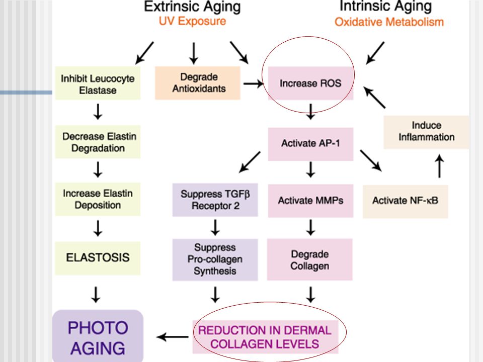
Molecular Consequences of Photoaging
UVR ROS TbRII receptor mRNA TGF ß ROS SMAD 7 mRNA SMAD 7 TGF ROS AP-1(via c-Jun) ROS EGFR Downregulate TGF Upregulate MMPs Decreased collagen, elastin, proteoglycan synthesis Increased matrix metalloproteinase synthesis
 ROS EGFR. Downregulate TGF Upregulate MMPs. Decreased collagen, elastin, proteoglycan synthesis. Increased matrix metalloproteinase synthesis.")
20
Molecular Consequences of Photoaging
UVR ROS ↑c-Jun ↑Activator protein 1 (AP-1) & NF-κB ↑ MMP ( AP-1 (c-Fos & c-Jun) MMPs ( collagenase, gelatinase, Stromelysin-1) Induces TIMP Increases elastin Increases polar gag Decreased collagen, elastin, proteoglycan synthesis
 & NF-κB ↑ MMP. ( AP-1 (c-Fos & c-Jun) MMPs ( collagenase, gelatinase, Stromelysin-1) Induces TIMP. Increases elastin. Increases polar gag. Decreased collagen, elastin, proteoglycan synthesis.")
21
Photoaging Free radical theory of aging & photoaging UVR ROS cellular damage Repair mechanisms: Redox enzymes Antioxidants

22
External Environmental Aging Ultraviolet Radiation
Photo-Redox Reactions UV chromophore → 1O2 or •O2— NADH-/NADPH Tryptophan Riboflavin Trans-urocanic acid 2 •O H+ → H2O2 + O2 H2O2 + Fe++ → OH• + OH-- + Fe+++ The generation of UV-induced reactive oxygen species has been well described. In the first reaction,UV absorbing chromophores present in the dermis, such as the NADH system, amino acids like tryptophan, B vitamins, and transurocanic acid to name a few, absorb energy and transfer it to molecular oxygen, generating a superoxide anion. The electrons are shuttled and the reaction self-propagates until, in the end, a highly reactive hydroxyl radical is generated.

23
External Environmental Aging Ultraviolet Radiation
Photo-Redox Reactions UV chromophore → 1O2 or •O2— NADH-/NADPH Tryptophan Riboflavin Trans-urocanic acid 2 •O H+ → H2O2 + O2 H2O2 + Fe++ → OH• + OH-- + Fe+++

24
External Environmental Aging Ultraviolet Radiation
Photo-Redox Reactions UV chromophore → 1O2 or •O2— NADH-/NADPH Tryptophan Riboflavin Trans-urocanic acid 2 •O H+ → H2O2 + O2 H2O2 + Fe++ → OH• + OH-- + Fe+++

25
External Environmental Aging Ultraviolet Radiation
Photo-Redox Reactions UV chromophore → 1O2 or •O2— NADH-/NADPH Tryptophan Riboflavin Trans-urocanic acid 2 •O H+ → H2O2 + O2 H2O2 + Fe++ → OH• + OH-- + Fe+++

26
External Environmental Aging Ultraviolet Radiation
Photo-Redox Reactions UV chromophore → 1O2 or •O2— NADH-/NADPH Tryptophan Riboflavin Trans-urocanic acid 2 •O H+ → H2O2 + O2 H2O2 + Fe++ → OH• + OH-- + Fe+++ Lipid peroxidation DNA damage

27
External Environmental Aging Ultraviolet Radiation
UVR ROS directly interact with the phosphorylation status of growth factor signal transduction cascades Specific growth factor functions altered ↓ cellular antioxidants Induces apoptosis Alters cross linking of collagen ↑ collagen degradation ↑ DNA damage The generation of the hydroxyl radical is important because it represents the first domino taking the plunge. The preferred path of least resistance for energy transfer alters the phosphorylation state of growth factors that are crucial to dermal matrix homeostasis, namely TGFbeta and EGFR.

28
Repair mechanisms: Redox enzymes -Superoxide dismutase -Catalase
-Glutathione reductase -GSH peroxidase Endogenous antioxidants

29
ANTIOXIDANTS Exogenous Vitamin A derivatives Vitamin C,B,E Co-enzyme Q
Endogenous Glutathione Vitamins C,E,A Ubiquinol Oxidative enzymes: catalase,superoxide dismutase,GSH peroxidases Exogenous Vitamin A derivatives Vitamin C,B,E Co-enzyme Q AHA Alpha lipoic acid Peptides: Cu; NH2

30
Treatment of Photoaged Skin
Reversal of photodamage -Replacement of antioxidants -Absorption of UVR by chromophores - Replacement of growth factors -Stimulation of new dermal matrix Upregulation of new collagen synthesis Inhibition of collagen breakdown

31
Treatment of Photoaged Skin
Replacement of antioxidants Topical formulations Vitamin A derivatives Vitamin C,B,E Co-enzyme Q AHA Alpha lipoic acid Peptides: Cu; NH2 Endogenous Glutathione Vitamins C,E,A Ubiquinol Oxidative enzymes: catalase,superoxide dismutase,GSH peroxidases

32
Treatment of Photoaged Skin
Effective Topical Antioxidants Agent must be in stable formulation Must penetrate/be absorbed in sufficient concentrations Must release reducing equivalents

33
Treatment of Photoaged Skin: Replacement of antioxidants
VITAMIN A DERIVATIVES Vitamin A ( retinoic acid; retinol; retinaldehyde) Tretinoin Adapalene Tazarotene
 Tretinoin. Adapalene. Tazarotene.")
34
Treatment of Photoaged Skin
VITAMIN A DERIVATIVES Antioxidant Antiinflammatory Upregulation of new collagen: Histologic restoration of photodamaged skin Normalization of cellular atypia Normalization of cellular architecture Stimulates new collagen/elastin Stimulates angiogenesis Normalizes gag synthesis Br J Dermatol 2007 Br J Dermatol 2007;157:

35
MOLECULAR MECHANISMS OF PHOTOAGING
Tretinoin effects UVR changes Induces c-Jun ÞMMP Induces TIMP Induces elastin mRNA Increases gag ( -SO4) Inhibits c-Jun induction: no ↑ MMP No effect on TIMP Inhibits elastin mRNA Normalizes gag
 Inhibits c-Jun induction: no ↑ MMP. No effect on TIMP. Inhibits elastin mRNA. Normalizes gag.")
36
TRETINOIN: Net Effects
Thin atrophic epidermis with atypical architecture & cellularity Altered dermis Net Effects: Molecular Inhibition of collagen breakdown New collagen synthesis Increased dermal ECM

37
TRETINOIN: Net effects
Net Effects: Clinical Smooth skin Less pigmentary alterations Less wrinkling/laxity of skin

38
Botanicals: Soy isoflavones Pycnogenol Green tea extracts Vitamin C
TREATMENT OF PHOTOAGED SKIN: Replacement of antioxidants ANTIOXIDANTS Botanicals: Soy isoflavones Pycnogenol Green tea extracts Vitamin C Vitamin E CoQ-10 Alpha lipoic acid

39
Antioxidants VITAMIN C Cofactor for collagen synthesis
VITAMIN E Naturally occurring Inhibits UVR induced lipid peroxidation Replenishes Vitamin C, GSH VITAMIN C Cofactor for collagen synthesis Reduces UVR induced changes Photoprotection Some formulations unstable Dermatologic Therapy 2007;20:

40
Treatment of Photoaged skin
Antioxidants Alpha Lipoic Acid Involved in oxidative metabolism ROS scavenger ( inhibits lipid peroxidation) ß NF-B Chelates metals Regenerates vitamins E & C Br J Dermatol 2003;149: 841-9
 ß NF-B. Chelates metals. Regenerates vitamins E & C. Br J Dermatol 2003;149:")
41
BOTANICALS Green tea extracts: (polyphenols) Isoflavones: Antioxidant
Anti-inflammatory; inhibits UVR damage Allantoin: Antiinflammatory promotes repair Isoflavones: Antioxidant Increases skin thickness May inhibit melanosome phago cytosis Pycnogenol: Reduces Vit. C Dermatologic Therapy 2007;20: Dermatplogic Therapy 2007;20: PhotodermatolPhotoimmunol Photomed 2007;23:

42
Replacement of Growth Factors/Stimulate cytokines
Treatment of Photoaged skin Replacement of Growth Factors/Stimulate cytokines Transforming growth factor Epidermal growth factor (receptor) Keratinocyte growth factor Fibroblast growth factor Platelet-derived growth factor Vascular endothelial growth factor Dermatologic Therapy 2007;20: Dermatologic Surgery 2006;32:
 Keratinocyte growth factor. Fibroblast growth factor. Platelet-derived growth factor. Vascular endothelial growth factor. Dermatologic Therapy 2007;20: Dermatologic Surgery 2006;32:")
43
Topical TGF- Products
Well tolerated Minimizes appearance of rhytides when compared to Vitamin C Few studies at molecular level of reversal of photodamage TNS Recovery Complex Bio-restoratiive Skin Cream Cell rejuvenation serum ( CRS) Dermatolog Surg 2006;32: Dermotologic Therapy 2007;20:
 Dermatolog Surg 2006;32: Dermotologic Therapy 2007;20:")
44
CLINICAL STUDIES WITHMULTIPLE GROWTH FACTOR COSMECEUTICALS
Study Design: 14 Female subjects 2 Months twice daily Facial wrinkle scoring (9-point score) Silicon replica Histology J Cosmetic & Laser Ther 2003, 5: 25-34 TNS Recovery Complex® (SkinMedica) 44
 Silicon replica. Histology. J Cosmetic & Laser Ther 2003, 5: TNS Recovery Complex® (SkinMedica) 44.")
45
CLINICAL STUDIES WITH MULTIPLE GROWTH FACTOR COSMECEUTICALS
Study Design: 18 Female subjects 2 Months twice daily, followed by 2 week wash-out Facial wrinkle scoring (5-point score) J Drugs Dermatol 2007, 6: BIO-RESTORATIVE SKIN CREAM WITH PSP® (NEOCUTIS) 45
 J Drugs Dermatol 2007, 6: BIO-RESTORATIVE SKIN CREAM WITH PSP® (NEOCUTIS) 45.")
46
Proposed Mechanism of Action of Growth Factor Cosmeceuticals
Large hydrophilic molecules unlikely to penetrate skin barrier but may penetrate through follicles, sweat glands and damaged skin Produce epidermal signaling cytokines which affect dermal fibroblasts Mechanism is not elucidated

47
CONCLUSIONS Human growth factor cosmeceuticals are effective for skin rejuvenation Multiple growth factor cosmeceuticals are particularly beneficial as demonstrated in several clinical studies Growth factor cosmeceuticals may be also helpful to improve outcome after cosmetic and dermatologic procedures Additional studies warranted to further elucidate promising technology: double blinded placebo controlled studies -both clinical and at molecular level 47

48
Topical treatment of Photoaged Skin
Sunscreens as protection against ongoing damage and combination of Tretinoin Alpha hydroxy acids Vitamin C Vitamin E Botanical antioxidants Growth factor cosmeceuticals

49
QUESTIONS TO BE ASKED OF COSMECEUTICALS
Can agent penetrate? Does agent have MOA in target tissue? Is there peer-reviewed data to support claim?

50
PERSPECTIVES FOR PHOTOAGING
Provide patient with sound reasonable advise about cosmeceuticals Prospective objective studies are needed

51
CONCLUSIONS Popularity of noninvasive treatments of photoaging ( cosmeceuticals) useful when care is taken to construct a treatment regimen based on understanding of the molecular mechanisms in place Patient needs to have realistic expectations and know limitations of products
 useful when care is taken to construct a treatment regimen based on understanding of the molecular mechanisms in place. Patient needs to have realistic expectations and know limitations of products.")
52
CONCLUSIONS Assess the patient relative to what bothers patient
Recognize that each of these therapies offers mild to modest effects but may prove more efficacious when used in combination

53
Pentapeptide( KTTKS) Lys-Thr-Thr-Lys-Ser ↑collagen synthesis
Copper Peptides Wound healing ↑collagen synthesis

54
Environemental Affects Upon Cutaneous Aging
Internal Environmental Factor Estrogen status Chronologic Aging Oxidative metabolism Chronologic aging, a bit of a redundant term, is the umbrella agent, the background upon which all additional contributions to aging occur. Chronologic aging describes the inherent unique genetic program carried by mammalian cells. There is an expected lifetime output of each cell that is tied to the pre-programmed cellular repair capacity. How cells handle free radical formation during oxidative metabolism provides a good example. These free-radical byproducts, which are a necessary consequence of generating energy for life, are, although not perfectly, in large part quenched by antioxidant enzymatic processes built-in to the cell. I will focus most of the discussion on a few factors encountered over a lifetime that accelerate the aging process by either exhausting cell-repair capacity, as we’ll see with external environmental factors, namely UV radiation and tobacco smoke; or by directly altering cell signaling capacity, which occurs with the internal environmental alteration that is secondary to the menopausal state, estrogen deprivation. My goal is to make clear that each of these independent environmental factors, whether external or internal, overlap pathophysiologically as they converge upon this. A final common pathway involved in maintaining dermal homeostasis. External Environmental Factors UVR Tobacco

Similar presentations


. Major Functions of Skin Barrier (excludes infectious agents & some chemicals; retains moisture, prevents dessication)>")


 and Activator Protein – 1 (AP-1) Brooke T. Mossman*>")
