Download presentation
Presentation is loading. Please wait.

1
ORDER: RODENTIA “to gnaw”
RAT DISSECTION ORDER: RODENTIA “to gnaw”

3
Day One Investigation External anatomy
Opening thoracic and abdominal cavities

4
Day Two Investigation Abdominal Cavity Thoracic Cavity
Digestive System Urogenital System (Excretory & Reproductive) Thoracic Cavity Circulatory System Respiratory System
 Thoracic Cavity. Circulatory System. Respiratory System.")
5
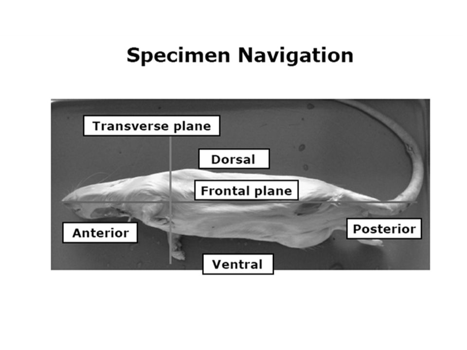
Observe External Anatomy
List 3 ways the rat is DIFFERENT from humans. List 3 ways the rat is SIMILAR to humans 1. 2. 3.

8
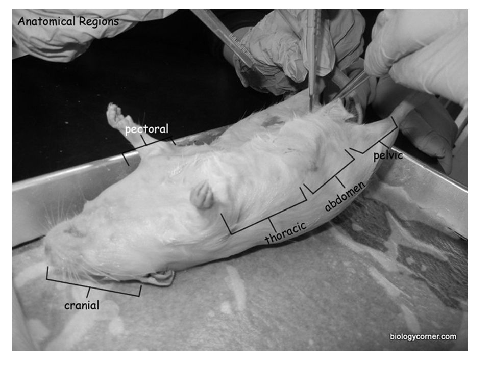
Part I. External Anatomy
Pre-Dissection Questions: Where do rats live? Would you typically see a rat during the day or night? What do rats eat? Do rats give birth to live young? What scientific term means to give birth to live young?

9
External Anatomy - Teeth
All mammals have teeth that correspond to the type of food they eat. The name RODENTIA comes from the Latin word meaning “to gnaw”. Rodents such as the rat have 2 pairs of CONTINUOUSLY GROWING INCISORS (front teeth) which are kept at the correct length by constant chewing.
 which are kept at the correct length by constant chewing.")
10
The mouth of the rat has a large cleft in the upper lip which exposes the large front INCISORS – locate them. How long are they? Are they easily seen? Describe them compared to the other teeth.

11
External Anatomy – Cranial Region
Notice the rat has a hairy coat that covers the entire body and sensory whiskers located on the rat’s face. VIBRISSAE (whiskers) enable rats to feel their way through tight spaces in the dark. Describe these whiskers:
 enable rats to feel their way through tight spaces in the dark. Describe these whiskers:")
12
External Anatomy – Cranial Region
Locate the EYES with the large pupil and the nictitating membrane found at the inside corner of the eye. This membrane can be drawn across the eye for protections similar to eyelids found in humans. What color eyes does your rat have?

13
External Anatomy – Cranial Region
Locate the external EARS in rats are surrounded by a PINNA, which is a flap of cartilage covered by skin that can rotate to direction of sounds heard by the ears. Locate the external NARES used for respiration and odor detection.

14
External Anatomy – Abdominal Region
Locate the TEATS (nipples) on the ventral (stomach) side of the rat. Regardless if your rat is male or female – does your rat have teats? ___________ Remember a mammal, male or female will have mammary glands, nipples.
 on the ventral (stomach) side of the rat. Regardless if your rat is male or female – does your rat have teats ___________. Remember a mammal, male or female will have mammary glands, nipples.")
15
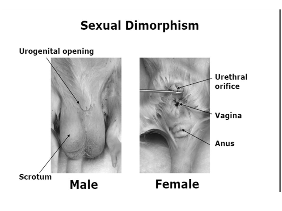
External Anatomy – Pelvic Region
Locate the exit openings for the digestive, excretory, and reproductive body systems. Like humans, male rats have 2 exit openings; females have 3 exit openings. In addition, females have a separate opening for reproduction and excretory waste (urine). Males have a SHARED urogenital opening that releases both sperm and urine.
. Males have a SHARED urogenital opening that releases both sperm and urine.")
17
What Sex Is It? Because mammals are endothermic animals creating lots of internal heat, and the ideal temperature necessary for sperm production is a few degrees cooler than body temperature, male mammals, must have their TESTES suspended outside their bodies in a sac (SCROTUM) to keep them cooler. Testes outside the body in a scrotum are called TESTICLES. Is your rat male or female? _____________
 to keep them cooler. Testes outside the body in a scrotum are called TESTICLES. Is your rat male or female _____________.")
18
External Anatomy - Tail
Examine the TAIL of your rat. What are some observations about the tail of your rat? Rats do not have hair on their tails unlike many other rodents that do like, gerbils, squirrels, and chipmunks.

19
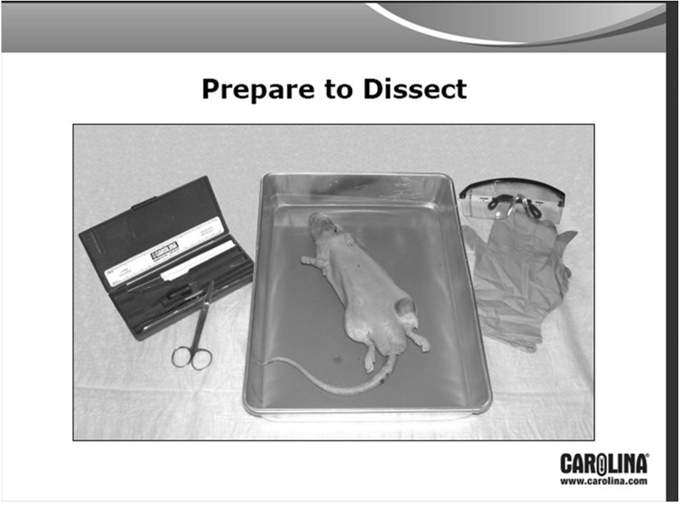
Part II: Dissection Preparation Exposing the Abdominal & Thoracic Cavities

20
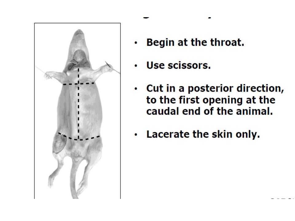
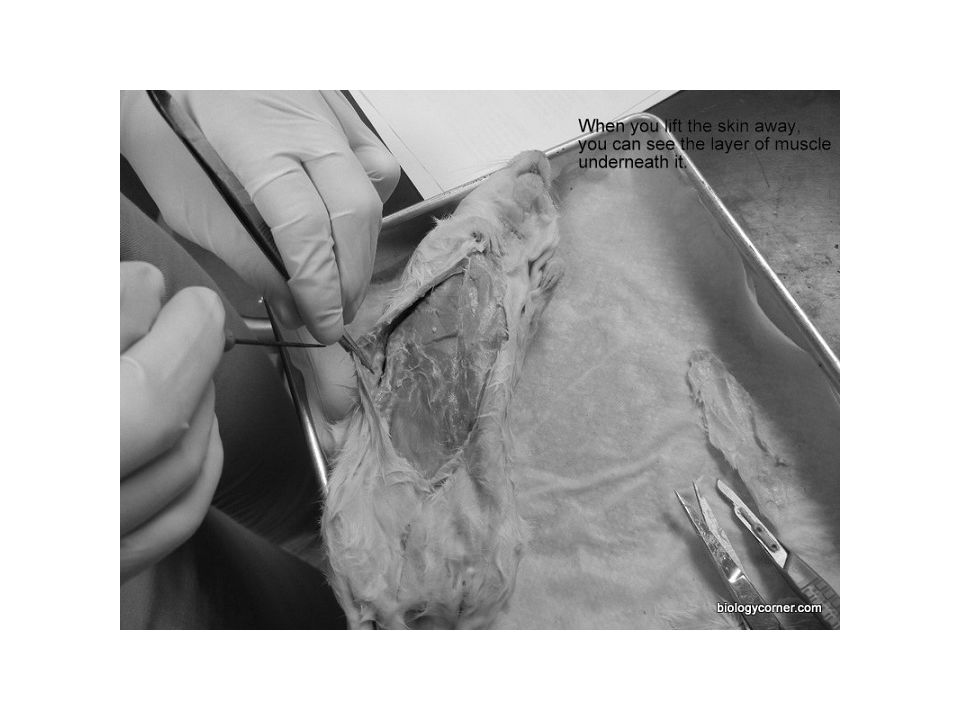
STEP ONE – SKIN LAYER You will need to separate the skin from muscles for your first cuts similar to the fish and amphibian dissections. This time – START AT THE NECK AREA. There should be an incision hole that you can use to begin. You will need to use your metal blunt ended probe to separate the connective tissue of the skin from the first layer of muscle. When you have gotten a section separated you can begin to cut carefully down towards the anus. YOU MUST CONTINUE USING THE PROBE TO TEASE AWAY THE SKIN FROM THE MUSCLE AS YOU CUT TO PREVENT CUTTING THE MUSCLE TISSUE! GO SLOWLY! DO NOT CUT THE MUSCLES!

24
STEP TWO – CUTTING THROUGH MUSCLES
Mammals are known as EUCOELOMATES. There is a space you see surrounding the internal organs that is called the “true coelom” – the body cavity is lined with a membrane. Mammals have a DIAPHRAGM. This is a sheet of muscle that separates the body cavity into two compartments: Thoracic Cavity – heart and lungs Abdominal Cavity – digestive, excretory, and reproductive organs For this reason the rat must be dissected in TWO STAGES!

25
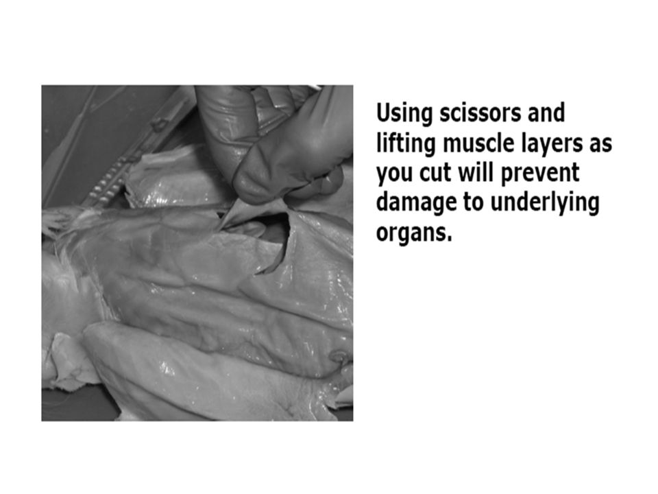
DIGESTIVE SYSTEM DISSECTION
Use your scissors to cut through the abdominal wall of the rat following the incision marks in the picture provided. Be careful not to cut too deeply and keep the tip of your scissors always points upwards. Start at the lower abdomen and cut up towards the diaphragm. To locate the diaphragm feels where the ribs are located and it will be just next to them. Then make two incisions towards the back of the rat like you see on your picture.

26
Abdomen cuts ONLY Be careful! Stop when get to the diaphragm!

28
This is what your cuts should look like – notice that the chest area is NOT CUT!

29
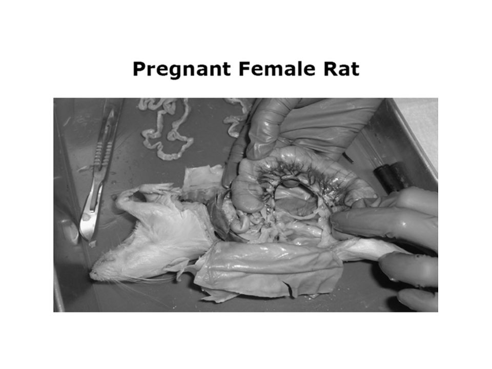
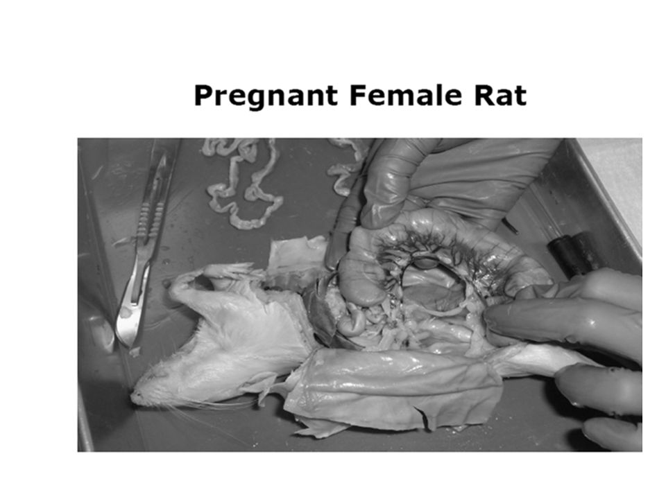
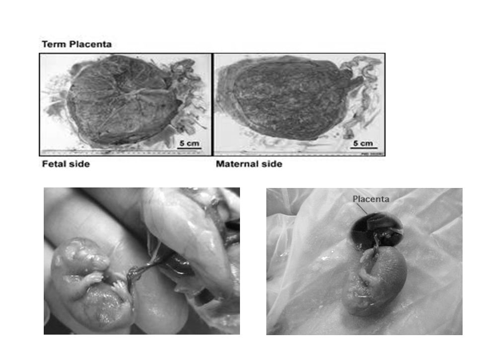
**PREGNANT FEMALES** Some of you may have the pregnant females to dissect. Please take care with them as your classmates will need to view these. Your TEACHER will carefully help you remove the babies and set them aside for the class to observe. Please do not attempt this without assistance!!

31
DIGESTIVE SYSTEM

32
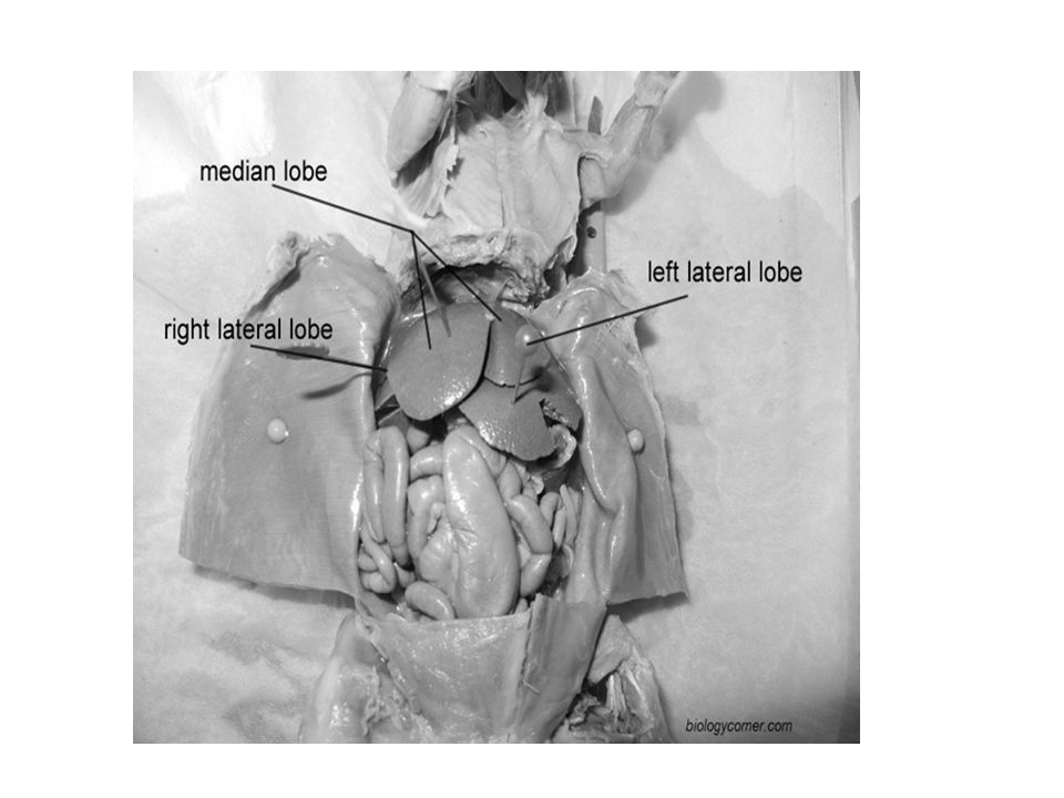
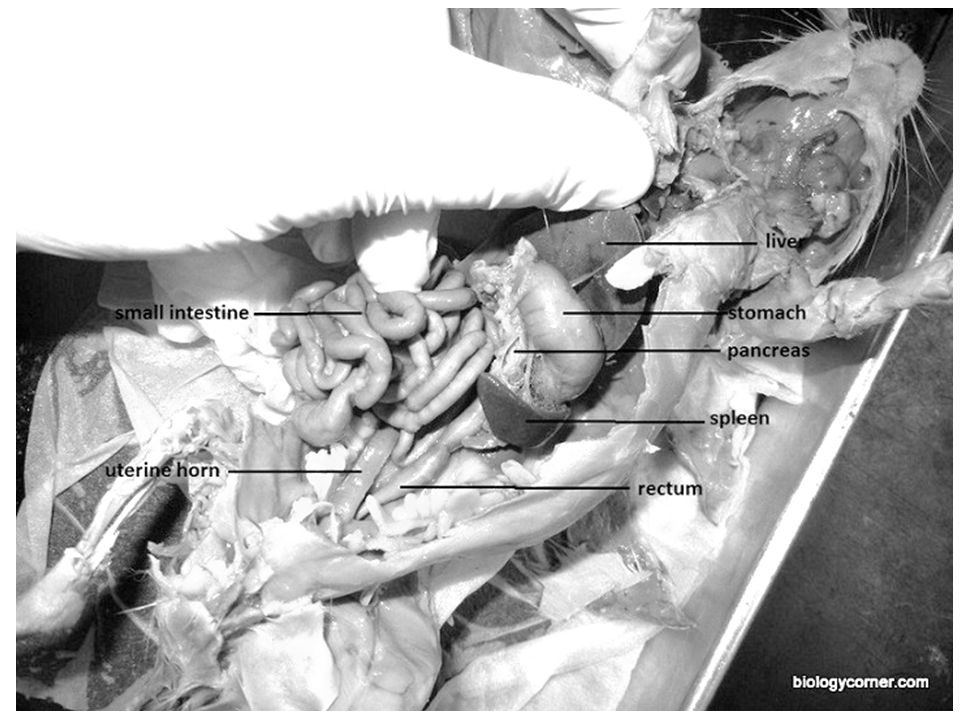
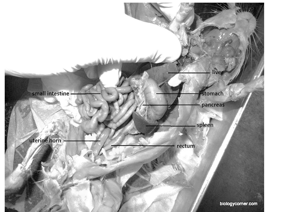
LIVER Locate the LIVER, which is a large, dark colored organ just under the diaphragm. (you can reference the photo on next slide). There are 3 lobes of the liver. The liver has many functions like digesting fats from the bile produced. It also stores glycogen for energy and transforms toxins. Rats do NOT have a GALLBLADDER because they do not eat anything that would contain fats that would require bile stored in the gallbadder.

34
STOMACH Locate the STOMACH.
It is a curved organ usually located on the right side (your right), just under the liver. Look at the top of the stomach and you should be able to see the ESOPHAGUS. You should be able to see how the esophagus pierces through the diaphragm muscle from thoracic cavity and joins the stomach. Where the esophagus and stomach meet is called the CARDIAC SPHINCTER. This controls the food going into the stomach and prevents digesting food from reentering back into the esophagus even if hanging upside down.
, just under the liver. Look at the top of the stomach and you should be able to see the ESOPHAGUS. You should be able to see how the esophagus pierces through the diaphragm muscle from thoracic cavity and joins the stomach. Where the esophagus and stomach meet is called the CARDIAC SPHINCTER. This controls the food going into the stomach and prevents digesting food from reentering back into the esophagus even if hanging upside down.")
35
Stomach

37
PANCREAS Next locate the PANCREAS. To find this you will need to lift up the stomach. It will look like a thin membrane or a bumpy cottage cheese like membrane (it won’t look like the nice leaf structure we have seen previously). It will be found towards the bottom of the stomach close to the beginning of the small intestines. It is always found in this location since it releases enzymes into the small intestines to further digest food. You can reference the photo on previous slide for assistance.
. It will be found towards the bottom of the stomach close to the beginning of the small intestines. It is always found in this location since it releases enzymes into the small intestines to further digest food. You can reference the photo on previous slide for assistance.")
38
SPLEEN Locate the SPLEEN. It will be about the same color of the liver and you will find it attached to the greater curvature (the outer margin of the curve) of the stomach. It is shaped like a banana and is associated with our circulatory system. You may again reference photo previously shown.
 of the stomach. It is shaped like a banana and is associated with our circulatory system. You may again reference photo previously shown.")
39
SMALL INTESTINES Now locate the SMALL INTESTINES.
It is a slender coiled tube that begins at the bottom of the stomach at the Pyloric Sphincter (a pinching will indicate it’s location). The term “small” refers to its diameter tube NOT its length. It consists of 3 sections: duodenum (mostly straight), jejunum and ileum (both curly).
. The term small refers to its diameter tube NOT its length. It consists of 3 sections: duodenum (mostly straight), jejunum and ileum (both curly).")
41
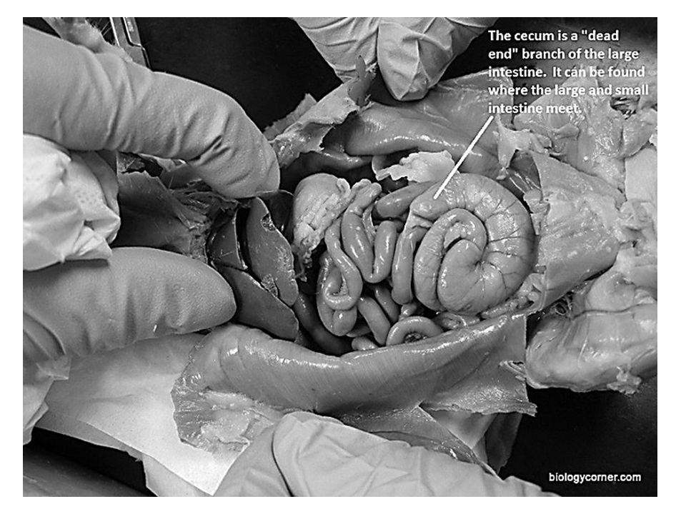
CECUM Following the Small Intestines, locate the CECUM.
This is a pouch that connects the small and large intestines together. Food is temporarily stored in the cecum so helpful bacteria can digest the cellulose found in the cell walls of plants. Since rats are considered a type of herbivore, what type of digestive system do they have? (you may need to look back in your notes, hint - focus on what digestive system has the enlarged cecum) ________________________________
 ________________________________.")
43
LARGE INTESTINES After locating the cecum, locate the LARGE INTESTINES (also known as the COLON). It is actually a large greenish tube that extends from the small intestines and leads to the anus. This is where the final stages of digestion and water absorption occur. It has 4 sections: Ascending colon (food moves up) Transverse colon (short section parallel to diaphragm) Descending colon (food moves back down) Rectum (short terminal section that leads to anus – this temporarily stores the feces before its expelled)
 Transverse colon (short section parallel to diaphragm) Descending colon (food moves back down) Rectum (short terminal section that leads to anus – this temporarily stores the feces before its expelled)")
44
CHECKPOINT – before moving on make sure to confirm identification of digestive organs with teacher! You will come back to this cavity at the end to identify two other systems.

45
THORACIC CAVITY DISSECTION

46
DISSECTING THE THORACIC CAVITY
Now use your scissors to cut up into the thoracic cavity. You will have to cut just above the diaphragm. Sometimes cutting from the left or right sides towards center helps. Be careful not to cut too deeply and keep the tip of your scissors always points upwards. Make incisions like you see in diagram. Cut up the neck area. You want to be able to see all the way up to the trachea.

47
DIAPHRAGM Locate the DIAPHRAGM. Notice how the muscle separates the two cavities. As discussed in class, why would this muscle separating the heart and lungs from the abdominal area be such a good adaption in mammals?

48
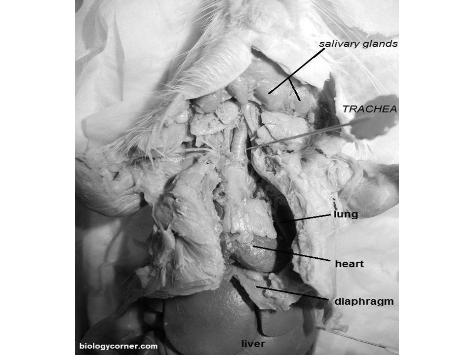
HEART Locate the HEART. It is centrally located in this cavity.
The THYMUS GLAND may be visible at the upper part of the heart. See photo on next slide for reference (use can use this for lungs as well).
.")
50
LUNGS Locate the LUNGS. They are spongy organs that lie on the left and right side of the heart and should take up most of the thoracic cavity. They lie closer to the back of the rat, you will need to push the ribs to the side to find them. Now locate the TRACHEA and see how they branch into the lungs. You may use the photo from the previous slide.

51
CHECKPOINT – before moving on make sure to confirm identification of circulatory & respiratory organs with teacher!

52
EXCRETORY SYSTEM

53
They many share the same “exit parts”, but have different functions.
The excretory and reproductive systems of mammals work closely together. They many share the same “exit parts”, but have different functions. Excretory system removes wastes Reproductive system produces gametes (sperm or eggs)
")
54
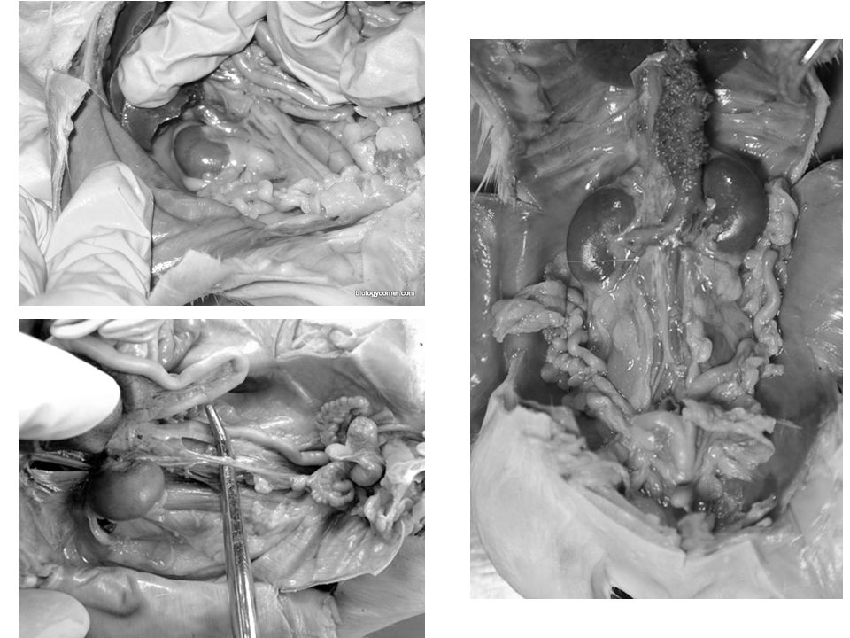
KIDNEYS The primary organ of the excretory system are the kidneys.
They are large bean shaped structures located toward the back of the abdominal cavity under the intestines and found on both sides of the spine. (lift up the intestines and lay them over to one side to view them) Locate the KIDNEYS. Note all the veins and arteries that connect the kidneys. Locate the URETERS attaching to the kidneys and trace them to the URINARY BLADDER.
 Locate the KIDNEYS. Note all the veins and arteries that connect the kidneys. Locate the URETERS attaching to the kidneys and trace them to the URINARY BLADDER.")
56
Remove ONE kidney and cut it lengthwise.
What does the kidney look like inside? The small yellowish glands in the fat around the kidneys are the ADRENAL GLANDS which secrete adrenaline into the blood during times of crisis (fight or flight).
.")
57
CHECKPOINT – before moving on make sure to confirm identification of excretory organs with teacher!

58
REPRODUCTIVE SYSTEM

59
MALE REPRODUCTION If you do not have male, make sure to view one!!
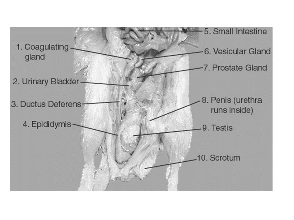
If you do have a male, the major reproductive organ is visible on the exterior – the TESTES are located in the SCROTAL SAC. Carefully cut through the scrotal sac, and locate the TESTICLES.

61
FEMALE REPRODUCTION Since the females are pregnant, identification of the female organs will be difficult since we will be removing the uterus that contain the babies. However, if you follow a short gray tube extending to the URINARY BLADDER from the exterior, this is the VAGINA. Following this up, the vagina divides into two UTERINE HORNS. This duplex uterus is found in mammals that can accommodate multiple embryos (babies - litters). In a simple uterus, like found in humans, there is only a single chamber allowing for development of a single embryo (although sometimes more in twins, etc).
. In a simple uterus, like found in humans, there is only a single chamber allowing for development of a single embryo (although sometimes more in twins, etc).")
64
Describe the little rat babies – describe all the detailed features you can see:
How many were found in the pregnant females?

66
CHECKPOINT – before moving on make sure to confirm identification the reproductive system with teacher! **MAKE SURE TO LOOK AT MALE & FEMALE RATS – AND MAKE SURE TO LOOK AT BABIES AND PLACENTAS!

Similar presentations


>")


