Download presentation
Presentation is loading. Please wait.

1
Gallbladder Disease in Infants and Children
George W. Holcomb III, MD, MBA Children’s Mercy Hospital Kansas City, Missouri

4
Biliary Disease Gallstones Biliary dyskinesia Acalculous disease
Hemolytic disease Non-hemolytic disease Biliary dyskinesia Acalculous disease

5
Risk Factors for Cholelithiasis in Infants and Children
Hemolytic Sickle cell disease Spherocytosis Thalassemia Nonhemolytic Total parenteral nutrition Gallbladder stasis Lack of enteral feeding Ileal resection (necrotizing enterocolitis and Crohn’s disease) Biliary tract anomalies Adolescent pregnancy Oral contraceptives
 Biliary tract anomalies. Adolescent pregnancy. Oral contraceptives.")
6
Biliary Dyskinesia Symptomatic biliary colic w/o stones
Reduced GBEF with CCK stimulation IU study – 37 pts – 71% resolution of symptoms GBEF < 15% successful resolution of symptoms (O.R. – 8.00) Chronic cholecystitis seen in histological examination of many specimens
 Chronic cholecystitis seen in histological examination of many specimens.")
7
Pilot Study

8
Pilot Study

9
Complicated Cholelithiasis
Acute cholecystitis Jaundice Pancreatitis

10
Timing of Cholecystectomy
Non-complicated – 2 weeks Complicated Jaundice – following work-up Cholecystitis – 2-4 days Pancreatitis – once resolved

11
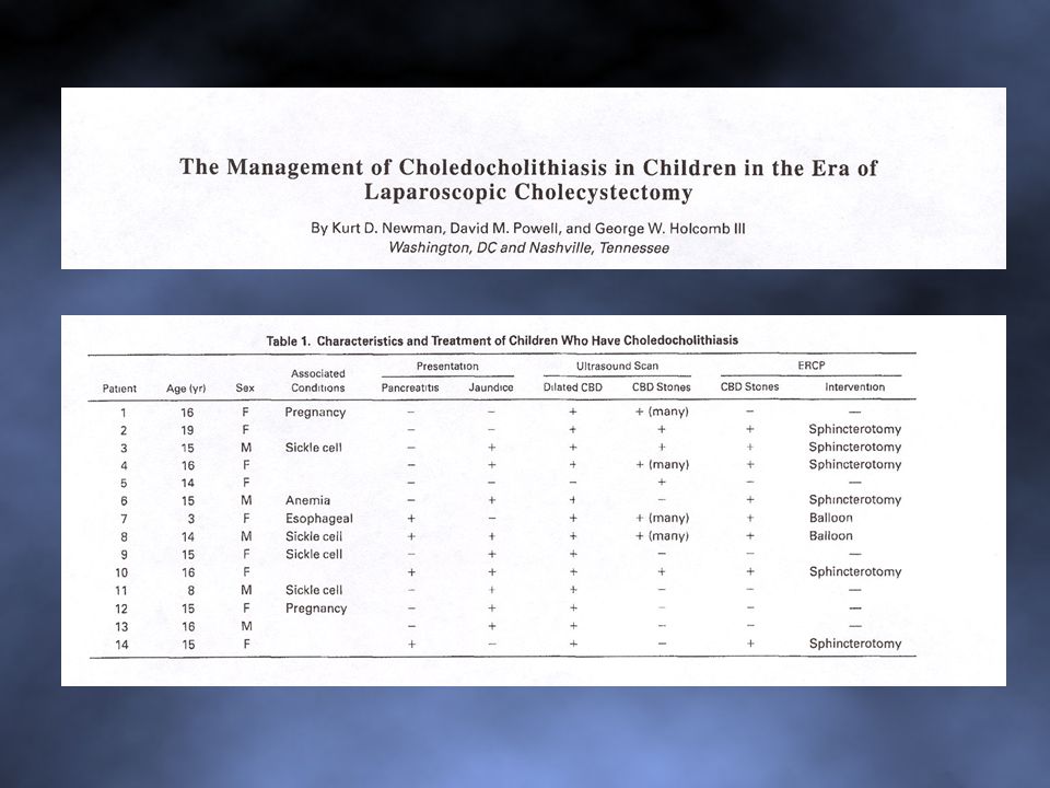
When to Suspect Choledocholithiasis?
Elevated bilirubin (jaundice) Elevated lipase, amylase (pancreatitis) Dilated CBD or stone(s) in CBD on ultrasound
 Elevated lipase, amylase (pancreatitis) Dilated CBD or stone(s) in CBD on ultrasound.")
12
SUSPECTED CHOLEDOCHOLITHIASIS (Pre-operatively)
Management Options

13
Management Options Pre-op ERCP, sphincterotomy, stone extraction
Laparoscopic or open CBD exploration at time of cholecystectomy Post-op ERCP, sphincterotomy, stone extraction

14
Factors Surgeon’s experience with laparoscopic CBD exploration
Availability of an endoscopist to perform ERCP in children

16
Algorithm Suspected Choledocholithiasis

17
Why? Surgeon knows at time of laparoscopic cholecystectomy whether CBD (laparoscopic or open) exploration needed Potentially avoids a third anesthesia and operation

18
Disadvantage A number of ERCPs will be performed in patients that do not have CBD stones

19
IS ROUTINE CHOLANGIOGRAPHY NEEDED?

20
Cholangiography : Reasonable to perform cholangiography to become facile with technique 2006: Most surgeons have become facile with this technique

21
Cholangiography To evaluate for CBD stones To define anatomy

22
One Surgeon’s Approach
Reserve cholangiography for cases where anatomy is unclear Use ultrasound pre-operatively to define CBD involvement

23
Pre-operative Ultrasound
Prior to laparoscopic cholecystectomy Confirm gallbladder stones, evaluate for CBD dilation or stones Cost-effective strategy

24
Immediate Pre-op Evaluation with US Intraoperative Cholangiography
Financial analysis of preoperative ultrasonography versus intraoperative cholangiography for detection of choledocholithiasis at Children's’ Mercy Hospital, Kansas City MO Immediate Pre-op Evaluation with US Charges ($) Intraoperative Cholangiography Ultrasound study (including radiologist fee) 307.67 15-minutes OR time C-Arm with radiologist fee 365.41 Sterile drape for C-Arm 20.00 Cholangiocatheter 83.50 Contrast for cholangiogram 40.00 TOTAL $307.67 $
 Intraoperative Cholangiography. Ultrasound study (including radiologist fee) minutes OR time C-Arm with radiologist fee Sterile drape for C-Arm Cholangiocatheter Contrast for cholangiogram TOTAL. $ $")
25
Cholangiography Cystic Duct Cannulation Kumar Clamp Technique

26
Kumar Clamp Technique Surg Endosc 8: , 1994

27
Where do I place the instruments/ports?

28
Port Placement

29
Stab Incision Technique
2 cannulas 2 stab incisions J Pediatr Surg 38: , 2003

30
The Use of Stab Incisions
PAPS 2003 JPS 38: , 2003

31
Cost Savings from Stab Incisions
PAPS 2003 JPS 38: , 2003

32
Key Steps in Operation Begin dissection high on gallbladder to expose triangle of Calot

33
Create 90 b/w cystic duct and CBD
Key Steps in Operation Create 90 b/w cystic duct and CBD

34
What Do I Do If I Cut the Common Bile Duct?

35
Options Ligate duct Repair laparoscopically Repair open
wait for it to enlarge transfer to experienced biliary surgeon Repair laparoscopically Repair open interrupted sutures T – tube choledochojejunostomy at second operation

36
CMH Experience 2000 - 2006 224 Pts (65% female) Indication
(12.9 yrs, 58.3 kg) Indication Symptomatic gallstones 166 Biliary dyskinesia 35 Gallstone pancreatitis 7 Gallstones/splenectomy 6 Calculous cholecystitis 5 Other 4 IPEG, 2007
 Indication. Symptomatic gallstones 166. Biliary dyskinesia 35. Gallstone pancreatitis 7. Gallstones/splenectomy 6. Calculous cholecystitis 5. Other 4. IPEG,")
37
CMH Experience 2000-2006 Mean operative time 77 min Cholangiogram –
Preoperatively (ERCP) 17 Stones 8 Intraoperatively 38 Stones 9 Cleared intraop 5 Cleared postop 4 Postoperatively (ERCP) 2 Stones 0 Ductal injuries 0 IPEG, 2007
 17. Stones 8. Intraoperatively 38. Stones 9. Cleared intraop 5. Cleared postop 4. Postoperatively (ERCP) 2. Stones 0. Ductal injuries 0. IPEG,")
38
Laparoscopy for Splenic Conditions
George W. Holcomb, III, M.D., MBA Children’s Mercy Hospital Kansas City, MO

39
Splenic Conditions ITP Spherocytosis Splenic cysts Wandering spleen
J Pediatr Surg 28: , 1993

40
Pre-Operative Preparation
Ultrasound Often done by pediatrician, hematologist Rarely needed for splenectomy, except may be useful for extremely large spleen CT Scan – Useful in planning splenic cystectomy WinRho Bone marrow stimulant Usually used to platelet count Useful pre-operatively to platelet count in ITP pt. Immunizations –Pneumococcus (Prevnar, Pneumovax)
")
41
Patient Positioning

42
Patient Positioning

43
Personnel Positions

44
Laparoscopic Splenectomy
ITP, spherocytosis Port placement (2) cannulas (5, 12) (2) stab (3 mm) incisions Instruments Harmonic scalpel (5 mm) Articulating stapler (12 mm)
 cannulas (5, 12) (2) stab (3 mm) incisions. Instruments. Harmonic scalpel (5 mm) Articulating stapler (12 mm)")
45
Laparoscopic Splenectomy
Operative Steps Divide spleno-colic ligament, then short gastrics Clip artery Autotransfuse pt Protects stapler malfxn

46
Laparoscopic Splenectomy
Operative Steps Divide spleno-renal lig. Articulating stapler across hilum Bag specimen, morcellate extracorporally

47
Laparoscopic Splenectomy

48
Issues How large is too large?
28 cm. – Splenic artery ligation helpful Can divide spleen (spherocytosis) with harmonic, if necessary
 with harmonic, if necessary.")
49
Issues Postoperative platelet ct. > 500,000
Reports of splenic vein/portal vein thrombosis following splenectomy (open and laparoscopic) Baby aspirin ( 81 mg) QD for 6 mos Re-check at 3 months & 6 months
 Baby aspirin ( 81 mg) QD for 6 mos. Re-check at 3 months & 6 months.")
50
Splenic Cysts Primary Pseudocysts (secondary) epithelial lining
no epithelial lining often develop after trauma

51
Laparoscopic Splenic Cystectomy
First step is decompression of cyst

52
Laparoscopic Splenic Cystectomy
Excise cyst as close as possible to splenic parenchyma with harmonic scalpel Coagulate lining with Argon beam coagulator ? Place omentum adjacent to exposed cyst lining

53
European Experience 3 European centers (Mainz, Mannheim, Hannover)
14 pts (median 8.5 yr) 10 recurrences (71%) APSA 2006
 10 recurrences (71%) APSA")
54
Wandering Spleen

55
Wandering Spleen

56
Laparoscopic Splenopexy
J Pediatr Surg 42:E23-27, 2007

57
I.U. Experience 1995 - 2006 231 patients Mean age 7.7 yrs
Lap splenectomy – 223 211 - total 12 - partial Lap splenic cystectomy – 6 Lap splenopexy - 2 Ann Surg, in Press

58
I.U. Experience 1995 – 2006 Complications
Ileus - 5 Bleeding - 4 Acute chest syndrome- 5 Pneumonia - 2 Portal vein thrombosis - 1 HUS - 1 Diaphragm perforation 2 Colon injury - 1 Port site hernia - 1 Total splenectomy after partial - 1 Recurrent cyst - 1 11% overall, 22% in SCD Ann Surg, in Press

Similar presentations