Download presentation
Presentation is loading. Please wait.

1
Cell block: Monokeratin – rare dot Tissue biopsy: Monokeratin – positive On Slide Control

3
Pre-Analytical (surgery → unstained section) Ischemic time Type of fixation Length of fixation Decalcification Processing Dehydration and clearing Paraffin impregnation Paraffin sectioning Storage Analytical (unstained section → coverslip) Antibody supplier Clone Epitope retrieval Dilution Amplification Chromogen Counterstain Detection Blocker Mounting Post-Analytical (interpretation) Design of controls Positive controls (internal and external) Negative controls Interpretation Critical indicators Reporting Changing the type of fixation results in downstream problems in the analytical and post-analytical phases !
 Ischemic time Type of fixation Length of fixation Decalcification Processing Dehydration and clearing Paraffin impregnation Paraffin sectioning Storage Analytical (unstained section → coverslip) Antibody supplier Clone Epitope retrieval Dilution Amplification Chromogen Counterstain Detection Blocker Mounting Post-Analytical (interpretation) Design of controls Positive controls (internal and external) Negative controls Interpretation Critical indicators Reporting Changing the type of fixation results in downstream problems in the analytical and post-analytical phases !")
5
This implies that a laboratory must have supply of control tissue developed for each of these situations with IHC protocols developed for each antibody on these tissues Let’s be honest – who actually does this? Am J Clin Pathol 2010;133:354-365

6
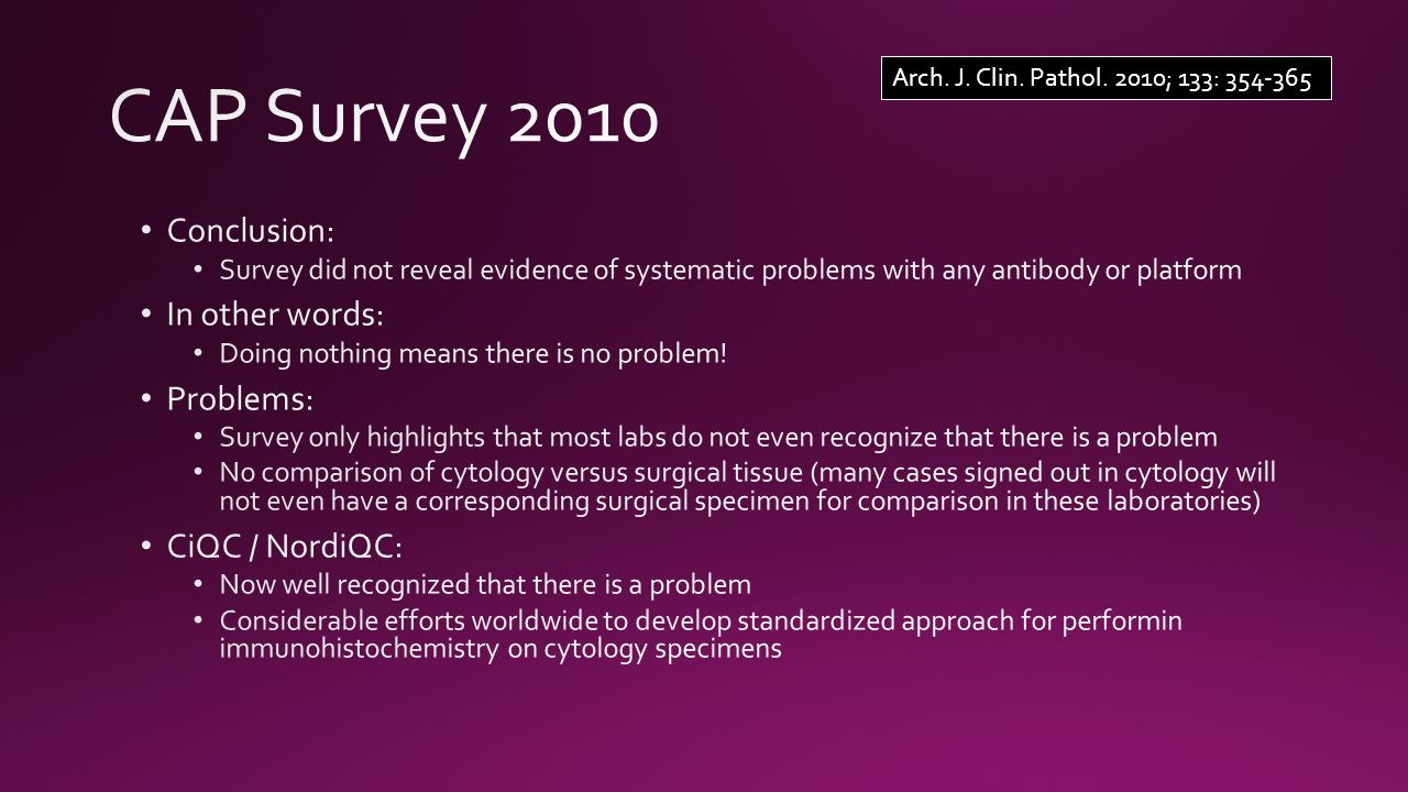
Immunohistochemical Practices of Cytopathology Laboratories: A survey of Participants in the College of American Pathologists Nongynecologic Cytopathology Education Program. Arch. J. Clin. Pathol. 2010; 133: 354-365

7
Arch. J. Clin. Pathol. 2010; 133: 354-365

9
TTF1 FFPE CytoLyt® fixation and decalcification pretreatments alter antigenicity in normal tissues compared to standard formalin fixation. Jennette R. Gruchy, Penny J. Barnes, Kelly A. Dakin Hache. Applied Immunohistochemistry and Molecular Morphology, 2014, in press. TTF1 CytoLyte™ TTF1 Expression in Normal Lung Tissue

10
CytoLyt® fixation and decalcification pretreatments alter antigenicity in normal tissues compared to standard formalin fixation. Jennette R. Gruchy, Penny J. Barnes, Kelly A. Dakin Hache. Applied Immunohistochemistry and Molecular Morphology, 2014, in press. CD20 Expression in Normal Appendix CD20 FFPECD20 CytoLyte™

11
D2-40 Expression in Normal Appendix CytoLyt® fixation and decalcification pretreatments alter antigenicity in normal tissues compared to standard formalin fixation. Jennette R. Gruchy, Penny J. Barnes, Kelly A. Dakin Hache. Applied Immunohistochemistry and Molecular Morphology, 2014, in press. D2-40 FFPED2-40 CytoLyte™

13
Monokeratin control appropriate on both cytology cell block slide and tissue biopsy slide Cell block: Monokeratin – rare dot Tissue biopsy: Monokeratin – positive

14
TTF-1 control appropriate on both cytology cell block slide and tissue biopsy slide Cell block: TTF-1 – negative Tissue biopsy: TTF-1 – positive

15
Synaptophysin control appropriate on both cytology cell block slide and tissue biopsy slide Cell block: Synaptophysin – focal weak Tissue biopsy: Synaptophysin – positive DX: small cell carcinoma

16
On-slide tissue control shows that the slide receives the antibody of interest On-slide tissue control cannot detect false negative reaction if control tissue is not fixed and processed in the same manner as the patient tissue Placing formalin fixed control tissue on the slide gives a false sense of security Formalin fixed controls are not valid for cytology specimens

Similar presentations


>")


 Lab 11. IHC IHC refers to the process of detecting antigens in cells of a tissue section by exploiting the principle of antibodies.>")

 Primary antibody Secondary antibody.>")

 histology is a lost art, especially if one is interested.>")


