Download presentation
Presentation is loading. Please wait.

1
Advanced Topics in STR DNA Analysis AAFS 2006 Workshop #6 Seattle, WA February 20, 2006 Dr. John M. Butler Dr. Bruce R. McCord DNA Quantitation with Real-time qPCR and Low Copy Number Issues mccordb@fiu.edu john.butler@nist.gov

2
Outline for This Section Why Quantify DNA? Slot blot vs. real-time qPCR qPCR theory qPCR assays available Low-copy number challenges Stochastic threshold DNA Quantitation with Real-time qPCR and Low Copy Number Issues

3
Purpose of Human-Specific DNA Quantitation All sources of DNA are extracted when biological evidence from a crime scene is processed to isolate the DNA present. Thus, non-human DNA such as bacterial, fungal, plant, or animal material may also be present in the total DNA recovered from the sample along with the relevant human DNA of interest. For this reason, the DNA Advisory Board (DAB) Standard 9.3 requires human-specific DNA quantitation so that appropriate levels of human DNA can be included in the subsequent PCR amplification. Multiplex STR typing works best with a fairly narrow range of human DNA – typically 0.5 to 2.0 ng of input DNA works best with commercial STR kits.
 Standard 9.3 requires human-specific DNA quantitation so that appropriate levels of human DNA can be included in the subsequent PCR amplification. Multiplex STR typing works best with a fairly narrow range of human DNA – typically 0.5 to 2.0 ng of input DNA works best with commercial STR kits..")
4
Calculation of the Quantity of DNA in a Cell 1. Molecular Weight of a DNA Basepair = 618g/mol A =: 313 g/mol; T: 304 g/mol; A-T base pairs = 617 g/mol G = 329 g/mol; C: 289 g/mol; G-C base pairs = 618 g/mol 2. Molecular Weight of DNA = 1.85 x10 12 g/mol There are 3 billion base pairs in a haploid cell ~3 x 10 9 bp (~3 x 10 9 bp) x (618 g/mol/bp) = 1.85 x 10 12 g/mol 3. Quantity of DNA in a Haploid Cell = 3 picograms 1 mole = 6.02 x 10 23 molecules (1.85 x 10 12 g/mol) x (1 mole/6.02 x 10 23 molecules) = 3.08 x 10 -12 g = 3.08 picograms (pg) A diploid human cell contains ~6 pg genomic DNA 4. One ng of DNA contains the DNA from 167 diploid cells 1 ng genomic DNA (1000 pg)/6pg/cell = ~333 copies of each locus (2 per 167 diploid genomes)
 x (618 g/mol/bp) = 1.85 x g/mol 3. Quantity of DNA in a Haploid Cell = 3 picograms 1 mole = 6.02 x molecules (1.85 x g/mol) x (1 mole/6.02 x molecules) = 3.08 x g = 3.08 picograms (pg) A diploid human cell contains ~6 pg genomic DNA 4. One ng of DNA contains the DNA from 167 diploid cells 1 ng genomic DNA (1000 pg)/6pg/cell = ~333 copies of each locus (2 per 167 diploid genomes).")
5
Why do you want to be in the DNA quantitation “sweet spot”? Higher quality data which results in easier data interpretation –Better balance across loci, –Peaks on-scale with no pull-up from dye bleedthrough –No split peaks from partial adenylation –No stochastic effects on amplification STR kits, especially those amplifying more loci, are optimized for a narrow range of input DNA

6
Impact of DNA Amount into PCR D3S1358 -A +A 10 ng template (overloaded) 2 ng template (suggested level) DNA Size (bp) Relative Fluorescence (RFUs) 100 pg template 5 pg template DNA Size (bp) We generally shoot for 0.5-2 ng
 2 ng template (suggested level) DNA Size (bp) Relative Fluorescence (RFUs) 100 pg template 5 pg template DNA Size (bp) We generally shoot for ng")
7
Current Quantification Methods UV 280/254 – not sensitive not human or DNA specific Yield gel – not human specific, tells sample quality, not sensitive Fluorescence – not human specific, sensitive Slot blot – Human specific, sensitive, poor dynamic range RtPCR- human specific, very sensitive, good dynamic range

8
Slot Blot vs Real-time qPCR Slot Blot (Quantiblot) 2-3 hours of rinses, incubations, pipettings, washes, exposures, and developments Involves comparison to standards run simultaneously Semiquantitative by manual comparison or through scanner Quantity obtained will not reflect PCR inhibitors (amount of “amplifiable” DNA) Real-time qPCR (Quantifiler or other assay) 1 hour setup time and 2 hour run time Involves comparison to standards run simultaneously Automated quantitation Quantity obtained reflects amount of “amplifiable” DNA
 2-3 hours of rinses, incubations, pipettings, washes, exposures, and developments Involves comparison to standards run simultaneously Semiquantitative by manual comparison or through scanner Quantity obtained will not reflect PCR inhibitors (amount of amplifiable DNA) Real-time qPCR (Quantifiler or other assay) 1 hour setup time and 2 hour run time Involves comparison to standards run simultaneously Automated quantitation Quantity obtained reflects amount of amplifiable DNA")
9
Sample Setup and Data Analysis with Slot Blot versus Real-time qPCR

10
Assay Sensitivity and Dynamic Range Quantiblot-ECL 40 pg - 2.0 ng ACES 2.0 40 pg - 4.0 ng No longer available (ACES tended to work better on degraded DNA) Real Time qPCR1.0 pg - 16 ng qPCR has lower detection limit and larger dynamic range
 Real Time qPCR1.0 pg - 16 ng qPCR has lower detection limit and larger dynamic range")
11
Quantitative PCR What is rtPCR or qPCR? How does it work? How does it compare to traditional methods of Human DNA quantitation? What techniques are available? What systems are available?

12
History RtPCR is a very recently developed technique –Developed by Higuchi in 1993 –Used a modified thermal cycler with a UV detector and a CCD camera –Ethidium bromide was used as intercalating reporter As [dsDNA] increased fluorescence increased First paper on qPCR: –Higuchi, R.; Fockler, C.; Dollinger, G.; Watson, R. “Kinetic PCR analysis: real-time monitoring of DNA amplification reactions” Biotechnology (N Y). 1993 Sep;11(9):1026-30 Warning: RT-PCR also means reverse transcriptase PCR which is used when working with RNA
![History RtPCR is a very recently developed technique –Developed by Higuchi in 1993 –Used a modified thermal cycler with a UV detector and a CCD camera –Ethidium bromide was used as intercalating reporter As [dsDNA] increased fluorescence increased First paper on qPCR: –Higuchi, R.; Fockler, C.; Dollinger, G.; Watson, R.](http://images.slideplayer.com/17/3369847/slides/slide_12.jpg "Kinetic PCR analysis: real-time monitoring of DNA amplification reactions Biotechnology (N Y) Sep;11(9): Warning: RT-PCR also means reverse transcriptase PCR which is used when working with RNA.")
13
PCR amplification Theoretically the quantity of PCR template T doubles with each cycle. After 2 cycles the quantity of product is 2T After N cycles the quantity of product is –Thus there is a exponential relationship between the original quantity of product and the amount of template

14
PCR Product Amount is Proportional to the Amount of Input DNA Template During the exponential expansion of the PCR the amount of product produced is proportional to the amount of template. Here we show the total amount of product following 32 cycles. 2ng template 1ng template 0.5ng template

15
What is qPCR? To use PCR as a quantitative technique, the reaction must be clearly defined In fact there are several stages to a PCR reaction –Baseline stage –Exponential stage –Plateau stage baseline exponential plateau

16
PCR plateaus PCR product can not double forever –Limited by –Amount of primer –Taq polymerase activity –Reannealing of product strands Reach plateau –No more increase in product End point detection –Run for fixed # cycles and then quantify on agarose gels

17
Problem #1: End point plateau does not depend on T Equal template in all tubes Even if same amount of template, different tubes will reach different PCR plateaus Karen Carleton Hubbard Center for Genome Studies and Department of Zoology

18
Problem #2: For endpoint detection, how many cycles should you do? Different wells reach plateau at different cycle numbers. When you look changes what you see. Karen Carleton Hubbard Center for Genome Studies and Department of Zoology

19
Issues for quantitation by non RT-PCR methods In spite of its use in mixture resolution, PCR is not technically a quantitative technique The time and rate at which plateau appears varies with temperature, tube position, inhibitors, matrix Once plateau appears, increase in product concentration is non linear Standards can be added but they must have the same primer binding sites and similar sequence to target

20
Solution Use data when still in exponential phase –PCR product proportional to initial template Need to look at PCR product each cycle –Use fluorescent detection, where fluorescence is proportional to PCR product Use real time PCR machine which records fluorescence for each well at each cycle Karen Carleton Hubbard Center for Genome Studies and Department of Zoology

21
Quantitation using the PCR Reaction PCR proceeds exponentially doubling each cycle: Y n = Y n+1 (1+E c ) Where E c is the efficiency (E c = 1 for a perfect amplification) and Y n is the yield of product for a particular cycle During the exponential stage of the reaction E c is relatively constant and the reaction yield Y is a function of the quantity of input DNA, X Y = X (1+ E c ) n
 Where E c is the efficiency (E c = 1 for a perfect amplification) and Y n is the yield of product for a particular cycle During the exponential stage of the reaction E c is relatively constant and the reaction yield Y is a function of the quantity of input DNA, X Y = X (1+ E c ) n")
22
Effect of efficiency on [DNA] E c is a function of: Hybridization efficiency Quantity of reactants/target DNA Temperature http://www.med.sc.edu:85/pcr/realtime-home.htm
![Effect of efficiency on [DNA] E c is a function of: Hybridization efficiency Quantity of reactants/target DNA Temperature](http://images.slideplayer.com/17/3369847/slides/slide_22.jpg "Effect of efficiency on [DNA] E c is a function of: Hybridization efficiency Quantity of reactants/target DNA Temperature")
23
Real Time PCR Quantitation of DNA is a based on the number of cycles required to reach a threshold intensity, C t. The greater the amount of starting DNA, the sooner this threshold value is reached. CtCt http://www.med.sc.edu:85/pcr/realtime-home.htm

24
Quantitation using C t The log of DNA template concentration vs C t is plotted using a series of stds yielding a calibration curve The unknown is then run and the number of cycles required to reach threshold, C t is compared to the calibration curve.

25
0.0 ng 5.0 ng 1.3 ng 0.31 ng 0.078 ng CtCt Development of a standard curve (reagent blank)
")
26
The output data is plotted on a log scale and the fractional # cycles required to reach C t is measured

27
Standard curve Plot the cycle # at threshold CT vs concentration Concentration = 10^(-0.297*CT+ 4.528) nanograms Cycle #
 nanograms Cycle #")
28
Detection Methods Fluorescent intercalating dye - SYBR Green –Fluorescence increases with concentration of dsDNA Taqman probes –Fluorescence increases as quenched probe is digested Molecular beacons –Fluorescence increases as quenched probe hybridizes to template

29
SYBR green product detection Easy –Fluorescence only with dsDNA –Use with existing PCR primers Generic, –Detects all double stranded products, including primer dimers –However, can be very specific with proper primer design Singleplexed –Multiple probes cannot be used dsDNA Intercalation http://www.probes.com/handbook/figures/1557.html

30
Molecular beacons –Consist of ssDNA with an internal complementary sequence that keeps reporter and quencher dyes close → No fluorescence –Following denaturation, beacon anneals to template, separating both dyes and yielding fluorescence proportional to PCR product concentration Reporter Quencher Molecular beacon

31
Molecular Beacons Improved specificity and multiplexing –Non-specific amplification will not produce a signal –Can multiplex several probes (quantify nuclear, Y, int std.) Can be tricky to design –Loop portion – binds to DNA template –Stem portion – must be complementary to other stem – Probe must denature from template below 72º so Taq polymerase does not chew it up during extension step T anneal < T m < T ext Above T m loop structure reforms and probe leaves template
 Can be tricky to design –Loop portion – binds to DNA template –Stem portion – must be complementary to other stem – Probe must denature from template below 72º so Taq polymerase does not chew it up during extension step T anneal < T m < T ext Above T m loop structure reforms and probe leaves template")
32
Taqman Probe also binds to PCR product during extension but is always quenched –5’-3’ exonuclease activity of Taq polymerase digests probe and frees reporter dye from quencher –Free dye accumulates with PCR product Taq R Q

33
Probes vs SYBR Green SYBR Green –Singleplex probes (Alu) –If no sample, amplification of contaminants occurs at high cycle # –If inhibition, no result or poor efficiency curve Probes (Taqman, Mol. beacons) –Multiplex targeted probes – Quant Y, nuclear DNA, int. std –Inhibition and no sample can yield no result (if single locus probe) –to check for inhibition, an internal std. is used Choice: Simplicity (SYBR green) vs Multiplexing (probes)
 –Multiplex targeted probes – Quant Y, nuclear DNA, int. std –Inhibition and no sample can yield no result (if single locus probe) –to check for inhibition, an internal std. is used Choice: Simplicity (SYBR green) vs Multiplexing (probes).")
34
Single vs Multilocus Targets SYBR Green – Multilocus Probe –Alu inserts occur at multiple locations throughout the genome - sensitive –If no sample, amplification of contaminants occurs at high cycle # –Syber green requires no special kit –Inexpensive Probes (Taqman, Mol. beacons) –Single location in genome –an internal std. is used to check for amplification and correct for changes in efficiency –Lower sensitivity due to noise at low copy number Choice: Sensitivity (SYBR green) vs Internal Standard Precision (probes)
 –Single location in genome –an internal std. is used to check for amplification and correct for changes in efficiency –Lower sensitivity due to noise at low copy number Choice: Sensitivity (SYBR green) vs Internal Standard Precision (probes).")
35
Effects of Inhibitors on Alu Assay Use Alu sequence, present at 1,000’s of copies/cell – Assay is sensitive to ambient human DNA in air and water – Normal Reagent blanks have a Ct at about 27-29 cycles If inhibitors are present – no amplification occurs or efficiency is altered – Thus low level ambient DNA serves as an internal control for inhibitors For non Alu based RtPCR, an internal standard is required to detect inhibition Reagent Blank

36
samplertPCRslot blotTho1 Allele blood on stick0.320.501880 blood on metal0.400.501890 blood on concrete0.400.501860 blood on leaves0.080.201540 blood on cardboard0.270.241450 blood on cloth0.040.05577 blood on denim0.251.001240 From validation work of Jan Nicklas and Eric Buel Nicklas, J.; Buel, E. (2003) J. Forensic Sci. 48(5): 936-944 Calibration studies in McCord lab with experimental primers Comparison Studies Slot Blot vs RT qPCR
 J. Forensic Sci. 48(5): Calibration studies in McCord lab with experimental primers Comparison Studies Slot Blot vs RT qPCR.")
37
Future Applications of qPCR Determination of Mt vs Nuclear DNA Determination of Y vs Nuclear DNA Determination of sample degradation Sample screening by melt curves http://pathmicro.med.sc.edu/pcr/realtime-home.htm

38
Sequential detection of mtDNA and nuclear DNA (Alu) Nuclear mt Reagent Blank
 Nuclear mt Reagent Blank")
39
Quality of data depends on technique! R-Value: Perfect 1.000 !! Rayna and Sarah

40
Work in FIU Laboratory- with assistance of Vermont Crime Lab Development of miniplex STRs for degraded DNA typical sizes 60-120 bp. Slot blot works poorly on these samples So develop a series of different primers to selectively amplify degraded dna

41
Determination of DNA Quality by qPCR AluYa5 Primers – Nicklas and Buel Primer design

42
Results using primers from Nicklas & Buel Vermont Crime Lab Lane A – X HaeIII Lane B – No digestion Lane C – 30 sec digestion Lane D – 1 min digestion Lane E – 2 min digestion Lane F – 3 min digestion Lane G – 4 min digestion Lane H – 8 min digestion Lane I – 12 min digestion Lane J – 16 min digestion Lane K – 24 min digestion Lane L – 32 min digestion Lane M – 48 min digestion

43
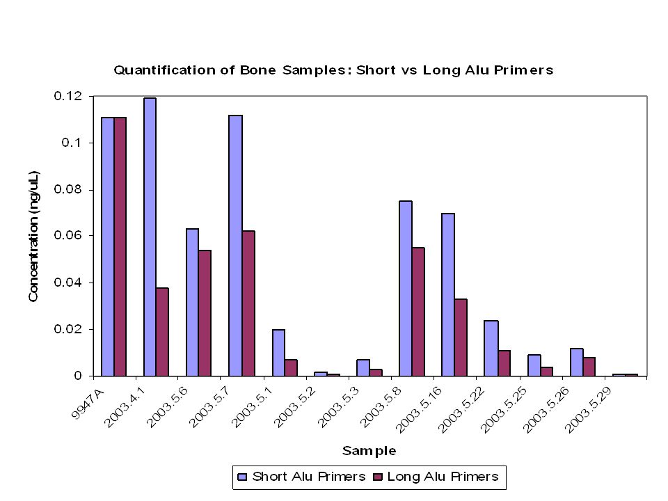
Quantitation of DNase I degraded DNA using 3 primer sets An example of the quantitation results obtained with a degraded DNA sample. Error bars represent 95% confidence interval.

46
RT-qPCR Instruments Cited Corbett Research Rotorgene –Phenix Research, Hayward, CA ABI 7000 or 7500 Sequence Detection System ABI 7700 (discontinued) ABI 7900HT Sequence Detection System –Applied Biosystems Foster City, CA Corbett Rotorgene ABI 7000
 ABI 7900HT Sequence Detection System –Applied Biosystems Foster City, CA Corbett Rotorgene ABI 7000")
47
Real-Time qPCR Efforts Marie Allen – nuclear and mtDNA assay (BioTechniques 2002, 33(2): 402-411) Eric Buel – Alu system (JFS 2003, 48(5):936-944) Centre for Forensic Sciences – nuclear; TH01 flanking region (JFS 2003, 48(5):1041-1046) John Hartmann – Alu system (SWGDAM Jan 2003) CA-DOJ – TH01 assay (NIJ DNA Grantees June 2003) SYBR Green assay – human-specific with right PCR Quantifiler kit (ABI) – separate nuclear and Y assays
: ) Eric Buel – Alu system (JFS 2003, 48(5): ) Centre for Forensic Sciences – nuclear; TH01 flanking region (JFS 2003, 48(5): ) John Hartmann – Alu system (SWGDAM Jan 2003) CA-DOJ – TH01 assay (NIJ DNA Grantees June 2003) SYBR Green assay – human-specific with right PCR Quantifiler kit (ABI) – separate nuclear and Y assays")
48
NIST Lessons Learned from Real Time-qPCR Assays Results are RELATIVE to standards used Single source and mixed source samples with same UV concentrations differ with RT-qPCR assays Need to keep instrument clean to avoid background fluorescence problems Assay reagent costs: –Quantifiler: $2.46/sample (only permits 2 µL/sample) –SYBR Green: $0.80/sample (up to 10 µL/sample) –QuantiBlot: $0.54/sample (5 µL/sample) Using ABI 7500 (early work with ABI 7000 and some Roche LightCycler) http://www.cstl.nist.gov/biotech/strbase/DNAquant.htm
 –SYBR Green: $0.80/sample (up to 10 µL/sample) –QuantiBlot: $0.54/sample (5 µL/sample) Using ABI 7500 (early work with ABI 7000 and some Roche LightCycler)")
49
Proceeding with Testing when “No DNA” Detected If the qPCR results indicate that there is no detectable DNA, will you stop testing or will you proceed with attempting STR typing? The practice of proceeding even with a “no result” Quantiblot was because the STR typing assay was more sensitive than the quantification method. What types of experiments might be done to satisfy you that “no result” from a qPCR assay is truly “no DNA”?

50
Difference in DNA Quantitation Capability vs. STR Typing Sensitivity 1 ng 100 pg 1 pg (less than a single cell) Real-time qPCR LOD Quantiblot Limit of Detection (LOD) STR typing (28 cycles) LOD LCN STR typing (34 cycles) LOD mtDNA possible due to higher copy # Nuclear DNA quantities Low Copy Number Realm This gap has kept labs proceeding with “no result” slot blot samples
 Real-time qPCR LOD Quantiblot Limit of Detection (LOD) STR typing (28 cycles) LOD LCN STR typing (34 cycles) LOD mtDNA possible due to higher copy # Nuclear DNA quantities Low Copy Number Realm This gap has kept labs proceeding with no result slot blot samples.")
51
DNA Quantitation Summary RT-qPCR is a homogeneous PCR based method that enables human specific quantification –Is easily automated, provides electronic storage of data –SYBR green or targeted probes can be used Results give quantity of amplifiable DNA – not necessarily overall quantity –PCR inhibition can be detected –Multiplexing can be used Big advantages are speed and dynamic range Commercial kits are now available

52
Acknowledgements Jan Nicklas and Eric Buel - Vermont Crime Laboratory Jiri Drabek Denise Chung, Kerry Opel Nancy Tatarek John Butler, Yin Shen Major support provided by The National Institute of Justice The OU Provost’s Undergraduate Research Fund Ohio University Research Incentive Fund

53
References http://www.med.sc.edu:85/pcr/realtime-home.htm http://www.realtimeprimers.org/ http://dna-9.int-med.uiowa.edu/realtime.htm http://dorakmt.tripod.com/genetics/realtime.htm In Print Nicklas, J.; Buel, E., J. Forens. Sci. 2003, 48(5) pp. 936-944 Andreasson, H; Gyllensten, U.; Allen, M. Biotechniques 2002, 33, pp. 402-411. Klein, D. “Quantification using rtPCR technology: applications and limitations” Trends in Molecular Medicine, 2002, 8(6) pp. 257- 260. Tyragi, S.; Kramer, F. “Molecular Beacons: Probes that fluoresce upon hybridization” Nat. Biotechnol. 1996, 14, pp. 303. Ginzinger, D. “Gene Quantification using real-time quantitiative PCR” Experimental Hematology, 2002, 30, pp. 503-512. Jordan, J. Real time detection of PCR products and microbiology, Trends in microbiology 2000, 12, pp. 61-66 On-line
 pp Andreasson, H; Gyllensten, U.; Allen, M. Biotechniques 2002, 33, pp Klein, D. Quantification using rtPCR technology: applications and limitations Trends in Molecular Medicine, 2002, 8(6) pp Tyragi, S.; Kramer, F. Molecular Beacons: Probes that fluoresce upon hybridization Nat. Biotechnol. 1996, 14, pp Ginzinger, D. Gene Quantification using real-time quantitiative PCR Experimental Hematology, 2002, 30, pp Jordan, J. Real time detection of PCR products and microbiology, Trends in microbiology 2000, 12, pp On-line.")
54
Low-Copy Number (LCN) Work Early work on touched objects and single cells : –van Oorschot, R. A. and Jones, M. K. (1997) DNA fingerprints from fingerprints. Nature. 387(6635): 767 –Findlay, I., Taylor, A., Quirke, P., Frazier, R., and Urquhart, A. (1997) DNA fingerprinting from single cells. Nature. 389(6651): 555-556 Application to routine forensic casework was pioneered by the Forensic Science Service : –Gill, P., Whitaker, J., Flaxman, C., Brown, N., and Buckleton, J. (2000) An investigation of the rigor of interpretation rules for STRs derived from less than 100 pg of DNA. Forensic Sci. Int. 112(1): 17-40 –Whitaker, J. P., Cotton, E. A., and Gill, P. (2001) A comparison of the characteristics of profiles produced with the AMPFlSTR SGM Plus multiplex system for both standard and low copy number (LCN) STR DNA analysis. Forensic Sci. Int. 123(2-3): 215-223 –Gill, P. (2001) Application of low copy number DNA profiling. Croatian Medical Journal 42(3): 229-32
 DNA fingerprints from fingerprints. Nature. 387(6635): 767 –Findlay, I., Taylor, A., Quirke, P., Frazier, R., and Urquhart, A. (1997) DNA fingerprinting from single cells. Nature. 389(6651): Application to routine forensic casework was pioneered by the Forensic Science Service : –Gill, P., Whitaker, J., Flaxman, C., Brown, N., and Buckleton, J. (2000) An investigation of the rigor of interpretation rules for STRs derived from less than 100 pg of DNA. Forensic Sci. Int. 112(1): –Whitaker, J. P., Cotton, E. A., and Gill, P. (2001) A comparison of the characteristics of profiles produced with the AMPFlSTR SGM Plus multiplex system for both standard and low copy number (LCN) STR DNA analysis. Forensic Sci. Int. 123(2-3): –Gill, P. (2001) Application of low copy number DNA profiling. Croatian Medical Journal 42(3):")
55
Stochastic Fluctuation Effects Unequal sampling of the two alleles present in a heterozygous individual can occur when low levels of input DNA are used (results in allele drop-out) PCR reactions with <100 pg (~17 diploid copies) Walsh et al. (1992) – propose avoiding stochastic effect by adjusting the number of PCR cycles in an assay so that the sensitivity limit is around 20 or more copies of target DNA (i.e., a full profile is obtained with ~125 pg) Walsh PS, Erlich HA, Higuchi R. Preferential PCR amplification of alleles: Mechanisms and solutions. PCR Meth Appl 1992; 1:241-250.
 – propose avoiding stochastic effect by adjusting the number of PCR cycles in an assay so that the sensitivity limit is around 20 or more copies of target DNA (i.e., a full profile is obtained with ~125 pg) Walsh PS, Erlich HA, Higuchi R. Preferential PCR amplification of alleles: Mechanisms and solutions. PCR Meth Appl 1992; 1:")
56
Stochastic Statistical Sampling True amount What might be sampled by the PCR reaction… >20 copies per allele 6 copies copies per allele (LCN) Resulting electropherogram OR Copies of allele 1 Copies of allele 2 Allele imbalanceAllele dropout
 Resulting electropherogram OR Copies of allele 1 Copies of allele 2 Allele imbalanceAllele dropout")
57
Allele Drop In 1ng 8pg Comparison of STR Kit Amplification SOP with LCN Using the Same DNA Donor Data from Debbie Hobson (FBI) – LCN Workshop AAFS 2003 Input DNA SOP LCN Allele Drop Out 50 µL PCR 5 µL PCR Heterozygote Allele Imbalance PHR = 87% PHR = 50%
 – LCN Workshop AAFS 2003 Input DNA SOP LCN Allele Drop Out 50 µL PCR 5 µL PCR Heterozygote Allele Imbalance PHR = 87% PHR = 50%")
58
Balance of Assay Sensitivity and Potential for Stochastic Effects One of the ways that assays can be made more sensitive is by increasing the number of PCR amplification cycles Optimal cycle number will depend on desired assay sensitivity The number of PCR cycles was set to 28 for ABI STR kits to limit their sensitivity for generating full profiles to ~125 pg or 20 cells Sensitivity is a combination of fluorescent dye characteristics (relative to the instrument and laser excitation used) and PCR amplification conditions such as primer concentration and amount of polymerase used Note that Promega STR kits use higher numbers of cycles to generate roughly equivalent sensitivity to ABI kits because they have less efficient dye labels and lower primer and polymerase concentrations
 and PCR amplification conditions such as primer concentration and amount of polymerase used Note that Promega STR kits use higher numbers of cycles to generate roughly equivalent sensitivity to ABI kits because they have less efficient dye labels and lower primer and polymerase concentrations")
59
Higher Sensitivity Raising the number of PCR cycles creates a higher potential of allele drop-in being detected (increased noise) Ideally an improved fluorescent dye could be used to improve detection sensitivity and thereby permit a lower number of PCR amplification cycles to be used (peak intensity does not always correlate with stochastic effect) Ju et al., Nature Medicine 2, 246 (1996) Hung et al., Anal. Biochem, 243, 15 (1996) Berti et al., Anal. Biochem. 292, 188 (2001) Medintz et al., BioTechniques 32, 270 (2002) High Sensitivity Energy Transfer Dye Labeling 6FAM ET dyes (example: LIZ) permit a 10-30X improvement in signal over non-ET dyes
 Berti et al., Anal. Biochem. 292, 188 (2001) Medintz et al., BioTechniques 32, 270 (2002) High Sensitivity Energy Transfer Dye Labeling 6FAM ET dyes (example: LIZ) permit a 10-30X improvement in signal over non-ET dyes.")
60
Challenge with Being Able to Go Lower In DNA Quantitation Measurements qPCR enables measurement of lower amounts of DNA but… Going into the low copy number realm introduces new challenges –Interpretation of mixtures –Defining thresholds for different dyes and amplification systems –Defining the difference between investigative data and reliable “court-worthy” data

61
New Interpretation Rules Required for LCN

62
Suggestions to Optimal Results with LCN At least two* PCR amplifications from the same DNA extract An allele cannot be scored (considered real) unless it is present at least twice in replicate samples Extremely sterile environment is required for PCR setup to avoid contamination from laboratory personnel or other sources *five is better; results are investigative
 unless it is present at least twice in replicate samples Extremely sterile environment is required for PCR setup to avoid contamination from laboratory personnel or other sources *five is better; results are investigative")
63
LCN Summary LCN often defined as <100-200 pg input DNA Typically involves increasing the number of PCR cycles when performing multiplex PCR to amplify DNA with conventional STR kits (e.g., 34 cycles instead of 28 cycles) Enables lower amounts of DNA to be detected with STR markers but is prone to contamination Cautious data interpretation rules must be adopted as allele drop-out and drop-in may occur due to stochastic amplification effects
 Enables lower amounts of DNA to be detected with STR markers but is prone to contamination Cautious data interpretation rules must be adopted as allele drop-out and drop-in may occur due to stochastic amplification effects")
Similar presentations


>")