Download presentation
Presentation is loading. Please wait.

1
Nanoparticle mediated Delivery and Imaging Sarah Fredriksson, PhD Founder of Genovis AB I am sorry that I missed your meeting. Here I have put together a few slides presenting our nanoparticles. Hope to see you in the near future! /Sarah Here I show a film were you can see the cell responding to a magnetic field. The cell is green since it has taken up millions of nanoparticles carrying fluorescent probes on their surfaces. I will attach the film separately.

2
GENOVIS IN BRIEF: Focus on Nanotechnology for Life Science Patent pending technologies Founded in 1999, R&D intense years 2000 – 2007 Starts marketing and building sales channels in 2008 Principal Business areas: – Biomolecule Delivery – Imaging Facility in Lund, Sweden 9 employees

3
The Competitive Edge of Genovis Team Application driven design of unique nanostructures for use in (combined) delivery and imaging applications Production skills Products on the market for in vitro use Strong technical and practical customer support Pipeline with focus on in vivo imaging and drug delivery
 delivery and imaging applications Production skills Products on the market for in vitro use Strong technical and practical customer support Pipeline with focus on in vivo imaging and drug delivery")
4
Diagnostics Drug delivery Imaging Novel therapeutics NANOPARTICLE APPLICATIONS IN BIOMEDICINE

5
Nanoparticles Nanoparticles in relation to micro o The surface to volume ratio increases with a factor of 1000 o The material characteristics as we know them can change o Production processes become a challenge Nanoparticle structures (< 300 nm) o Shell structures o Solid core particles o Polymer based formulations o Lipid based formulations o and more …..
 o Shell structures o Solid core particles o Polymer based formulations o Lipid based formulations o and more …..")
6
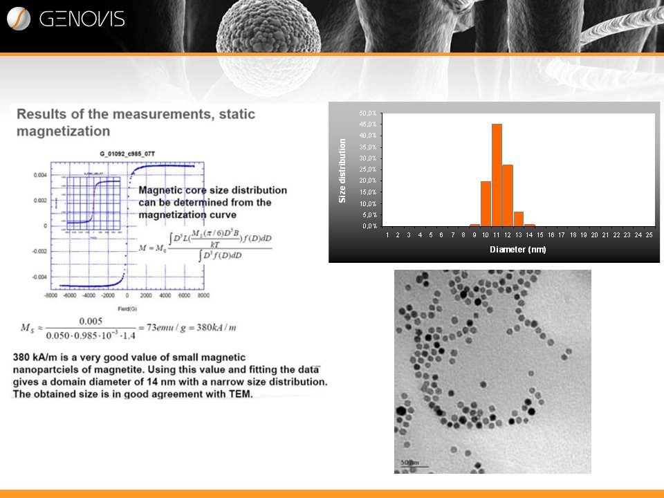
Genovis has developed NIMT FeOdots – nanoparticles for Life Science Applications Hi, I am a FeOdot. NIMT FeOdots are superparamagnetic nanoparticles specially designed for use in the fields of biomedicine and biotechnology. FeOdots are engineered nanoparticles of solid iron oxide cores composed mainly of Fe3O4 (Magnetite). Due to non classical synthesis routes size and shape can be controlled. The nanoparticles have a high magnetic saturation (Ms) of about 80 emu/g and are monodispersed (11 nm ± 1 nm core size).
. Due to non classical synthesis routes size and shape can be controlled. The nanoparticles have a high magnetic saturation (Ms) of about 80 emu/g and are monodispersed (11 nm ± 1 nm core size)..")
7
Solid homogenous iron oxide cores Spherical shape Possibility to vary core diameter Superparamagnetic iron oxide cores

8
Superparamagnetism = no ”magnetic memory” when M-field is removed Particle cores < approx. 20 nm generally exhibits superparamagnetic behaviour

10
Particle coating objectives: Surface for binding/protecting the cargo molecule (DNA/siRNA/peptide etc) during transport Cargo release Suitable for bioconjugation Promote colloidal stability Possibility of fluorescence Prevent unspecific interactions Particle coating
 during transport Cargo release Suitable for bioconjugation Promote colloidal stability Possibility of fluorescence Prevent unspecific interactions Particle coating")
11
Particles designed from inside and out: Two coating procedures were developed: PEG based & Lipid based Iron Oxide H2NH2N NH 2 PEG Phospholipid Phospholipid-Texas Red Iron Oxide H2NH2N NH 2 FeOfection| PURPLE FeOfection| YELLOW FeOfection| PINK

12
Fig 1. Particles incubated for 48 hours in different pH. Even under extreme salt concentrations (Fig 2) the particles remain in solution with no observed agglomeration. This has been further confirmed by size measurements using dynamic light scattering (DLS). Fig 3. NIMT FeOdots incubated with 1-5 M NaCl for 48 hours. The extreme stability of NIMT FeOdots are of importance for in vivo applications or application were complex medium is used. The magnetic nanoparticles remain stable in serum and other high protein concentration solutions for a extended period of time. No agglomeration or precipitations are observed upon incubation in human serum (Fig 4). This has been further confirmed by size measurements using dynamic light scattering (DLS). Fig 2 Colloidal stability is important
 the particles remain in solution with no observed agglomeration. This has been further confirmed by size measurements using dynamic light scattering (DLS). Fig 3. NIMT FeOdots incubated with 1-5 M NaCl for 48 hours. The extreme stability of NIMT FeOdots are of importance for in vivo applications or application were complex medium is used. The magnetic nanoparticles remain stable in serum and other high protein concentration solutions for a extended period of time. No agglomeration or precipitations are observed upon incubation in human serum (Fig 4). This has been further confirmed by size measurements using dynamic light scattering (DLS). Fig 2 Colloidal stability is important.")
13
Colloidal stability of PEG-Biotin particles

14
Nanoparticles in combined delivery and imaging Spinning Endosomes – this is a film clip showing how the interior of each endosome can follow an external magnetic field. The endosomes have taken up nanoparticles carrying a flourescent probe on their surface. I am not sure whether I can attach the film in the e-mail. Otherwise I hope that I will get an opportunity later. Cell nucleus

15
siRNA delivery –product application The nanoparticles consists of a stable iron-oxide core By varying the lipids and coatings of the particles the particles can be tailor-made to promote uptake and release of siRNA in different cell lines. FeOfection| is coated with cationic lipids to facilitate siRNA binding and cellular uptake

16
siRNA Delivery Using NIMT FeOfection Gene Silencing w/ NIMT ® FeOfectionPURPLE HEK cells transfected with Eg5 siRNA Cells lacking Eg5 cannot divide and therefore do not stay in their rounded up stage. Cell growth is arrested and the cells eventually die. A. B. siRNA Eg5 treated cells. 48 h post transfection siRNA negative control treated cells. 48 h post transfection

17
Simplify - siRNA delivery – NIMT® FeOfection

18
Gene Delivery Att tillföra en ny gen i en cell FeOfection is a solution of nanoparticles with an iron oxide core. The core is stable and the magnetic properties can be used e.g. in tracking of cells with MRI. The surface of the particles are modified to promote binding of DNA to the particles and facilitate transport of the resulting particle/DNA complexes into cells. FeOfection can be used for both transient (temporary expression) and stable (incorporated in the genome) transfection.
 and stable (incorporated in the genome) transfection..")
19
Imaging using magnetic nanoparticles Marknaden drivs av ett medicinskt behov av effektivare och känsligare diagnostik

20
Iron Oxide NH 2 Phospholipid Amino-PEG NHS-Alexa 647 Iron Oxide NH 2 U-2 OS cell incubated with Alexa-647 magnetic nanoparticles for 1 hour FeOdots incubated with cells and exposed to a magnetic field NH 2

21
Imaging - Regenerativ medicin Stamceller märks med Genovis magnetiska nanopartiklar ex vivo och injeceras i mus T2* Map Prussian blue positive cells at edge of tumor C6 glioma FeOlabeled cells were injected i.v. in C6 glioma in mouse flank 14 days prior to 3T MRI Cells labeled with FeOlabel can easily be visualised with MRI. Mesenchymal stem cells were labeled with FeOlabel and then injected into a mouse with a C6 glioma. After 14 days the cells are visible with MRI. Particles can also be visualised by Prussian Blue iron staining.

22
Genovis R&D project Targeted delivery of siRNA in vivo Multimodal imaging (MRI/SPECT, MRI/Ultrasound ) Targeting lymph nodes, microglia cells (brain) and glioma tissue. Developing nanoparticles for systemic delivery Design of an inner fluorescent coating layer for multimodal imaging

23
Thank you! Visit our web-site to learn more about the FeOdot adventures. www.genovis.com

Similar presentations