Download presentation
Presentation is loading. Please wait.

1
UTMB – Dept of Otolaryngology
Mandible Fractures Jacques Peltier MD Matthew Ryan MD UTMB – Dept of Otolaryngology May 2004

2
History Edwin Smith Papyrus 1650 described Hx, Phy, Diagnosis. Often fatal disease Hippocrates – Described monomaxillary dental fixation and binding Sulicetti – 1492 Described “tie teeth of jaw to teeth of uninjured jaw”

3
History Schede 1888 – Bone plate of steel secured with 4 screws
Luhr 1960 – Developed mandibular compression plates Michelet and Champy 1970’s – Placement of small bendable non-compression plates

5
Epidemiology Mandible most common after nasal fractures
Mandible : Zygoma : Maxilla 6:2:1 Ellis 4711 facial fractures, 45% with mandible fractures Assault>MVA>Fall>Sports

6
Epidemiology Sites of weakness Third molar (esp. impacted)
Socket of canine tooth Condylar neck

7
Epidemiology Boole et al (laryngoscope) 5196 fractures
Young military men Angle 35%, Symphysis 20%, Body 12%, Condylar 9%, Subcondylar 4%, Ramus 4%, Alveolar 3%, Coronoid 1% 70% 1 fracture, 30% 2 fractures, .2% more than 2 Facial lacs 30%, other facial fx. 16%, C-spine 0.8%

8
Haug et al

9
Fischer et al

10
Favorable vs. Unfavorable
Masseter, Medial and Lateral Pterygoid, and Temporalis tend to draw fractures medial and superior Almost all fractures of angle unfavorable

12
Evaluation Stabilization via ATLS protocol Part of secondary survey
Pain, malocclusion, trismus, V3 sensory deficit History of TMJ (earlier mobilization) Blow to face favors parasymphyseal fracture and contralateral angle fracture Fall to chin (bilateral condylar fractures)
 Blow to face favors parasymphyseal fracture and contralateral angle fracture. Fall to chin (bilateral condylar fractures)")
14
Evaluation Previous occlusion (Class I-III)
Psychiatric, nutritional, gastrointestinal, seizure disorders Previous facial trauma Other injuries (c-spine, intra-abdominal, likely prolonged intubation)
")
15
Physical Exam Complete Head and Neck exam Palpable step off
Tenderness to palpation Malocclusion Trismus (35 mm or less) FOM hematoma Altered sensation of V3 Crepitus
 FOM hematoma. Altered sensation of V3. Crepitus.")
16
Physical Exam Dental Exam Lost, fractured, or unstable teeth
Dental Health Relation to fracture Quantity

17
Physical Exam Unilateral fractures of Condyle
Decreased translational movement, functional height of condyle Deviation of chin away from fracture, open bite opposite side of fracture Bilateral fractures of condyle - Anterior open bite

18
Picture of open bites

20
Evaluation Panorex, mandible series CT scan
Not as diagnostic as plain films for nondisplaced fractures of mandible. Most useful for coronoid and condylar fractures, associated midface fractures

21
Physiology Primary Healing In rigid fixation techniques
Lag screws, compression plates, Recon plate, external fixation, Wire fixation, Miniplate fixation No callus formation Question of bone resorption

22
Physiology Secondary bone healing Callus formation
Remodeling and strengthening MMF, Wire fixation, Miniplate fixation

23
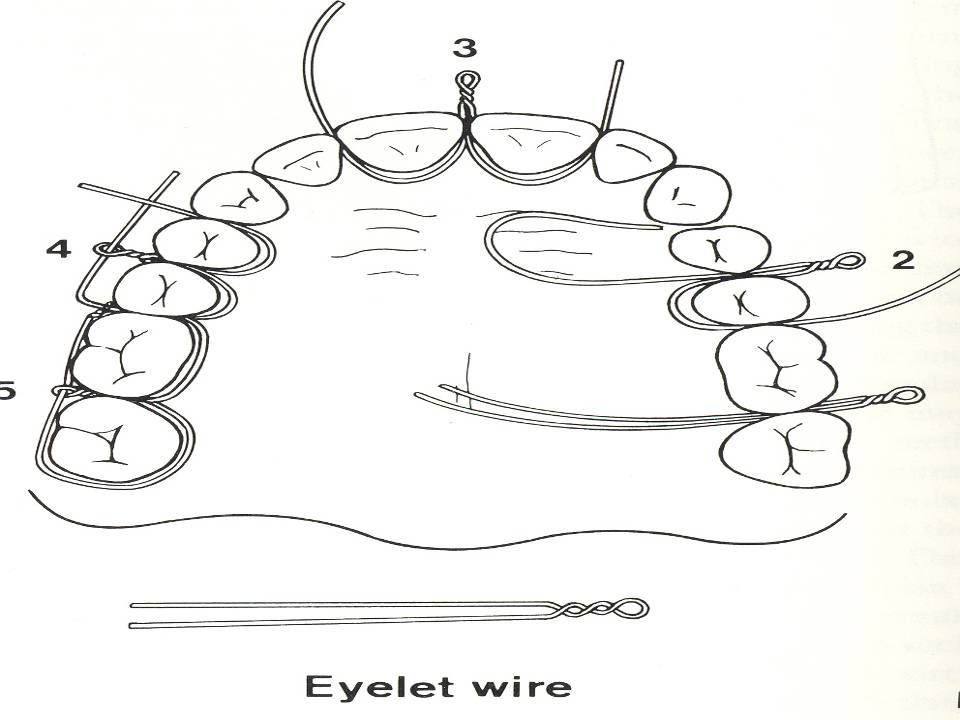
Closed Reduction Favorable, non-displaced fractures
Grossly comminuted fractures when adequate stabilization unlikely Severely atrophic edentulous mandible Children with developing dentition

24
Closed Reduction Length of MMF
De Amaratuga – 75% of children under 15 healed by 2 weeks, 75% young adults 4 wks Juniper and Awty – 82% had healed at 4 wks Longer period for edentulous fractures 6-10wks

25
Closed Reduction Edentulous fractures
Bradley found absent inferior alveolar artery in 40% yo’s Periosteal blood supply disturbed by stripping Up to 20% non-union despite type of treatment May consider Gunning Splints

29
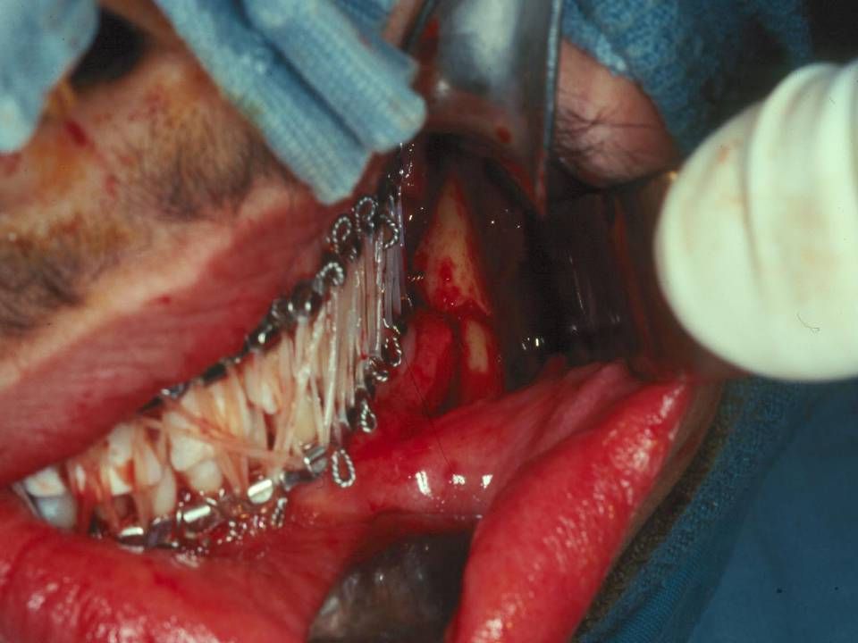
Open Reduction Displaced unfavorable fractures
Mandible fractures with associated midface fractures When MMF contraindicated or not possible Patient comfort Facilitate return to work

30
Open Reduction Contraindications General Anesthetic risk too high
Severe comminution and stabilization not possible No soft tissue to cover fracture site Bone at fracture site diffusely infected (controversial)
")
31
Open Reduction Associated condylar fracture
Associated Midface fractures Psychiatric illness GI disorders involving severe N/V Severe malnutrition To avoid tracheostomy in patients who need postoperative intubation

32
Open Reduction Intraosseous wiring Semirigid fixation Cheap
Technically difficult Primary and Secondary bone healing

34
Open Reduction Lag Screws Rigid fixation (Compression)
Good for anterior mandible fractures, Oblique body fractures, mandible angle fractures Cheap Technically difficult Injury to inferior alveolar neurovascular bundle

35
Open reduction Ellis 41 patients with anterior lag screw technique
4.9% infection rate No malocclusion No Non-union

36
Lag Screw Technique

37
Lag Screw Technique

38
Lag Screw Technique

39
Rigid Fixation Compression plates Rigid fixation
Allow primary bone healing Difficult to bend Operator dependent No need for MMF

42
Rigid Fixation Miniplates Semi-rigid fixation
Allows primary and secondary bone healing Easily bendable More forgiving Short period MMF Recommended

43
Rigid Fixation Schierle et al studied experimental model, then applied in patients. Model suggested two plates more stable Patients divided into two groups with equal complication rates, equal functional results

45
Miniplates, Champy technique

46
Rigid Fixation Reconstruction Plates Good for comminuted fractures
Bulky, palpable Difficult to bend Locking plates more forgiving

47
External Fixation Alternative form of rigid fixation
Grossly comminuted fractures, contaminated fractures, non-union Often used when all else fails

49
Edentulous Fractures Chalmers and Lyons 1976 – Recommended closed reduction to preserve periosteal blood supply Chalmers and Lyons 1995 167 fractures in edentulous mandibles ORIF 82% 15% complications 12% Fibrous union

50
Edentulous Fractures ORIF
Inferior alveolar canal more superior in location Vertical height 20mm compatible with standard plating systems Vertical height 10mm or less, likely need rib graft Plate removal after fracture healing if interferes with denture placement

51
Teeth in line of fracture
Keep teeth if Previously healthy Peridontal plexus intact No major structural injury Tooth does not interfere with reduction of fracture

52
Teeth in line of fracture
Neal and associates 32% incidence of morbidity with teeth in line of fracture No statistical difference if tooth was removed

53
Teeth in line of fracture
Amaratunga 16% complication rate in retained teeth 13% in removed teeth Retain teeth for 4-6 weeks if important for MMF

54
Condylar and Subcondylar
Lindhal and Hollender Closed reduction in children, teens, adults Intracapsular fractures Higher incidence of postoperative sequelae in adults Children and Teens with less sequelae, more remodeling

55
Condylar and Subcondylar
Norholt Children 5-20 with intracapsular condylar fractures Increased dysfunction with increasing age

56
Condylar and Subcondylar
Closed reduction with arch bars MMF 2-3 weeks mainstay for youths Ankylosis of TMJ and facial asymmetry most feared complication Less effective for increasing age decreased ramus height more displaced

57
Condylar and Subcondylar
ORIF, Absolute indications Displacement into middle cranial fossa Inability to achieve occlusion with closed reduction Foreign body in joint space

58
Condylar and Subcondylar
Relative indications Bilateral condylar fractures to preserve vertical height Associated injuries that dictate earlier function Soft tissue swelling causing airway compromise with MMF Intracapsular fracture on opposite side where early mobilization important

60
Immediate Mobilization
Kaplan et al. Studied ORIF in two groups, one with MMF for 2 weeks, one with immediate mobilization No statistical difference in rates of complications, postoperative pain, dental health, nutritional status

61
Bioabsorbable Plates Plating can relieve stress, no bone remodeling
Bulky plates, thermal sensitivity, palpable Absorbable plates expensive Better in children? Use of poly-L-lactide in 69 fractures by Kim et al 12% complication 8% infection No malunion

62
References Kim et al “Treatment of Mandible Fractures using Bioabsorbable plates”, Plastic and Reconstructive Surgery, vol 110, july 2002, 25-31 Bailey, Byron J. Head and Neck Surgery - OtolaryngologyThird Edition. Lippincott Williams and Wilkins, 2001. Ellis, E. “Treatment Methods for Fractures of the Mandibular Angle." Journal of Craniomaxillofacial Trauma, vol. 28. 1999: Ellis, E., et. al. “Lag Screw Fixation of Mandibular Angle Fractures.” Journal of Oral Maxillofacial Surgery, vol. 49. 1991: Kim et. al. "Treatment of Mandible Fractures Using Bioabsorable Plates." Journal of Plastic and Reconstructive Surgery, vol 2002: Boole et. al. "5196 Mandible Fractures Among 4381 Active Duty Army Soldiers, 1980 to 1998." Laryngoscope, 111(10). Oct. 2001: , Kaplan et al. "Immediate Mobilization Following Fixation of Mandible Fractures, A Prospective Randomized Study." Laryngoscope, vol. 111(9). Sept 2001: Spina and Marciani. Mandibular Fractures, pages Schierle et. al. "One or Two Plate Fixation of Mandible Fractures?" Journal of Cranio-Maxillofacial Surgery. Vol. 25, 1997:
. Oct. 2001: , Kaplan et al. Immediate Mobilization Following Fixation of Mandible Fractures, A Prospective Randomized Study. Laryngoscope, vol. 111(9). Sept 2001: Spina and Marciani. Mandibular Fractures, pages Schierle et. al. One or Two Plate Fixation of Mandible Fractures Journal of Cranio-Maxillofacial Surgery. Vol. 25, 1997:")
Similar presentations


>")


