Download presentation
Presentation is loading. Please wait.

1
Dr Prasanth S

2
3D Echo Basics Concept - Early 1980s. Conventional 2D echo requires cognitive 3D reconstruction of cardiac structures. Real time 3D echo provides anatomically realistic visualization of structures. Decrease the time required for complete image acquisition. 3D Echo can be viewed from various projections by rotation of images. A limitation- the information acquired as 3D dataset, must be displayed as 2D image.

5
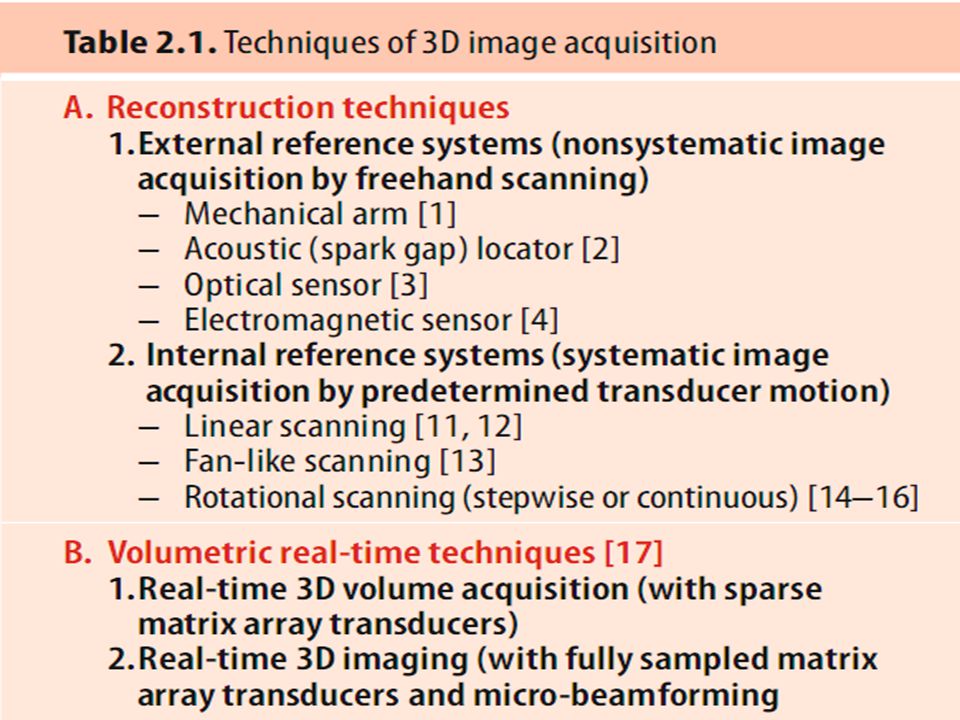
Linear scanning approach Earliest approach to dynamic 3D echo. Based on 3D concepts used in CT and MR imaging. Acquiring parallel and equidistantly placed 2D images using transducer mounted on a sliding carriage. Movement of transducer by a computer controlled stepper motor. Fan- like scanning approach Transducer moved in a fan like arc at prescribed angles.( manually or by stepper motor)
.")
6
Hardware used for fanlike acquisition

7
Rotational scanning approach Transducer is rotated in a semicircle around the central axis of imaging plane.

8
Disadvantages of Initial models – Time consuming – High Cost – Large Machines – Slow processor RT3DE in 2002 – Dense Matrix Array Transducer – 3000 elements – Fast processor

9
Real Time 3D – Pyramidal Volume

10
3D volumetric probe- Pyramidal Volume

11
Technical Issues in Real Time 3D Full volume images are typically obtained at frame rate of 20-40 Hz.( vary as a function of depth & size of the volume.) Either sacrifice frame rate for image quality (spatial resolution) or spatial resolution for frame rate Min Frame rate required 20 Hz – 20 frames per sec Each frame contains beams 1 beam requires.2ms for scanning If 100 beams / frame – 20 ms time – frame rate – 50Hz 250 beams / frame – 50 ms time – 20 Hz.
 Either sacrifice frame rate for image quality (spatial resolution) or spatial resolution for frame rate Min Frame rate required 20 Hz – 20 frames per sec Each frame contains beams 1 beam requires.2ms for scanning If 100 beams / frame – 20 ms time – frame rate – 50Hz 250 beams / frame – 50 ms time – 20 Hz.")
12
Innovations to overcome the limitations 1. Increase the distance between beams. Blurring of structures that are not adequately sampled. 2. Parallel receive beam processing – Increase frame rate 4 or more simultaneous receive beams for each transmitted beam 3. Limit the total number of beams by scanning a smaller volume - 3D zoom mode 4. Use the patient’s ECG signal to acquire smaller subvolumes for each R-R interval, and then stitch these subvolumes to produce the larger volume -full volume dataset 5. Broaden the transmit beam

13
Transducers Phased array transducer Sparse Matrix Array Transducer Microbeam Former Dense Matrix Array Transducer

14
Sparse Matrix Array Transducer: Only a small percentage of 2500 (50х50) elements are electrically connected or acoustically active. Usually strategically placed 256 elements will be active. Loss of signal to noise ratio. THI not supported- incapable of creating sufficient MI necessary to create tissue harmonic image.

15
Micro-beam forming Electrically group, small arrays of elements (patches). Typical patch contain approx 25 elements(5х5). 128 micro-beamformer with 128 wires connecting the transducer to mainframe beamformer- provide a fully sampled array of 3200 elements.
. 128 micro-beamformer with 128 wires connecting the transducer to mainframe beamformer- provide a fully sampled array of 3200 elements..")
17
Matrix Array TEE Transducer distal tip

18
Modes

19
Real time Mode Narrow angle 60 0 :30 0 Higher Resolution(high line density & narrow scan volume) No stitching Artifact Visualisation of valves,small masses, vegetations. Real time Interventional guidance

20
Zoom Mode Focused – 30 0 : 30 0 Enface view of MV Masses- Thrombi,vegetations Eliminate need for cropping. Low spatial resolution due to low line density.

21
Full volume mode Covers wider region 90 0 :90 0 upto 104 0. 4 or 7 live 3D subvolumes stitched together. Near real time Higher temporal resolution Spatial relationship of cardiac structures Chamber Quantification

22
Colour Flow Mode Limited angle 60:60 Limited Temporal resolution Shape & extention of jets

24
Basic 3D analysis 3D orientation – manual free rotation of the dataset to provide the best perspective of the structures of the heart Cropping – manually moving a cutting plane from outside the 3D volume towards its center- provide a view from the cutting plane – cropping can be performed either before (during) or after data acquisition Slicing – extraction of image planes from the 3D volume in different modes
 or after data acquisition Slicing – extraction of image planes from the 3D volume in different modes")
25
Full Volume

27
Artifacts

28
Stitch artifact Lines of disagreement between two neighboring subvolumes.

29
Dropout artifacts Losses of 3D surface information due to poor echo signal intensity. Structures- too thin to reflect. Appropriate gain settings, colour doppler can identify false defects.

30
Blurring and blooming artifacts Blurring refers to unsharp or hazy representation of thin structures- appear thicker. Eg: Mitral leaflets, mitral valve apparatus. Blooming: thickened or excessive representation of high echo density structures like mechanical prosthesis, pacemaker leads. Strongly related to the line density.

31
Blurring and blooming artifacts

32
Gain artifacts

33
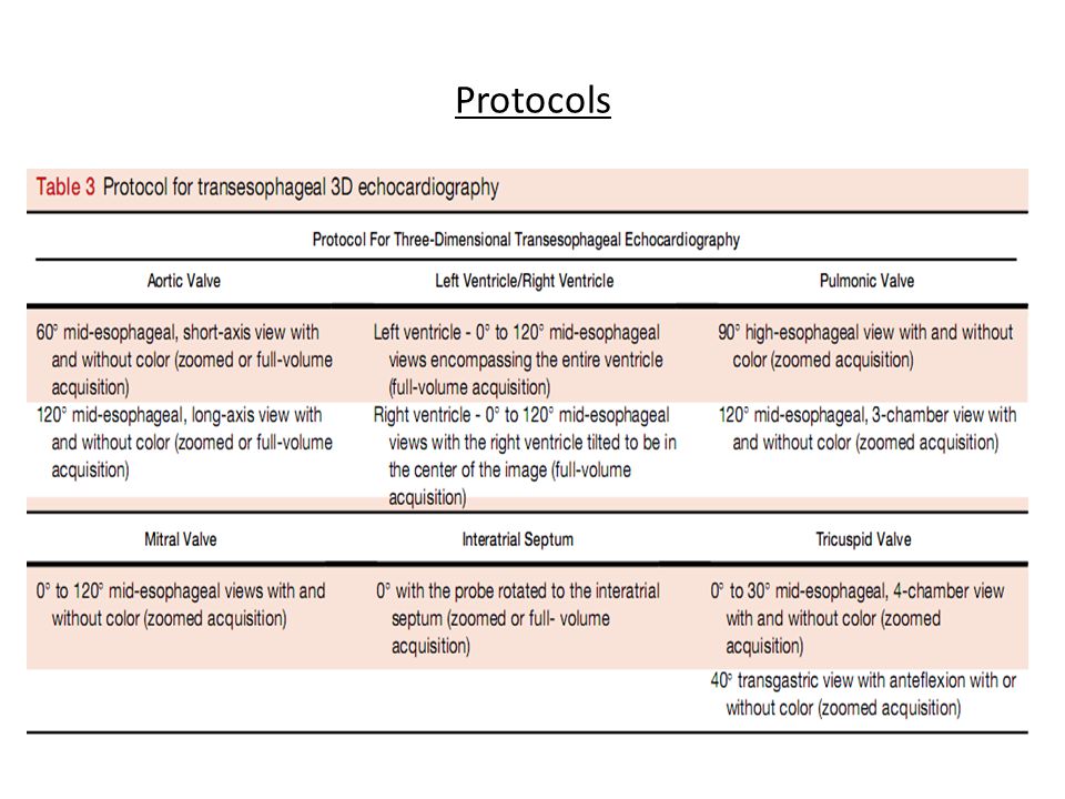
Protocols

35
Applications

36
Left ventricular function Unlike 2D echo, there is no geometrical assumptions. Left ventricular volume can be calculated by, 1. Method of discs 2. Directly sum the volumetric picture elements enclosed by endocardial borders of 3D structure.

37
Global LV function - EDV measurement

38
Global LV function - ESV measurement

39
LV Mass

40
Currently, 3D TTE and TEE assessment of LV volumes and ejection fraction is recommended over the use of 2D echocardiography, as it has been clearly demonstrated to provide more accurate and reproducible measurements. European Heart Journal – Cardiovascular Imaging (2012) 13, 1–46.(EAE/ASE RECOMMENDATIONS)
 13, 1–46.(EAE/ASE RECOMMENDATIONS).")
41
Parametric imaging Assessing segmental contraction of LV. 800 endocardial data points - to develop a polar map of endocardial surface of LV. Endocardial motion is displayed as shades of – Blue - positive excursion values - inward motion – Red - negative excursion values - outward motion – Black - no motion Akinetic or dyskinetic myocardium - black or red color Normal or hypokinetic segment - shades of light to dark blue

42
Parametric imaging NormalApical MI

43
LA Volume

44
RV volume & function RV has a complex geometrical shape. RV inflow, outflow and apex do not align in a single 2D plane. More heavily trabeculated. Normal reference values Indexed RV EDV49±10 ml/m² Indexed RV ESV16±6 ml/m² RV EF67±8%

46
Valvular Disease

47
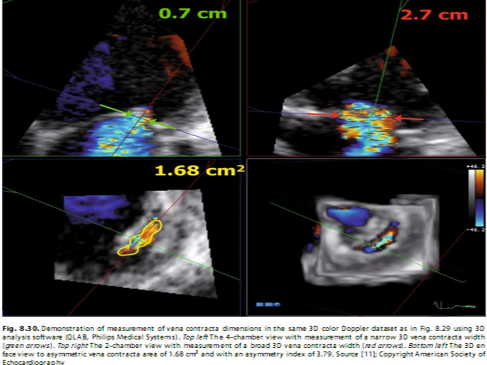
MVP - Mitral Regurgitation Accurate anatomy – Scallop Surgical View Vena contracta area – direct measurement TTE is good enough

48
Important parameters derived by RT3DE before MV repair 1.Anterior-posterior diameter (DAP) 2.Anterolateral-posteromedial diameter (DAlPm) 3.3D curvilinear length of posterior and anterior leaflet 4.Exposed area of leaflets (A3DE) 5.Minimal area of leaflets within the saddle-shaped annulus (A3Dmin) 6.Volume of leaflet prolapse (VProl) 7.Maximal prolapse height (HProl) 8.Length of antero-lateral and postero-medial chordae tendinae
 2.Anterolateral-posteromedial diameter (DAlPm) 3.3D curvilinear length of posterior and anterior leaflet 4.Exposed area of leaflets (A3DE) 5.Minimal area of leaflets within the saddle-shaped annulus (A3Dmin) 6.Volume of leaflet prolapse (VProl) 7.Maximal prolapse height (HProl) 8.Length of antero-lateral and postero-medial chordae tendinae")
52
MVP Multiple jets

54
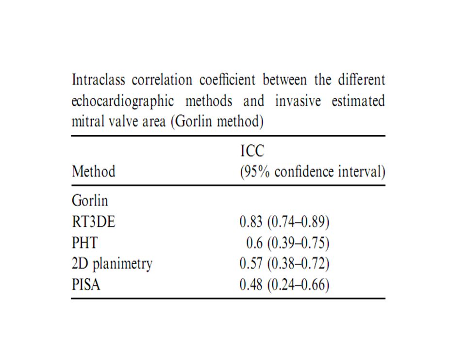
Mitral Stenosis MVA at smallest orifice Better assessment for BMV Planimetry from Apical window More accurate than Gorlin New GOLD STANDARD

55
Localizing smallest orifice

59
Aortic Regurgitation TEE is better Vena contracta area Better delineation of Etiology

60
Aortic Regurgitation

62
Aortic Stenosis AVA measurement Assess the noncircular LVOT area

63
BCAV DEGENERATIVE RHEUMATIC NORMAL

65
Other Applications

66
Dysynchrony

67
VSD

68
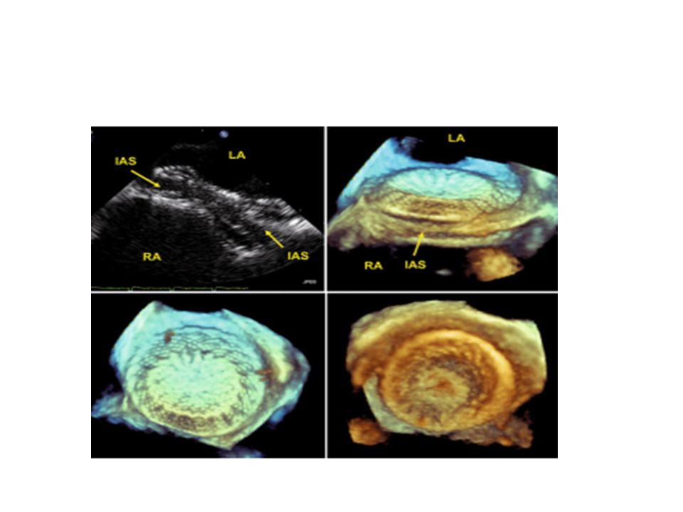
ASD

69
Thrombus

70
Myxoma

71
Intervention

74
References Feigenbaum’s Echocardiography Clinical Echocardiography;Otto 3D Echo Thomas Buck, Andreas Franke,Mark J. Monaghan ASE 2012 Guidelines ESC – 3D Echobox Cardiology clinics 2007 vol 25

Similar presentations


 Continuous Wave 2) Pulse Wave 3) Color Flow DOPPLER ULTRASOUND.>")
. - a spherical occluder is contained by metal.>")


 FOR MITRIAL REGURGITATION>")
