Download presentation
Presentation is loading. Please wait.

1
The Dementias William S. Woodfin, M. D
The Dementias William S. Woodfin, M.D. Neurology Specialists of Dallas Clinical Assoc. Prof. of Neurology UT Southwestern Medical School

2
Definition Dementia:The development of multiple cognitive deficits suffficiently severe to cause impairment in occupational or social functioning Mild Cognitive Impairment: usually refers to a single cognitive domain

3
Classification Alzheimer’s Disease: Sporadic v. Familial
Parkinson Syndromes Fronto-Temporal Lobar Vascular Infectious Metabolic Pseudodementia Others

4
Differential Diagnosis of Dementia
Keywords: Diagnosis, Incidence Differential diagnosis of dementia Alzheimer’s disease (AD) is the most common cause of cognitive decline in the elderly.1 A substantial number of diseases can present with dementia and should be included in the differential diagnosis when evaluating a patient. Numerous disorders mirror Alzheimer’s-type dementia, and dementia syndrome may have more than 1 etiologic factor in the geriatric patient. It is estimated that within the diagnosis of dementia, approximately 65% of patients present with AD. Vascular dementia (VaD) is the second most common cause of dementia. Other potential causes include Parkinson’s disease, Lewy body disease, and frontal lobe dementia.2,3 AD and VaD are easily confused because many symptoms are similar. One primary difference is that the onset of AD is gradual, while VaD begins abruptly and progresses in a stepwise manner. Also, in AD, deterioration in a broad range of intellectual abilities can occur without affecting motor abilities until advanced stages of the disease. In addition, in AD, focal signs and symptoms are generally absent. On the other hand, while AD may present a normal brain CT, a VaD patient will show evidence of stroke or stroke-related change. VaD patients frequently have an underlying vascular disorder while AD patients do not. In addition, there are a few causes of dementia that are reversible such as depression, effects of medication, vitamin B12 deficiency, and hypothyroidism. These causes need to be ruled out and treated appropriately.2,4 References: 1. Small GW, Rabins PV, Barry PP, et al. Diagnosis and treatment of Alzheimer disease and related disorders: consensus statement of the American Association for Geriatric Psychiatry, the Alzheimer’s Association, and the American Geriatrics Society. JAMA. 1997;278: Morris JC. Differential diagnosis of Alzheimer’s disease. Clin Geriatr Med. 1994;10: Zurad EG. New treatments of Alzheimer’s disease: a review. Drug Benefit Trends. 2001;137: Corey-Bloom J, Thal LJ, Galasko D, et al. Diagnosis and evaluation of dementia. Neurology. 1995;45:
 is the most common cause of cognitive decline in the elderly.1 A substantial number of diseases can present with dementia and should be included in the differential diagnosis when evaluating a patient. Numerous disorders mirror Alzheimer’s-type dementia, and dementia syndrome may have more than 1 etiologic factor in the geriatric patient. It is estimated that within the diagnosis of dementia, approximately 65% of patients present with AD. Vascular dementia (VaD) is the second most common cause of dementia. Other potential causes include Parkinson’s disease, Lewy body disease, and frontal lobe dementia.2,3. AD and VaD are easily confused because many symptoms are similar. One primary difference is that the onset of AD is gradual, while VaD begins abruptly and progresses in a stepwise manner. Also, in AD, deterioration in a broad range of intellectual abilities can occur without affecting motor abilities until advanced stages of the disease. In addition, in AD, focal signs and symptoms are generally absent. On the other hand, while AD may present a normal brain CT, a VaD patient will show evidence of stroke or stroke-related change. VaD patients frequently have an underlying vascular disorder while AD patients do not. In addition, there are a few causes of dementia that are reversible such as depression, effects of medication, vitamin B12 deficiency, and hypothyroidism. These causes need to be ruled out and treated appropriately.2,4. References: 1. Small GW, Rabins PV, Barry PP, et al. Diagnosis and treatment of Alzheimer disease and related disorders: consensus statement of the American Association for Geriatric Psychiatry, the Alzheimer’s Association, and the American Geriatrics Society. JAMA. 1997;278: Morris JC. Differential diagnosis of Alzheimer’s disease. Clin Geriatr Med. 1994;10: Zurad EG. New treatments of Alzheimer’s disease: a review. Drug Benefit Trends. 2001;137: Corey-Bloom J, Thal LJ, Galasko D, et al. Diagnosis and evaluation of dementia. Neurology. 1995;45:")
5
Corticobasalganglionic
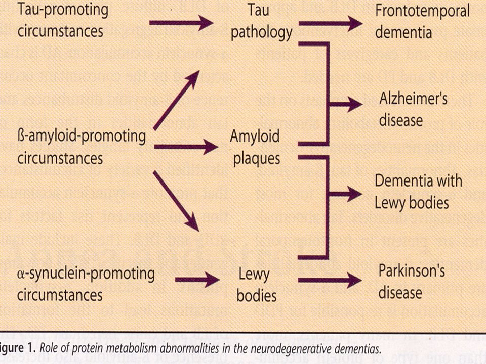
DEMENTIA SYNDROMES Normopressure Hydrocephalus Vascular Parkinsonism Multiple System Atrophy FXTAS Alzheimer’s Disease Lewy Body Parkinson’s Disease Supranuclear Palsy Diffuse Lewy Body Disease Corticobasalganglionic Degeneration Fronto-temporal Dementia Amyloid and Tau α-SYNUCLEINOPATHIES TAUOPATHIES

6
Alzheimer’s History: Alois Alzheimer 1906
Epidemiology: * 4 million pts. * A disease of advancing age but not normal aging. Loss vs. shringage of neurons * Age % Age % * Underdiagnosed * Women more than men * Cost $110 billion

7
DSM-IV Definition of Alzheimer’s Disease
Development of multiple cognitive deficits manifested by both memory impairment (amnesia) and 1 or more of the following cognitive disturbances: aphasia, apraxia, agnosia, or disturbance in executive functioning (abstractions) Cognitive deficits cause significant impairment in social functioning and represent a significant decline from a previous level of functioning Course is gradual in onset with continuing cognitive decline Deficits are not due to any other CNS disorder, systemic illness, or substance-induced condition Deficits do not occur exclusively during the course of delirium Purpose: To list the DSM-IV criteria for defining AD Key Points: The DSM-IV definition of Alzheimer’s disease is listed here. Memory loss alone is not sufficient—at least 1 other cognitive disturbance such as apraxia or agnosia is required to fulfill the DSM-IV criteria for AD. Additionally, observed cognitive decline must impair social functioning to a significant degree and be gradual in onset. Finally, all other potential causes for dementia should be ruled out first. Source: Diagnostic and Statistical Manual of Mental Disorders. 4th ed. Washington, DC: American Psychiatric Association; 1994:85-86.
 and 1 or more of the following cognitive disturbances: aphasia, apraxia, agnosia, or disturbance in executive functioning (abstractions) Cognitive deficits cause significant impairment in social functioning and represent a significant decline from a previous level of functioning. Course is gradual in onset with continuing cognitive decline. Deficits are not due to any other CNS disorder, systemic illness, or substance-induced condition. Deficits do not occur exclusively during the course of delirium. Purpose: To list the DSM-IV criteria for defining AD. Key Points: The DSM-IV definition of Alzheimer’s disease is listed here. Memory loss alone is not sufficient—at least 1 other cognitive disturbance such as apraxia or agnosia is required to fulfill the DSM-IV criteria for AD. Additionally, observed cognitive decline must impair social functioning to a significant degree and be gradual in onset. Finally, all other potential causes for dementia should be ruled out first. Source: Diagnostic and Statistical Manual of Mental Disorders. 4th ed. Washington, DC: American Psychiatric Association; 1994:")
8
Alzheimer’s Disease (AD): More Than Just Memory Loss
AD is a progressive, degenerative disease involving: A Decline in ability to perform activities of daily living B Changes in personality and behavior C Loss of memory and other cognitive functions D Eventual nursing home placement, death $ Increases in resource utilization AD affects all aspects of life for both the patient and the caregiver

9
Progression of Alzheimer's Disease
Early Diagnosis Mild-Moderate Severe 30 Cognitive Symptoms 25 20 Loss of ADLs MMSE score 15 Behavioral Problems 10 Nursing Home Placement 5 Death 0.5 1 1.5 2 2.5 3 3.5 4 4.5 5 5.5 6 6.5 7 7.5 8 8.5 9 Years Feldman H, Gracon S. In: Clinical Diagnosis and Management of Alzheimer’s Disease. 1996,

10
Risk Factors for Alzheimer’s Disease
Keywords: Risk Factors Risk factors for Alzheimer’s disease The greatest risk factor for Alzheimer’s disease (AD) is age.1 It has been noted that after age 65, the prevalence of AD doubles approximately every 5 years until age 84.2 Various genetic patterns play a role in the expression of AD. AD may be inherited on chromosomes 14 and 21.1 History of a first-degree family member with AD increases one’s risk of developing the disease.1,3 Female gender is an additionally known risk factor for AD.4 Other potential risk factors include previous head injury,3,5 history of depression, and hypothyroidism.3 References: 1. Corey-Bloom J, Galasko D, Thal LJ. Is it Alzheimer’s? A strategy for diagnosis. Intern Med. 1995;16: Ritchie K, Kildea D. Is senile dementia “age-related” or “aging-related”?—evidence from meta-analysis of dementia prevalence in the oldest old. Lancet. 1995;346: Henderson AS. Alzheimer’s disease in its epidemiological context. Acta Neurol Scand. 1993;149(suppl): Jorm AF, Korten AE, Henderson AS. The prevalence of dementia: a quantitative integration of the literature. Acta Psychiatr Scand. 1987;76: Small GW, Rabins PV, Barry PP, et al. Diagnosis and treatment of Alzheimer disease and related disorders: consensus statement of the American Association for Geriatric Psychiatry, the Alzheimer’s Association, and the American Geriatrics Society. JAMA. 1997;278:
 is age.1 It has been noted that after age 65, the prevalence of AD doubles approximately every 5 years until age Various genetic patterns play a role in the expression of AD. AD may be inherited on chromosomes 14 and 21.1 History of a first-degree family member with AD increases one’s risk of developing the disease.1,3. Female gender is an additionally known risk factor for AD.4. Other potential risk factors include previous head injury,3,5 history of depression, and hypothyroidism.3. References: 1. Corey-Bloom J, Galasko D, Thal LJ. Is it Alzheimer’s A strategy for diagnosis. Intern Med. 1995;16: Ritchie K, Kildea D. Is senile dementia age-related or aging-related —evidence from meta-analysis of dementia prevalence in the oldest old. Lancet. 1995;346: Henderson AS. Alzheimer’s disease in its epidemiological context. Acta Neurol Scand. 1993;149(suppl): Jorm AF, Korten AE, Henderson AS. The prevalence of dementia: a quantitative integration of the literature. Acta Psychiatr Scand. 1987;76: Small GW, Rabins PV, Barry PP, et al. Diagnosis and treatment of Alzheimer disease and related disorders: consensus statement of the American Association for Geriatric Psychiatry, the Alzheimer’s Association, and the American Geriatrics Society. JAMA. 1997;278:")
11
Apolipoprotein E ε 4 allele: a “susceptibility gene” on chromosome 19
single copy→ 2-3 x risk, double copy→ 5 x risk lowers age of onset may be assoc. c clearing of Aβ- increased plaques, but no increase in NFTs ε 2 allele appears protective “Not necessary or sufficient”- ½ of AD pts. don’t have the allele, 10-20% of normal older adults carry one or two

12
Gross Pathology Temporal lobes esp. hippocampus & entorhinal cortex. Olfactory bulbs and tracts Parietal lobes Subcortical nuclei that project to the cortex: Nucleus Basalis of Meynert (AcH) Locus ceruleus (NE) Raphae nuclei (Serotonin)
 Locus ceruleus (NE) Raphae nuclei (Serotonin)")
13
Neuropathologic Changes Characteristic of Alzheimer’s Disease
Normal AD NORMAL ALZHEIMER’S AP NFT Gross, diffuse atrophy with resulting enlargement of the ventricular system is seen in brains from AD patients, especially in the late stages of the disease. It is the neurofibrillary tangles and amyloid plaques that are the most distinctive features of AD. The amyloid plaques begin in a diffuse form and develop into a compact, fibrillar form surrounded by dystrophic neurites and glial cells. In the photomicrograph of the left, two large plaques with light staining cores and dense staining halos are visible in the upper left-hand corner. The photomicrograph on the right shows neurofibrillary tangles that contain abnormal cytoskeletal elements and appear as dark staining “flames” within the neurons. Acetylcholine, serotonin, and norepinephrine are neurotransmitters that are decreased as a result of these neurologic changes. Immunoreactivity to substance P is reduced also. AP = amyloid plaques. NFT = neurofibrillary tangles. Courtesy of Albert Enz, PhD, Novartis Pharmaceuticals Corporation. EXTRACELLULAR INTRACELLULAR 6

14
βaptists v Tauists

15
Microscopic Pathology
Amyloid (Senile) Plaques: Extraneuronal Aβ Dystrophic axons and dendrites Astrocytes Microglia Neurofibrillary Tangles: Intraneuronal- predominantly axonal, longer axons Hyperphosphorylated tau protein Neuronal loss Vascular change: Cerebral Amyloid Angiopathy Aβ 40
 Plaques: Extraneuronal. Aβ. Dystrophic axons and dendrites. Astrocytes. Microglia. Neurofibrillary Tangles: Intraneuronal- predominantly. axonal, longer axons. Hyperphosphorylated tau protein. Neuronal loss. Vascular change: Cerebral Amyloid Angiopathy. Aβ 40.")
16
Etiology: Amyloid Hypothesis
Cleavage of transmembranous APP by secretases Aβ 40 & Aβ 42 Insoluble oligomers Insoluble fibrils Diffuse plaque Mature plaque- due to inflammatory reaction with astrocytes and microglia Neuronal and synaptic injury NFTs and Neuronal death Loss of neurotransmitters

17
Evidence for the Amyloid Hypothesis
Aβ neurotoxic in vitro Overexpression of APP in transgenic mice=disease Mutations in APP = early onset disease All known mutations= increased Aβ Downs Syndrome with 3 copies of APP gene Apolipoprotein E € 4 accelerated Amyloid deposition Amyloid antibodies in mice and men slows disease

18
Familial Alzheimer’s Chromosome 14 c presenilin 1 gene
Both code for a portion of γ-secretase Chromosome 21 c APP mutations Onset of sxs. In 40s & 50s

19
PET Imaging of Amyloid

20
Tau Association With Microtubules
Hyperphosphorylated tau subunits Tau bound to microtubule Microtubule PHF composed of tau subunits PHF = paired helical filaments.

22
Prevention Anti-Inflammatories Hormones Vitamins and Herbs
Diet and Antioxidants Alcohol and Smoking Exercise Basic Medical Care Blood Pressure Lipids Homocystine Specific Agents Cholinesterase Inhibitors NMDA Receptor Blockers

23
Anti-Inflammatories Dutch study, NEJM 2001: RR 0.95 < 1 month
months 0.20 > 2 years No benefit with trials of: Prednisone Diclofenac Rofecoxib Naproxen

24
Estrogens Mechanisms: Estrogen receptors associated with NGF receptors
May enhance neurotransmitter function, esp. Ach May diminish excitatory effect of Aβ May alter APP resulting in less Aβ PET shows increased blood flow and glucose metabolism in hippocampus Early studies mixed: Prior to 1999, 4 impairment, 7 improvement WHIMS: Estrogen & Progesterone: Mild increase in stroke and dementia Estrogen alone: stopped this year, risk of dementia about the same Would earlier institution of estrogens or longer duration of treatment be useful? Cache Co. Utah study

25
Vitamins and Herbs Vitamin E- No help
Potential toxicity: bleeding, HA,N,V,diarrhea, bone pain, hair loss Vitamin C- No compelling evidence Folic Acid- Increasing evidence for protection for AD and VaD Would use more than 400 mcgm/day Ginko Biloba- several studies suggest some improvement St. Johns Wort- caution

26
Diet & Antioxidants Fats: Diets high in unsaturated, unhydrogenated fats and low in saturated/transunsaturated fats may protect against dementia and coronary disease. Cholesterol: Mixed findings. Dietary cholesterol has less impact on serum cholesterol than does saturated fat intake. Dietary Flavinoids: May diminish risk Caloric Intake: Animal studies show all degenerative diseases associated with aging diminish with reduced caloric intake. Increased oxidative stress and accumulation of free radicals.

27
Alcohol & Smoking Red Wine: 250-500 ml/day may protect
May be due to flavanoids, also found in tea,fruit, and vegetables. Beer: May worsen odds with low intake of thiamine and other B vitamins Dangers in the elderly: Lean body mass Trauma Interactions with medications Smoking: Accelerates microvascular cerebral disease

28
Exercise Physical: Decreases glucose and LDL levels, raises HDL
Aerobic vs. anaerobic and frontal lobe function Mental: Educational attainment Ongoing cognitive efforts: Nun study- Top 10% were % less likely than bottom 10% to become demented. Sensory support: eyeglasses, hearing aids

29
Basic Medical Care Control of blood pressure and glucose
Statins: Inhibit activity of β and γ secretase May limit effects of APO € 4 allele Endothelial remodeling Increase e NOS Decrease endothelin-1 PROSPER study (Pravastatin) HPS study (Simvastatin) Atorvastatin
 HPS study (Simvastatin) Atorvastatin.")
30
Specific Pharmaceutical Intervention
Cholinesterase Inhibitors: Donepezil (Aricept) Rivistigmine (Exelon) Galantamine (Reminyl→Razadyne)) NMDA Inhibitors: Memantine (Namenda) Considerations in their use: Cognition Behavior Activities of Daily Living Efficacy, Safety, Side Effects and Cost (~$140/mo.)
 Rivistigmine (Exelon) Galantamine (Reminyl→Razadyne)) NMDA Inhibitors: Memantine (Namenda) Considerations in their use: Cognition. Behavior. Activities of Daily Living. Efficacy, Safety, Side Effects and Cost (~$140/mo.)")
31
Parkinson Syndromes Idiopathic PD: Up to 40% c dementia; 65% by age 85
3rd leading cause overall Increases c age at dx., early hallucinosis & advanced motor signs, presence of depression

32
Pathological changes are those of Alzheimer’s dis
Pathological changes are those of Alzheimer’s dis. In addition to the typical pathology of Lewy bodies and neuronal loss in the substantia nigra. Lewy bodies- intraneuronal, eosinophilic inclusions containing misfolded α-synuclein

33
2) Diffuse Lewy Body Disease
Motor sxs, dementia c often striking fluctuation and prominent haullucinosis Lewy bodies are diffusely distributede in the cerebral cortex

34
Fronto-Temporal Lobar
Fronto-Temporal Dementia: Characterized by behavioral & executive function changes % is familial % of all dementias. Earlier age of onset. Atrophy of frontal & temporal poles. Pick bodies- argyrophilic round intraneuronal inclusions composed mainly of abnormal tau proteins. Unresponsive to AChI- tret c SSRIs & ? Memantine.

35
2) Primary Progressive Aphasia
Predominantly expressive Other cognitive domains essentially intact Focal atrophy seen on imaging Eventual dementia

36
3) Semantic Aphasia Predominantly a receptive aphasia
Atrophy seen more in parietal and posterior temporal regions

37
4) Other Tauopathies Progressive Supranuclear Palsy (Steele-Richardson-Olsewski Syn.) Corticobasal ganglionic degeneration
 Other Tauopathies Progressive Supranuclear Palsy (Steele-Richardson-Olsewski Syn.) Corticobasal ganglionic degeneration.")
39
Vascular 2nd leading cause of dementia
Subtypes: Cortical- large vessel & embolic stroke. Stepwise progression. More severe aphasia. Sensoro-motor abnormalities. Subcortical- small vessel. Pseudobulbar palsy, gait impairment ( marche à petit pas), urinary incontinence
, urinary incontinence.")
40
Treatment of Vascular Dementia
Attempt to limit progression: Hypertension, diabetes, homocysteine, lipids, cardiac Cognitive: possibly AChIs & memantine Behavioral: above + SSRIs

41
Visuospatial Impairment
Typical Differential Points of Common Dementias at Initial Presentation Memory Loss Impaired Language Visuospatial Impairment Motor Signs Abnormal Behavior Vascular Event _ _ Alzheimer’s Disease FTD Dementia with Lewy Bodies Ischemic vascular dementia NPH + _ ++ – + – _ _ + + _ ++ + + ++ + _ _

43
Infectious Cruetzfeldt-Jacob Disease Familial Sporadic New Variant
Fatal familial insomnia Gerstmann-Sträussler-Scheinker

44
Other Infectious HIV GPI Lyme disease Fungal Tuberculous PML

45
Metabolic Thyroid B 12 Folate Thiamine Hepatic

46
Other Huntington’s disease: irritability, apathy, impulsive behavior, poor personal hygeine, psychosis. Etiol. unclear until chorea appears esp. c spontaneous mutation. HD gene on chrom. 4 contains trinucleotide repeats,CAG, encoding for glutamine preventing normal turnover of protein, huntingtin, in cytoplasm and nuclei. Abn. aggregation of this protein may→ pathology, cortical 7 subcortical

47
Other continued Tumor Subdural hematoma Hydrocephalus Demyelinating

48
Pseudodementia Probably the most common etiology in the patient population Stress Depressive disorders Anxiety disorders

49
Clinical Evaluation History Neurological Exam N-P testing Blood work
Imaging & EEG

50
Counseling for Patients & Families
Prognosis re. rate of decline & life expectancy Patient & family goals for treatment Review of finances & power of attorney Medical advance directives Driving Home safety Wandering ( Long term care Resources for family and caregiver includ. Alz. Assoc. & ABA Commission on Legal problems for the Elderly

Similar presentations