Download presentation
Presentation is loading. Please wait.

1
Pulmonary Anatomy and Physiology

2
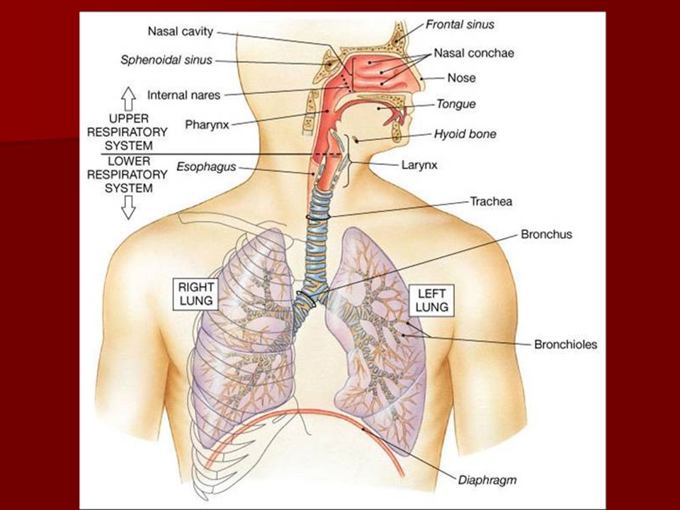
The Respiratory System
Functions to supply the body with O2 and remove CO2 There are actually 4 distinct processes: Ventilation – Movement of air into & out of the lungs External Respiration – Gas exchange between blood and air-filled chambers of the lungs Transport of Gases – Accomplished by Cardiovascular system Internal Respiration – Gas exchange between systemic blood and the tissue cells

4
Organs include: nose, nasal cavity, pharynx, trachea, bronchi, bronchioles, and the alveoli.
Divided into Respiratory and Conducting Zones. Gas exchange with the blood occurs in the respiratory zones. It does NOT occur in the conducting zones. The conducting zones transport, cleanse, warm and humidify the incoming air. Functional Anatomy

5
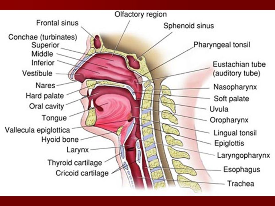
The Upper Airway Nose Oral Cavity Pharynx Larynx

7
The Nose Only externally visible part of the respiratory system.
Functions include: Providing an airway for respiration- Conduct Moistening and warming air- Warm and Humidify Filtering inspired air- Protect Serving as a resonating center for speech Housing the olfactory receptors.

8
Skeletal Framework of External Nose
Fashioned by the: Nasal and frontal bones superiorly Maxillary bones laterally Plates of hyaline cartilage (lateral, septal, and alar cartilages) inferiorly.
 inferiorly.")
9
Nasal Cavity Lies in and posterior to the external nose
Divided by a midline nasal septum – formed anteriorly by septal cartilage and posteriorly by the vomer bone and perpendicular plate of the ethmoid bone. Continuous with the nasopharynx via the internal nares. Roof is formed by the sphenoid & ethmoid bones.

10
Nasal Cavity Floor is formed by the palate.
Hard palate contains portions of the maxillary and palatine bones. Soft palate lacks bone, a flexible mass of collagen fibers

11
Nasal Cavity Lined by 2 types of epithelium.
Slit-like superior region is lined by olfactory epithelium. What does it do? The rest is line by respiratory epithelium (pseudostratified ciliated columnar with goblet cells) It rests on a connective tissue layer richly supplied with mucous and serous glands.
 It rests on a connective tissue layer richly supplied with mucous and serous glands.")
12
Nasal Cavity Produces 1 quart of mucus per day.

13
Mucus vs Sputum Sputum is matter that is coughed up from the respiratory tract, such as mucus or phlegm, mixed with saliva and then expectorated from the mouth. Mucus is a slippery secretion of the lining of the mucous membranes in the body.. Mucus is produced by goblet cells in the mucous membranes that cover the surfaces of the membranes.

14
Mucus Mucus is produced by goblet cells in the mucous membranes that cover the surfaces of the membranes. It is made up of mucins and inorganic salts suspended in water. Contains Lysosomes. Phlegm is a type of mucus that is restricted to the respiratory tract, while the term mucus refers to secretions of the nasal passages as well.

15
Mucus High H2O content of mucus humidifies inward air
Ciliary current moves mucus to pharynx for swallowing. Cold temps disable these cilia; runny nose Rich plexuses of capillaries and veins underlie the nasal epithelium and warm incoming air

16
Nasal Cavity Protruding medially from each lateral wall of the nasal cavity are 3 scroll-like, mucosa-covered projections: the superior, middle, and inferior conchae or turbinates They increase the mucosal surface area exposed to air The groove inferior to each concha is a meatus. Nasal cavity is surrounded by a ring of paranasal sinuses located in the frontal, sphenoid, ethmoid and maxillary bones.

19
Anatomy and Physiology Revealed
CUT AWAY OF UPPER AIRWAY

20
How Does this System function..
Air first enters the nares into a slightly dilated area called the vestibule. The vestibule is lined with hairs called vibrissae, a protective mechanism against foreign particles. PROTECT The anterior 1/3rd of the nasal cavity is lined with stratified squamous epithelium, posterior 2/3rd lined with pseudostratified ciliated columnar epithelium

21
How Does this System function..
Air then travels through the turbinates (conchae) where the function is to separate inspired air into separate streams. This allows for an increase of surface area. This increased surface area increases the temperature of the air and adds moisture. WARM and HUMIDIFY
 where the function is to separate inspired air into separate streams. This allows for an increase of surface area. This increased surface area increases the temperature of the air and adds moisture. WARM and HUMIDIFY.")
22
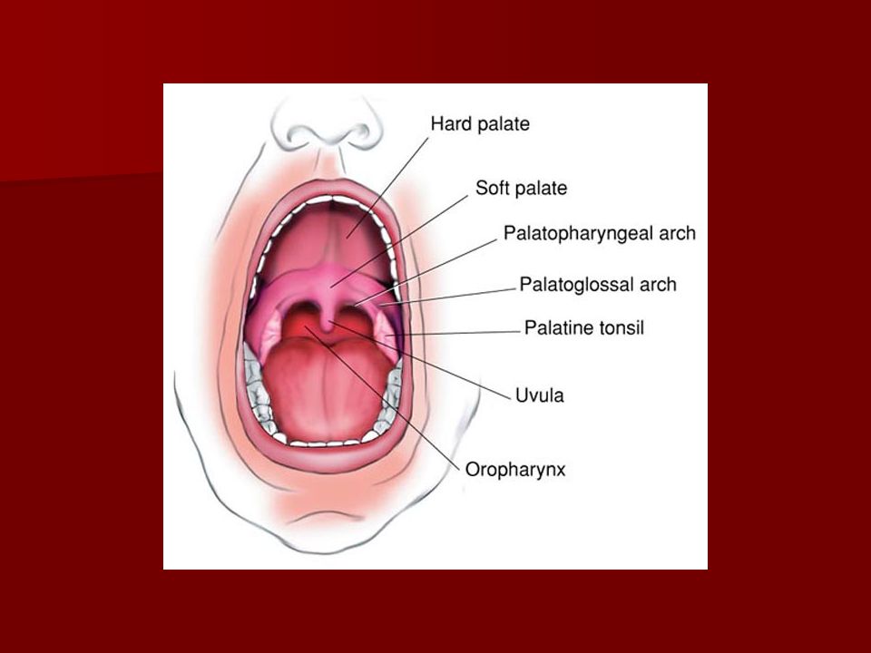
Oral Cavity Accessory Respiratory Passage
Lined with stratified squamous epithelium Air enters the vestibule (small outer portion between gums and lips) and large opening that extends to the back of the oropharynx Roof of the oral cavity Hard Palate Soft Palate Uvula
 and large opening that extends to the back of the oropharynx. Roof of the oral cavity. Hard Palate. Soft Palate. Uvula.")
23
Oral Cavity Soft palate rises, shutting off the passage between the nasal and oral cavity Levator veli palatinum muscle draws up and back Palatopharyngeal muscle draws down and forward Palantine Arches Palatopharyngeal Arch Palatoglossal Arch Contains the tonsils, adenoids and lymph tissue; Front line protection

25
Pharynx Funnel-shaped.
Connects the nasal cavity and mouth superiorly to the larynx and esophagus inferiorly 3 regions. From superior to inferior: Nasopharynx Oropharynx Laryngopharynx

26
Nasopharynx Posterior portion of the nasal cavity, superior portion of the soft palate, contains adenoids. Only an air passage. During swallowing, the soft palate and its uvula move superiorly and close it off. Lined by pseudostratified ciliated columnar epithelium. High on its posterior wall is the pharyngeal tonsil (adenoids) which traps entering pathogens. The eustachian tubes open into its lateral walls. They connect the middle ear to the nasal cavity; pressure release function
 which traps entering pathogens. The eustachian tubes open into its lateral walls. They connect the middle ear to the nasal cavity; pressure release function.")
27
Oropharynx Lies posterior to the oral cavity
Extends from the soft palate to the base of the tongue – hyoid bone Lined by stratified squamous epithelium Paired palatine tonsils lie in the lateral walls while the lingual tonsil covers the base of the tongue

28
Laryngopharynx Also called “hypopharynx”
Extends from the base of the tongue to the entrance of the esophagus. Common passage for both food and air Lined by stratified squamous epithelium

30
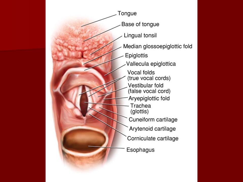
Larynx Superiorly attached to the hyoid bone and opens into the laryngopharynx. Inferiorly, it’s continuous with the trachea Main tasks are: Provision of a patent airway for air and food. Routing of air and food to proper pathways. Voice production.

32
Larynx Consists of an intricate arrangement of 9 cartilages connected by membranes and ligaments. 3 single cartilages Epiglottis, Cricoid, and Thyroid 3 paired cartilages Cuneiform, corniculate and arytenoid

33
The large, shield-shaped thyroid cartilage is formed by the fusion of 2 cartilage plates.
The fusion point is the laryngeal prominence (adam’s apple). The ridge is called the thyroid notch Inferior to the thyroid cartilage is the cricoid cartilage. Signet ring shape with increased size to the posterior. Cricoid membrane is the site for emergent airways. The first “C” shaped tracheal ring lies below the cricoid cartilage 3 pairs of small cartilages, the arytenoid, cuneiform, & corniculate cartilages form part of the lateral & posterior walls of the larynx The important arytenoids anchor the vocal cords
. The ridge is called the thyroid notch. Inferior to the thyroid cartilage is the cricoid cartilage. Signet ring shape with increased size to the posterior. Cricoid membrane is the site for emergent airways. The first C shaped tracheal ring lies below the cricoid cartilage. 3 pairs of small cartilages, the arytenoid, cuneiform, & corniculate cartilages form part of the lateral & posterior walls of the larynx. The important arytenoids anchor the vocal cords.")
34
The 9th cartilage (epiglottis) is spoon-shaped & composed of elastic cartilage.
Epiglottis is covered almost entirely by a taste-bud containing mucosa. During swallowing, the larynx is pulled superiorly and the epiglottis tips to cover the laryngeal inlet. If anything other than air enters the larynx – a cough/gag reflex is initiated by the sensory nerve; glossopharyngeal and the motor nerve; vagus.

36
Swallowing

38
Epiglottis and Vallecula
The space between the base of the tongue and the epiglottis is called the vallecula This is an important landmark in the airway While intubating, if using a macintosh blade the tip of the blade slides into the vallecula causing the epiglottis to lift. If using a miller blade, the epiglottis is directly lifted up to allow access to the airway.

41
Lying under the laryngeal mucosa on each side are the vocal ligaments These ligaments (made mostly of elastic fibers) form the core of mucosal folds called the vocal folds or true vocal cords Vocal cords vibrate, producing sounds as air rushes up from the lungs. Superior to the true vocal cords is a similar pair of mucosal folds called the vestibular folds or false vocal cords. The superior portion of the larynx is lined by stratified squamous epithelium, while below the vocal cords, it’s a pseudostratifed ciliated columnar epithelium.

42
The medial opening between them thru which the air passes is the rima glottidis or GLOTTIS.
In an adult, the glottis is the narrowest point of the adult larynx. In an infant and small child, the cricoid cartilage is the narrowest point. Subglottic swelling in an infant or small child, due to infection or trauma can cause stridor during inspiration

43
Epiglottitis vs Croup (LTB)
")
44
Croup – Steeple Sign

45
Epiglottitis – Thumb Sign

46
The larynx is closed by the epiglottis during swallowing.
In addition to opening and closing the glottis for speech, the vocal folds can act as a sphincter during conditions such as coughing, sneezing or straining

50
The Arytenoid cartilage is shaped like a pyramid which rests on the posterior portion of the cricoid cartilage At the base of the arytenoid cartilage a projection called the vocal process, the vocal ligaments attach vocal process and the thyroid cartilage The cuneiform and corniculate are accessory cartilages at the superior portion of the arytenoids

53
Animated Airways..

55
Laryngeal Musculature
Extrinsic Infrahyoid (below the hyoid) Pulls the larynx and hyoid down the neck Sternohyoid, sternothyroid, throhyoid and omohyoid Suprahyoid (above the hyoid) Pulls the hyoid bone forwards, upwards and backwards Stylohyoid, mylohyoid, digastric, geniohyoid and stylopharyngeus
 Pulls the larynx and hyoid down the neck. Sternohyoid, sternothyroid, throhyoid and omohyoid. Suprahyoid (above the hyoid) Pulls the hyoid bone forwards, upwards and backwards. Stylohyoid, mylohyoid, digastric, geniohyoid and stylopharyngeus.")
56
Laryngeal Musculature
Intrinsic They all deal with the arytenoid cartilage and vocal cord movement. Posterior cricoarytenoid, lateral cricoarytenoid, transverse, thyroarytenoid and cricothyroid.

58
Ventilatory Function of the Larynx
Ensures a free flow of air to and from the lungs During inspiration, vocal cords move apart; abduct, and widens glottis for improved airflow Forced expiration against a closed glottis, “Valsalva’s maneuver” causes massive adduction preventing air from escaping during cough, vomiting, urination, defecation and parturition. Forced inspiration against a closed glottis, “Mueller maneuver” --- missed sputum bowl question!

60
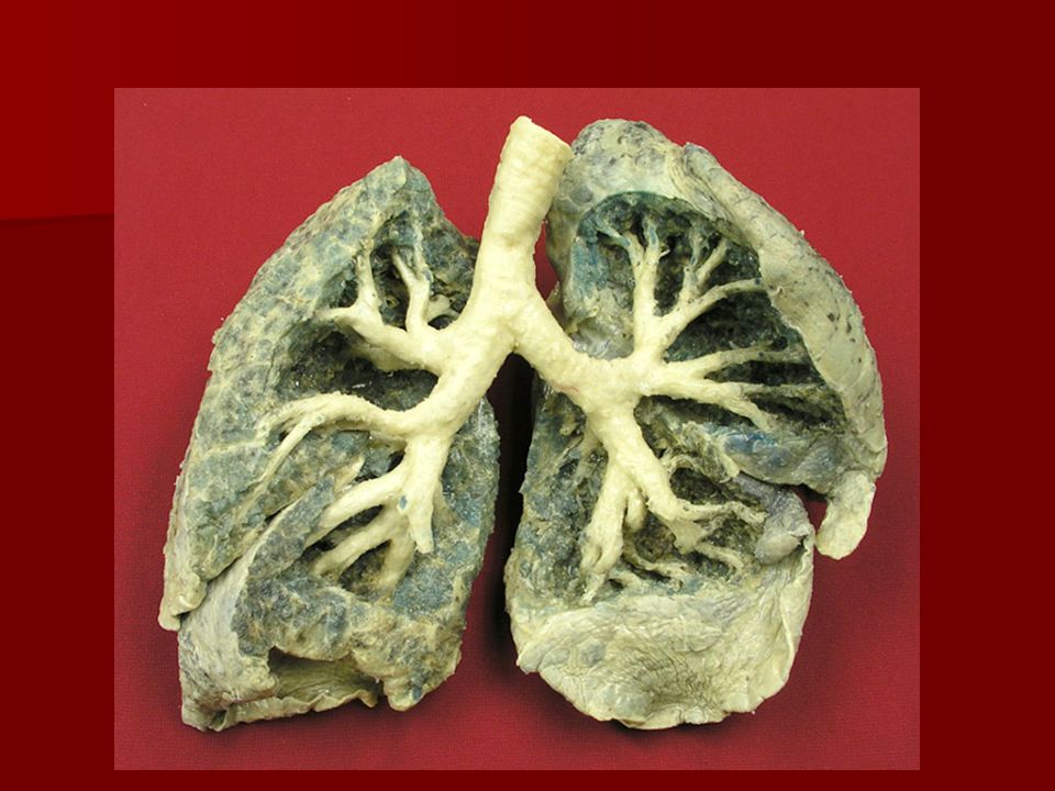
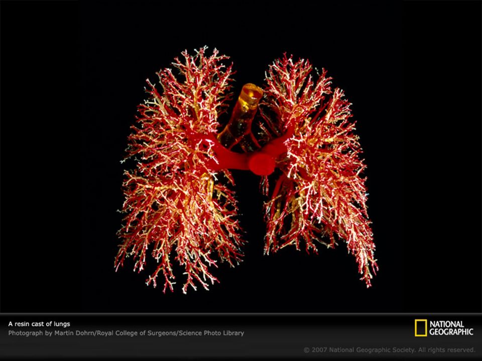
The Lower Airways After passing through the larynx, inspired air enters the Tracheobronchial Tree The traceobronchial tree consists of a series of branching airways called “orders” or “generations” It is believed that there are 28 generations or orders of the tracheobroncial tree

61
Dichotomous Branching

63
Tracheobronchial Tree
The tracheobronchial tree is divided into two general zones Conducting Zone or Cartilaginous Airways No Gas Exchange occurs in this zone Respiratory Zone or Non Cartilaginous Airways The site of Gas Exchange There a transition zone where no cartilage surrounds the airway, yet no gas exchange occurs

64
Histology of the Tracheobroncial Tree
Three layers Epithelial Lining Lamina propria Cartilaginous Layer

66
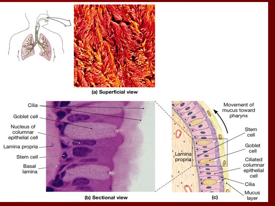
Epithelial Lining Psuedostratified ciliated columnar epithelium
Numerous Mucous glands interspersed Anchored to a basement membrane that contains basal cells (reserve cells and replenish mucus glands and ciliated cells) 200 cilia per cell Cells move from columnar to cuboidal and cilia disappear as you move down the tree
 200 cilia per cell. Cells move from columnar to cuboidal and cilia disappear as you move down the tree.")
68
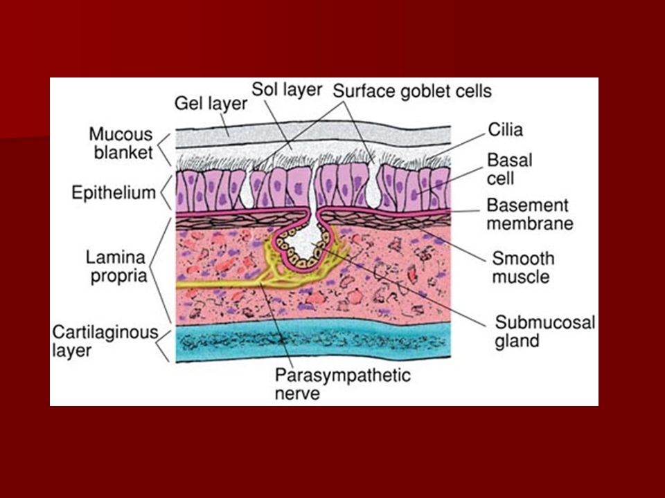
Epithelial Lining A mucus layer, or “mucous blanket” covers the epithelial lining of the tracheobronchial tree. Produced by goblet cells and submucosal /bronchial glands Goblet cells are located between the epithelial cells Submucosal glands extend into the lamina propria and are innervated by the parasympathetic nervous system. Composed of 95% water and the remainder is carbohydrates, glyocproteins, lipids, DNA, cellular debris and foreign particles.

70
Mucous Blanket The body produces about 100ml of secretions per day.
The viscosity of the secretions increase as you move from the lining to the lumen. Two distinct layers SOL Layer, adjacent to the epithelial lining Less viscous GEL Layer, adjacent to the inner lumen More viscous

71
Mucous Blanket Cilia move in a wavelike fashion, beating 1500 times per minute through the less viscous sol layer and strike the inner layer of the more viscous gel layer. This action propels the mucus layer, along with any foreign particles attached to the “sticky” gel layer towards the larynx at 2cm per minute The cough mechanism moves the secretions above the larynx and into the oropharynx

72
Mucous Blanket This cleansing process is called Mucociliary Transport or the Mucociliary Escalator What slows the rate down: Cigarette smoke, Dehydration Positive Pressure Ventilation Endotracheal Suctioning High FiO2 Hypoxia Atmospheric pollutants General anesthetics Parasympatholytics

73
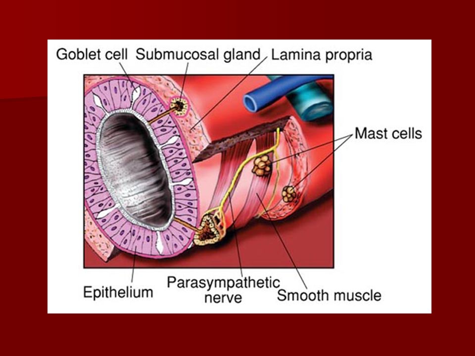
Lamina Propria This is a submucosal layer
Loose fibrous tissue containing blood vessels, lymphatic vessels, vagus nerve innervation Two sets of smooth muscle that wrap in spirals both clockwise and counterclockwise The smooth muscle fibers extend to the alveolar ducts The lamina propria is surrounded by a thin connective tissue layer called the peribronchial sheath

75
Lamina Propria Mast Cells are located in the lamina propria
Their cytoplasm is loaded with granules containing mediators of inflammation. Histamine, heparin, SRS-A (slow reacting substance of anaphylaxis), PAF (platelet activating factor), ECF-A (eosinophilic chemotaxic factor of anaphylaxis) Their surface is coated with a variety of receptors which, when engaged by the appropriate antigen trigger exocytosis of the granules. Destablization of mast cells in the lungs can be extremely dangerous, and is what we see in patients with an allergic asthmatic episode
, PAF (platelet activating factor), ECF-A (eosinophilic chemotaxic factor of anaphylaxis) Their surface is coated with a variety of receptors which, when engaged by the appropriate antigen trigger exocytosis of the granules. Destablization of mast cells in the lungs can. be extremely dangerous, and is what we see in patients with an allergic asthmatic episode.")
77
Cartilaginous Layer Outermost layer of the tracheobronchial tree
This layer progressively diminishes in size as the airway extend into the lungs. Cartilage is absent in bronchioles less than 1 mm in diameter

79
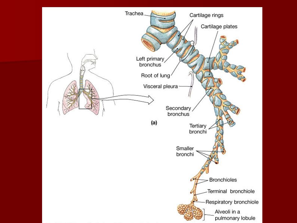
Lower Airway Cartilaginous Airways
Trachea Main stem Bronchi Lobar Bronchi Segmental Bronchi Subsegmental Bronchi THE CONDUCTING ZONE

80
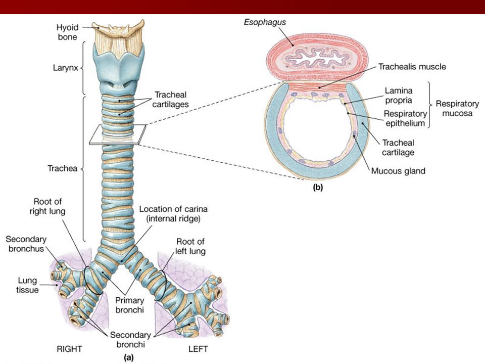
Trachea In the adult, it is 11 to 13 cm long, 1.5 – 2.5 cm in diameter
Descends from the cricoid to the 2nd costal cartilage ANGLE OF LOUIS It bifurcates, divides into the right and left main stem bronchi, this division is called the carina

81
There are C-shaped rings of cartilage that support the trachea, keeping the airway patent and prevent its collapse. The open posterior parts of the rings are adjacent to the the esophagus and are connected by fibers of the trachealis muscle, which is involved in coughing. .

83
Main Stem and Lobar Bronchi
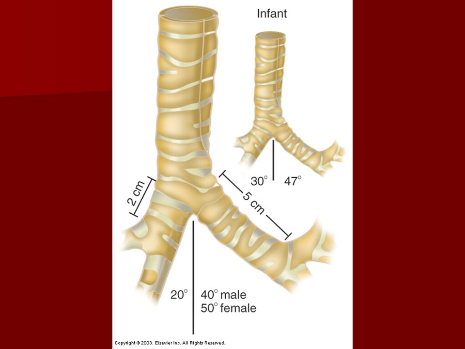
The Main Stem Bronchi are the 1st generation of the tracheobronchial tree The right main stem bronchus branches off the trachea at a 25 degree angle, the left bronchus forms a 40 – 60 degree angle The right bronchus is wider, more vertical, and 5 cm shorter than the left.

86
Main Stem and Lobar Bronchi
Main Stem bronchi are supported by ‘C’ shaped cartilage Each bronchus runs obliquely into the mediastinum before plunging into the medial depression (hilus) of the lung on its own side. Inside the lungs, the main stem or primary bronchi divide into lobar or secondary bronchi, 3 on the right and 2 on the left, each of which supplies one lung lobe. 3 lobes on the right, 2 lobes on the left (room for heart)
 of the lung on its own side. Inside the lungs, the main stem or primary bronchi divide into lobar or secondary bronchi, 3 on the right and 2 on the left, each of which supplies one lung lobe. 3 lobes on the right, 2 lobes on the left (room for heart)")
90
Bronchi and Subdivisions
The lobar bronchi become segmental; third generation. 10 in the right lung and 8 in the left lung Subsegmental bronchi range in diameter from 1 – 4 mm. Peribronchial sheaths containing nerves, vessels and lymphatic tissue surround the subsegmental bronchi down to the 1 mm These are the 4th – 9th generation.

91
Non Cartilaginous Airways
Bronchioles When the diameter decreases to less than 1mm, they are no longer surrounded by a connective sheath, cartilage is absent, rigidity is absent-airway patency can be compromised A muscle sheath surrounding the bronchioles Columnar epithelial becomes cuboidal Generations

92
Terminal Bronchioles 16th – 19th Generation;
Diameter is about 0.5 mm Cilia and mucous glands progressively disappear Epithelium is cuboidal and thin Channels called “Canals of Lambert” appear Connect the surface of terminal bronchioles to adjacent alveoli Thought to aid in collateral ventilation in individuals with respiratory disorders such as COPD Presence of Clara Cells Function unknown, they have a thick protoplasmic extensions that bulge into the bronchial lumen - perhaps they secrete an enzyme that detoxifies inhaled substances 16th – 19th Generation; TERMINAL- END: Structures beyond this point are not part of the Tracheobronchial tree. Structures distal to this point are the sites of gas exchange; referred to as the Respiratory Zone

96
As conducting tubes become smaller…
The cartilage support changes. It goes from rings in the trachea to irregular plates in the bronchi to none in the bronchioles. Why? The epithelium changes. It goes from respiratory to simple columnar to simple cuboidal. The number of cilia and goblet cells present decrease. The amount of smooth muscle increases.

97
Gas Flow Once in the Respiratory Zone, the cross sectional area of the lung increases exponentially. Forward motion of gas flow stops-no bulk flow, no laminar flow The movement of gas becomes molecular

100
Bronchial Blood Supply
Tracheobronchial tree requires blood flow Arteries follow the tracheobronchial tree as far as the terminal bronchioles Beyond the terminal bronchioles, this vascular system merges with the pulmonary vascular system. RA – RV– PA - Lungs – LA – LV –Aorta - Systemic

101
Bronchial Blood Supply
Approximately 1% of Cardiac Output serves the tracheobronchial tree. Of that 1%, 1/3rd of it returns to the Right Atrium as unoxygenated venous blood. The vessels responsible for this return Azygos Hemiazygos Intercostal veins RA – RV – PA - Lungs – LA – LV –Aorta - Systemic

102
via bronchopulmonary anastomoses
The remaining 2/3rd of the bronchial venous blood drains into the pulmonary circulation, into the pulmonary arteries and capillaries via bronchopulmonary anastomoses This results in a mixture of low oxygenated and high carbon dioxide blood from the tracheobronchial tree with highly oxygenated low carbon dioxide blood returning to the left atrium for systemic circulation RA – RV – PA - Lungs – LA – LV –Aorta – Systemic 2/3rd of blood returning from bronchical circulation This is an example of an anatomical shunt, and called venous admixture

103
The Respiratory Zone Distal to the terminal bronchioles are the functional units of gas exchange Consists of: 3 generations of Respiratory Bronchioles 3 generations of Alveolar Ducts Ending in grapelike clusters called Alveolar Sacs These units, Respiratory Bronchioles, Ducts and Alveoli are called a primary lobule or acinus, or terminal respiratory unit or lung parenchyma or functional units

105
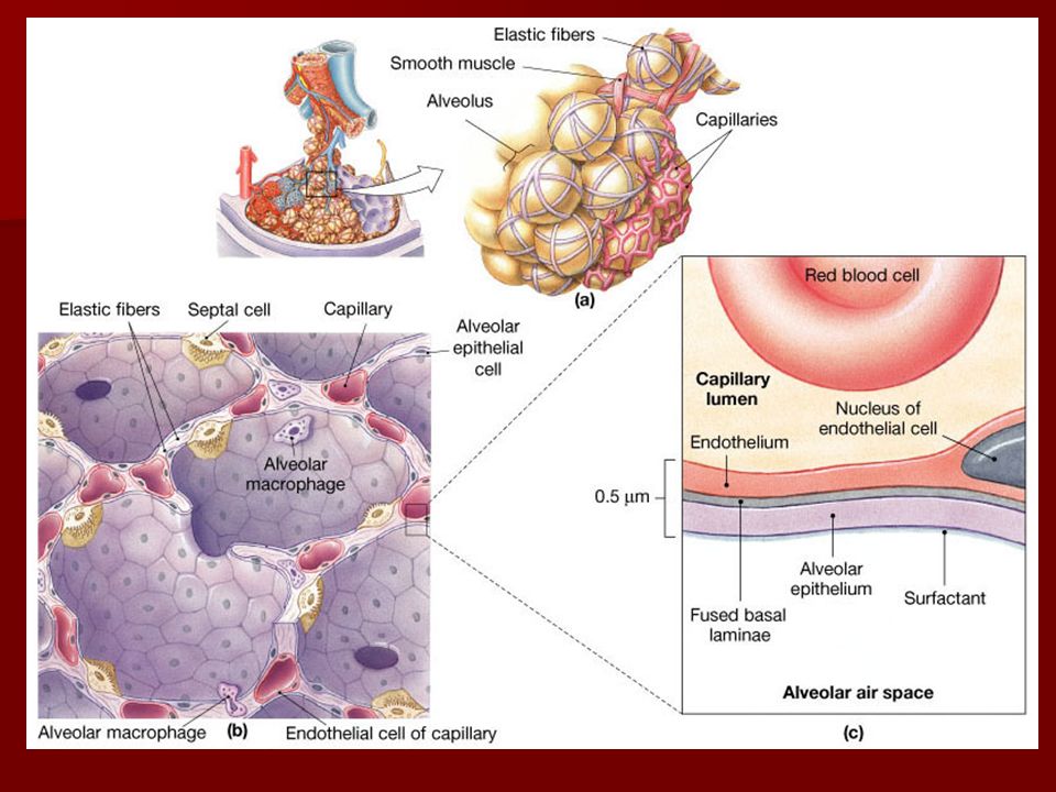
Alveoli Composed of smooth muscle fiber
Approximately 300 million alveoli which are 90% covered with capillaries. The surface area of the alveoli is 70 square meter, the surface of a tennis court Each primary lobule, 130,000 each stem from a single terminal bronchiole and contains about 2000 alveoli

107
Alveolar Epithelium Alveoli are consist of 3 cell types:
Type I cells; Squamous Epithelium Cover 95% of the alveolar surface 0.1 – 0.5 micrometers thick Major site of gas exchange Type II cells; Granular pneumocytes Have microvilli Cuboidal in shape Produce pulmonary surfactant Form the remaining 5% of the alveolar surface Type III cells; Alveolar macrophages Migrate through the blood stream and are embedded in the extracellular lining of the alveoli

109
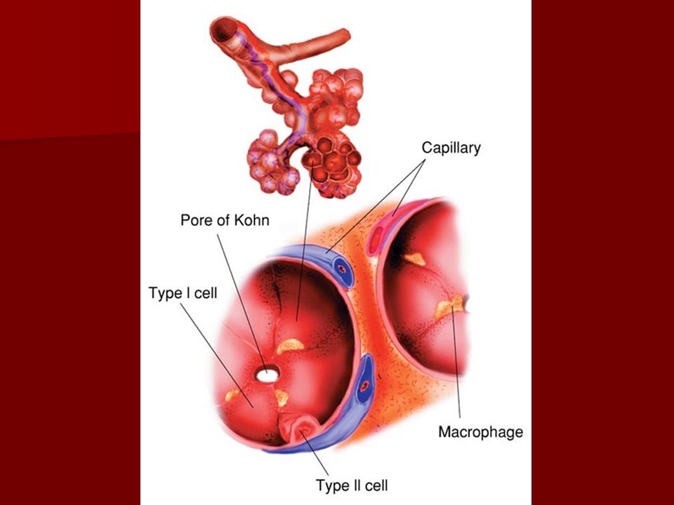
Pores of Kohn Pores of Kohn
Small holes in the alveolar wall or interalveolar septa Allow gas to move between alveoli Formed by Shedding of cells – desquamation Normal degeneration due to age Movement/detachment of macrophages

110
Scattered among the Type I’s are Type II cells which secrete surfactant.
Pores of Kohn connect adjacent alveoli. Alveolar macrophages (dust cells) crawl along the internal alveolar surfaces
 crawl along the internal alveolar surfaces.")
111
Interstitium Surrounds, supports and shapes the alveolar capillary clusters Gel like substance of hyaluronic acid molecules bound together by a network of collagen fibers Two compartments Tight Space; the area between the alveolar epithelium and the endothelium of the pulmonary capillaries-Site of gas exchange Loose Space: the area the surround the bronchioles, respiratory bronchioles, alveolar ducts and sacs. Lymphatic vessels and neural fibers are in this area

114
The Pulmonary Vascular System
Function is to deliver blood to and from the lungs for gas exchange It also supplies nutrition to portions of the lung distal to the terminal airways

117
Pulmonary Vascular System
Arteries Arterioles Capillaries Venules Veins

118
Pulmonary Arteries Three Layer
Tunica Intima; inner layer Endothelium and a thin layer of connective tissue Tunica Media; middle layer Thickest layer of the vessel Elastic connective tissue in large arteries, smooth muscles in smaller and medium arteries Tunica Adventitia: outer layer Connective tissue Contains small vessels that nourish all layers Stiff vessels, capable of carrying blood under high pressures.

119
Arterioles Endothelial layer Elastic layer Smooth muscle fibers
Called “resistance vessels” due to the ability of the smooth muscles to regulate the blood flow

120
Capillaries Surround 90% of the alveoli
Composed of endothelial layer; single layer of squamous epithelial cells Essentially an extension of the inner lining of the larger vessels This is where gas exchange occurs Some prostaglandins are produced here, some biological substances are destroyed here

121
Veins and Venules 3 Layers, same as arteries, 2 layers in the smaller veins-no tunica adventitia Middle layer is poorly developed and contain less smooth muscle and elastic tissues Due to less elastic and smooth muscle tissue, veins can hold a greater volume of blood with little pressure changes. Because of this, veins are called “capacitance vessels” Veins return to the heart in a more direct route out of the lungs

122
The Lymphatic System Function is to remove excess fluid and protein that leak out of the capillaries Located in a dense connective tissue sheath around the bronchioles, also in the loose space of the interstitium More lymphatic channels located on the left side, increased incidence of right sided pleural effusions due to less drainage Like veins, they have one way valves/flaps Bronchopulmonary lymph nodes, end of the line, are located outside the lung parenchyma No lymph vessels in the alveoli, they are located immediately adjacent to the alveoli called juxta-alveolar lymphatics

124
Neural Control of the Lungs
Controlled by the autonomic nervous system Regulates involuntary vital functions Cardiac muscles Smooth muscles Glands Two divisions Sympathetic Parasympathetic

125
Neural Control of the Lungs
Sympathetic Accelerates heart rate Constricts blood vessels Relaxes bronchial smooth muscles Raises blood pressure Parasympathetic Slows heart rate Constricts bronchial smooth muscles Increases intestinal peristalsis and gland activity

126
Neural Control of the Lungs
Sympathetic neural transmitters Epinephrine Norepinehrine These agents stimulate the Beta 2 receptors in the bronchial smooth muscles; causing airway muscle relaxation Alpha receptors in the arteriole smooth muscles, causing the pulmonary vascular system to contrict

127
Neural Control of the Lungs
Parasympathetic neural transmitters Acetylcholine Causes constriction of the bronchial smooth muscles Inactivity of either portion allows the other one to dominate the bronchial smooth muscles There must be careful attention to the role of pharmacological agents Beta blockers- causes parasympathetic to dominate Atropine-a parasympathetic blocker allows sympathetic to dominate

129
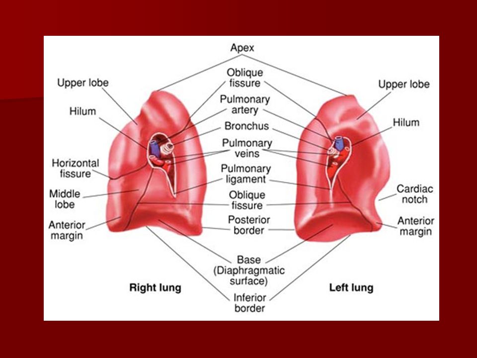
Lung Gross Anatomy Occupies all of the thoracic cavity except the mediastinum. Each lung is within its own pleural cavity Anterior, lateral, and posterior surfaces are costal, adjacent to ribs Rises above the clavicle to the level of the 1st rib The concave bases sit on the diaphragm. The mediastinal border is concave to heart and the other mediastinal structure The hilum is at the center of the mediastinal border, and is where the main stem bronchi, blood and lymph vessels, and nerves enter and exit the lungs

134
Lung Gross Anatomy The right lung is larger and heavier than the left. The Right lung has 3 lobes; upper, middle, and lower The lobes are divided by the oblique fissure which divides the upper and middle lobe from the lower lobe Horizontal fissure, divides the upper from the lower lobe. .

135
Lung Gross Anatomy The left lung is smaller than the right, contains 2 lobes; upper and a lower lobe and has an indentation (cardiac notch) where the heart sits. The lobes are divided by the oblique fissure
 where the heart sits. The lobes are divided by the oblique fissure.")
136
Lung Segments All Lobes are further divided into bronchopulmonary segments 10 on the right 8 on the left Careful with the numbering systems!

137
Mediastinum A cavity that contains the organs and tissues in the center of the thoracic cage between the right and left lung Bordered anteriorly by the sternum, and posteriorly by the vertabrae Contains the trachea, heart, the great vessels the major vessels that enter and exit the heart, the esophagus, thymus gland, lymph nodes, and nerves

139
The Pleural Membranes Two moist, slick surfaced membranes
Parietal pleura covers the thoracic wall, superior diaphragm and lateral portion of the mediastinum Visceral pleura firmly attached and covers the external lung surface, extends into the interlobar fissures The potential space between visceral and parietal pleurae is called the pleural cavity The two membranes are held together by a thin film of serous fluid. The fluid allows the membranes to glide over each other during inspiration and exhalation The pleural membranes hold the lung tissue to the inner surface of the thorax and diaphragm, allowing for lung expansion during inspiration

141
Because the lungs have a natural tendency to collapse and the thorax has a natural tendency to expand, a negative pressure normally exists between these two layers. Should air enter this space, the pleural membranes would separate causing a condition known as a pneumothorax

143
The Diaphragm The diaphragm is the major muscle of inspiration
Dome-shaped musculofibrous partition located between the thoracic cavity and the abdominal cavity Two separate muscles; the right and left hemidiaphragm, joined at midline by the central tendon Pierced by the esophagus, aorta, nerves and the inferior vena cava Innervated mainly by the phrenic nerve, the lower thoracic nerves contribute to some motor innervation

145
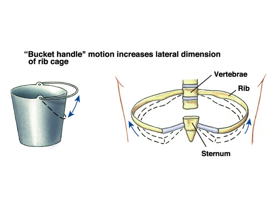
Inspiration When the diaphragm is stimulated to contract, it moves downward and the lower ribs move upward and outward This increases the thoracic volume Which causes the lung volume to increase The increased lung volume causes lung pressure; intrapleural and intra alveolar to decrease As a result, gas from the atmosphere flows into the lungs

149
During expiration, the diaphragm relaxes and moves upward into the thoracic cavity
This increases the intrapleural and intra-alveolar pressures and causes gas to flow out of the lungs Quiet expiration is a passive process that is due to the elasticity of the lungs. Forced expiration is an active process due to contraction of oblique and transverse abdominus muscles, internal intercostals, and the latissimus dorsi. Expiration

Similar presentations


. 2.Production of sound (vocal cords). 3.Pulmonary ventilation. 4. Inspiration (intercostals muscles lift.>")


