Download presentation
Presentation is loading. Please wait.

1
Procedures in Dermatology
Rich Callahan MSPA, PA-C ICM I Summer 2009

2
Overview Shave biopsy Punch biopsy Incision & Drainage (I&D)
Electrodessication & Curettage (ED&C) Excisions Cryotherapy
 Excisions. Cryotherapy.")
3
What is a skin biopsy? A skin biopsy is a diagnostic procedure in which a portion of skin (and/or subcutis) is submitted to the pathology lab. This specimen is fixed, sectioned and placed on slides for histologic analysis Special stains can be used to detect fungus, bacteria, immune complexes, lymphocytes, inflammatory mediators, arthropods, etc. The hope is that the pathologist can provide more information to aid in diagnosing the disease.

4
The biopsy is only as good as the specimen you provide
An inadequate specimen usually results in a biopsy report including the dreaded words: “small quantity of tissue provided precludes a definitive diagnosis.” or “superficial representation of dermis in specimen inadequate for full characterization of disease process.”

5
Skin Biopsy A good skin biopsy is one that provides an adequate specimen for the pathologist to review while at the same time using the utmost care and knowledge of anatomy to minimize the potential morbidity of the procedure. Also involves post-biopsy wound care, knowledge of anatomical danger zones, patient education.

6
Why do a skin biopsy? Skin biopsies usually provide diagnostic information that adds to the clinical picture already at hand. Many skin diseases have characteristic findings on routine histology that are highly diagnostic Biopsy results which support the clinical diagnosis tend to confirm it. Biopsy results that don’t support the clinical diagnosis cast it into doubt Biopsy results that don’t make sense at all should be viewed with skepticism – (the lab makes mistakes too!) You might have to biopsy a lesion several times prior to definitive diagnosis.
 You might have to biopsy a lesion several times prior to definitive diagnosis.")
7
Why do a skin biopsy? Ascertain benign vs. malignant, infectious vs. autoimmune, exogenous vs. endogenous process, etc. If strongly suspect skin cancer, biopsy can generate information such as subtype, differentiation, depth of invasion, type of spread, etc, which guides appropriate choices for treatment

8
Keep in mind, skin biopsy not necessary if….
The clinical picture is entirely diagnostic. If patient history and PE findings strongly point to a specific diagnosis, and you feel comfortable in diagnosing on clinical grounds alone, don’t do a skin biopsy. If the disease doesn’t respond to treatment or doesn’t follow the expected clinical course, then biopsy may be necessary.

9
Biopsy by shave technique
Removal of representative piece of skin by tangential incision with a blade. Can use scalpel or Dermablade I almost always use a Dermablade – basically a sharp, thin, extremely flexible razor blade. Idea is to sample both lesional and normal-appearing perilesional skin Depth needs to get down to at least superficial upper dermis – biopsies of epidermis only usually unsatisfactory. Some skin diseases require sampling of mid to deep dermis for diagnosis.

11
When to do a shave Patient who isn’t as concerned with scarring – by definition any defect which reaches down to mid-dermis will scar. When you know it’s skin cancer – it’s going to get removed anyway! Shave provides a quick and accurate specimen and there will be a scar from treatment anyway.

12
When to do a shave In sensitive anatomic locations where the depth of a punch biopsy puts nerves/blood vessels at risk (anatomic danger zones.) The highly active patient: Shave biopsy wounds have no limitation on activity. The patient who can’t/doesn’t want to come back for suture removal from punch biopsy.

13
How to do a shave Inform patient of potential for scarring!
Anesthetize the area for biopsy, starting with the subcutis and working you way up to the dermoepidermal junction (bleb or peau d’orange.) Map in your mind or with a surgical pen the specimen you are trying to collect beforehand (as with many things in life/work, it helps to have a plan first!) Create skin tension with hands or an assistant’s hands.
 Map in your mind or with a surgical pen the specimen you are trying to collect beforehand (as with many things in life/work, it helps to have a plan first!) Create skin tension with hands or an assistant’s hands.")
14
How to do a shave Grasp blade between thumb and index finger, place edge against skin and rotate hand in a gentle back-and-forth motion which allows the blade to saw through the tissue. Point the blade slightly downwards until you are under the middle of your planned specimen, then point slightly upwards until finished Goal is for a saucer-shaped specimen providing adequate representation of the skin lesion.

15
Shave biopsy – Wound Care
Resulting defect is usually a circular to ovular extending down into papillary to mid-reticular dermis. Hemostasis with aluminum chloride/Monsel’s solution for minimal bleeding/Electrocautery for moderate bleeding. After hemostasis achieved, ointment and occlusive dressing are applied. Important that patient educated on keeping would moist and occluded until healed. Wound bed is populated by granulation tissue and fibroblasts hours post procedure. These cells thrive in a moist, low-oxygen environment! Scab formation greatly slows down the wound healing process. Scab formation greatly slows healing process

16
Biopsy by punch technique
Removal of a representative piece of skin and subcutis with a trephine, or punch Best way to look at it is like a little cylindrical cookie-cutter which punches all the way through the skin Usually a more involved procedure than shave needing extra time for anesthesia, hemostasis and would closure

18
When to do a punch Punch superior for any skin diseases where a picture of the deep dermis/subcutis is diagnostic. Tends to provide more information for inflammatory skin disorders, as they tend to involve greater depth of dermis Usually a better choice for the scar-averse patient, although it is no guarantee as even the best punch biopsy closure can dehisce. Better choice for deeply-seated lesions in dermis and subcutis.

19
How to do a punch Plan/map out the specimen you want
Anesthetize the area, with particular care in the subcutis and deep dermis. Carefully align the trephine with the skin, and then gently push down/twist in one direction. Gently pinch the skin around the area with your free hand.

20
How to do a punch You will feel considerable resistance throughout the dermis The trephine will then punch through to the subcutis, which feels to the hand like a sudden decrease in resistance to the trephine’s blade. Gently grab the specimen with pickups and lift it out (a crushed specimen is an inferior specimen) Usually specimen lifts right out with small amount of subcutis attached. If not, trim with scissors to include small amount adherent fat.
 Usually specimen lifts right out with small amount of subcutis attached. If not, trim with scissors to include small amount adherent fat.")
21
How to do a punch Hemostasis then obtained with combination of manual pressure, electrocautery or aluminum chloride solution. Never forget: Pressure is the King of Hemostasis! Wound then closed with sutures, or can be left to heal by second intention (warn patient extended wound care for 1-4 weeks in these cases) Anatomical Danger zones Superficial motor nerves - skin is loaded with sensory nerves If you transect a motor nerve you lose all function to muscles - Funny bone (ulnar nerve) - Coffee table nerve (fibular nerve/head of fibula) - Herb’s point (11th cranial nerve – spinal accessory nerve) – lose levator scapulae /draw a line from ear lobe to mandibular angle, go down ~6cm. Anywhere within a 1cm radius Temporal branch of facial nerve Mandibular – lateral jaw line Can pinch and raise dermis to do punch which separates dermis from subc
 Anatomical Danger zones. Superficial motor nerves. - skin is loaded with sensory nerves. If you transect a motor nerve you lose all function to muscles. - Funny bone (ulnar nerve) - Coffee table nerve (fibular nerve/head of fibula) - Herb’s point (11th cranial nerve – spinal accessory nerve) – lose levator scapulae /draw a line from ear lobe to mandibular angle, go down ~6cm. Anywhere within a 1cm radius. Temporal branch of facial nerve. Mandibular – lateral jaw line. Can pinch and raise dermis to do punch which separates dermis from subc.")
22
Incision and Drainage (I&D)
Treatment of choice for abscess, furuncle and carbuncle – inflammatory collections of pus and damaged tissue secondary to infection Drainage of these lesions tends to lead to quick resolution and provides material for culture should specific antimicrobial treatments become important

23
Incision and Drainage (I&D)
Local anesthesia is obtained, and then a moderate incision is made immediately adjacent to the head, or “point” of the lesion. Contents can occasionally be under significant pressure! Majority of abscess contents then squeezed out with bimanual pressure.

24
Incision and Drainage (I&D)
Pus drainage/necrotic tissue can be collected for culture Abscess can then be explored with a small curette to free up any loculations and adherent debris Wound is then flushed several times with saline solution. Wound left to heal by second intention with/without sterile packing Do no stitch as they need to drain

25
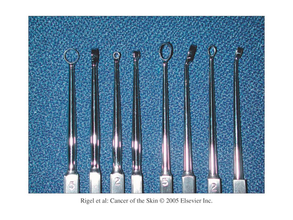
Electrodessication and Curettage (ED&C)
Essentially a process whereby superficial cancerous and pre-cancerous growths are removed from the skin by repeated scraping and burning. An effective, safe, expedient means of treating certain skin cancers in certain locations. Scrape and burn

29
Electrodessication and Curettage (ED&C)
Indicated for SCC in situ, superficial and selected nodular BCC. Works best on trunk and extremities in non- hairbearing areas Extreme caution on scalp, neck and high-risk areas of the face. Do it 3x

30
Electrodessication and Curettage (ED&C)
After appropriate regional anesthesia is obtained, a curette is passed over the lesion with firm pressure in back and forth strokes. Curettage is alternated with passes with electrocautery for hemostasis. Technique is guided by feel – skin cancer yields easily to the blade, whereas healthy dermis is quite tough and leathery. When you reach firm dermis with regular pinpoint bleeding, you are done. Do it 3x

31
Excision Procedure whereby a full thickness specimen of skin is removed either for therapeutic or diagnostic purposes. Excisions usually in elliptical shape oriented along skin tension lines (Langer’s lines.) Can be left to heal by second intention or closed by simple or intermediate repair. Procedure learned by seeing/doing. We will only cover basic concepts here!
 Can be left to heal by second intention or closed by simple or intermediate repair. Procedure learned by seeing/doing. We will only cover basic concepts here!")
32
Why do an excision? Usually done to completely remove a lesion for therapeutic reasons: Skin cancer Dysplastic nevus (abnormal mole) Epidermal inclusion cyst
 Epidermal inclusion cyst.")
33
Why do an excision? Can also be used for excisional biopsy, which is when the larger, full-thickness specimen obtained by excisional technique is needed for diagnostic purposes. Example: Initial punch biopsy shows features suggestive of CTCL (cutaneous T-cell lymphoma) but pathology needs substantially more tissue for gene rearrangement studies to further classify the malignancy.
 but pathology needs substantially more tissue for gene rearrangement studies to further classify the malignancy.")
34
How to do an excision First of all, know what you are getting in to.
Excisions on the face, fingers, genitals out of the scope of practice of most PA’s. These regions have superficial blood vessels, motor nerves, sensitive anatomical features requiring advanced training/familiarity to work with.

35
How to do an excision Scalp is richly vascularized – one can quickly get into bleeding that is difficult to control Lower legs/feet – slowest healing parts of body and also more prone to infection. Careful in the elderly as concomitant diabetes/stasis disease can predispose to complications such as poor healing, wound infection and dehiscence.

36
How to do an Excision Plan your surgical margins and orientation of your ellipse with skin tension lines Most excisions have elliptical shape to diminish wound tension after closure Sterilize/Prep/Anesthetize skin and subcutis and surrounding areas Cut specimen out in fusiform fashion holding scalpel perpendicular to skin surface Blade is turned purely by rotation of blade handle to keep its downwards track as vertical as possible

37
Surgical Excision After specimen is removed, hemostasis achieved by pressure, electrocautery and ligature – important to minimize chances of hematoma formation, infection and necrosis Undermining of surrounding dermis may be necessary to reduce wound tension, reduce healing time, better cosmetic outcome and minimize necrosis and/or wound dehiscence

38
Closure Simple Repair: Wound is closed with single layer of top stitches: Non-absorbable suture material tied in interrupted or running fashion – penetrates both epidermis/dermis Intermediate repair: Wound closed with buried layer of interrupted/ absorbable sutures which encompass entire dermis and up to dermoepidermal junction

39
Wound Care Minimize activity at least until top stitches are out as more activity = more risk for fluid accumulation, hematoma formation, infection, wound dehiscence and gaping scars Keep surface of wound moist and occluded with petrolatum or polysporin/bacitracin ointment and non-adherent wound dressings

Similar presentations