Download presentation
Presentation is loading. Please wait.

1
Welcome Back!! The SL material you learned last year is important to review before each unit this year… Today’s Opener: Draw and label a simple diagram of the molecular structure of DNA. (4 marks)
")
3
Happy Monday! 8/23/2010 The structure of the DNA double helix was described by Watson and Crick in Explain the structure of the DNA double helix, including its subunits and the way in which they are bonded together. (Total 8 marks)
.")
4
subunits are nucleotides;
one base, one deoxyribose and one phosphate in each nucleotide; description / diagram showing base linked to deoxyribose C1 and phosphate to C5; four different bases – adenine, cytosine, guanine and thymine; nucleotides linked up with sugar-phosphate bonds; covalent / phosphodiester bonds; two strands (of nucleotides) linked together; base to base; A to T and G to C; hydrogen bonds between bases; antiparallel strands; double helix drawn or described; Accept any of the points above if clearly explained in a diagram. [8]
 linked together; base to base; A to T and G to C; hydrogen bonds between bases; antiparallel strands; double helix drawn or described; Accept any of the points above if clearly explained in a diagram. [8]")
5
IB Topics: DNA, Transcription, Translation
3.3, 3.4, 3.5 7.1, 7.2, 7.3, 7.4

6
DNA Structure

7
7.1.1 Describe the structure of DNA, including the
antiparallel strands 3¢–5¢ linkages H bonding between purines (A, G) and pyrimidines (C, T) A/T 2 H bonds G/C 3 H bonds Major and minor grooves, direction of the “twist”, alternative B and Z forms, and details of the dimensions are not required.
 and pyrimidines (C, T) A/T 2 H bonds. G/C 3 H bonds. Major and minor grooves, direction of the twist , alternative B and Z forms, and details of the dimensions are not required.")
8
7.1.2 Outline the structure of nucleosomes.
Nucleosome = DNA wrapped around 8 histone proteins; held together by another histone protein

9
A Human chromosome can be 4 cm long!
7.1.3 State that nucleosomes help to supercoil chromosomes and help to regulate transcription. A Human chromosome can be 4 cm long! DNA wraps twice around 8 core histones (DNA - , histone +) No transcription when packaged so tightly—regulation!
 No transcription when packaged so tightly—regulation!")
10
7.1.4 Distinguish between unique or single-copy genes & highly repetitive sequences in nuclear DNA.
Highly repetitive sequences constitutes 5–45% of the genome sequences typically base pairs per repeat, and may be duplicated as many as 105 times per genome “satellite DNA” = clustered regions of repeats centromeres Most is dispersed throughout genome Probably don’t code TOK: once classified as “junk DNA”, showing a degree of confidence that it had no role; research has been sparse. This addresses the question: To what extent do the labels and categories used in the pursuit of knowledge affect the knowledge we obtain? Transposable elements...can move around w/in the genome (McClintock, 1950)
")
11
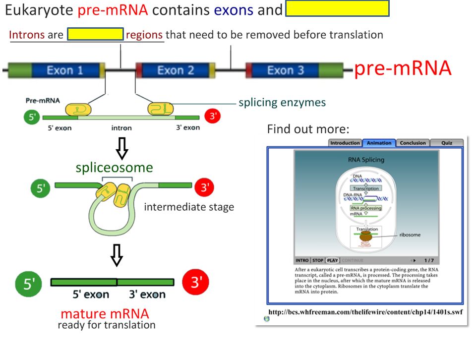
7.1.5 State that eukaryotic genes can contain exons and introns.
Less than 2% of human genome is genes that code for proteins HGProject, mid-70s – 2001 Introns, exons (expressed)
")
19
Happy Thursday!!!! 9/15 Living organisms use DNA as their genetic material. Explain how DNA is replicated within the cells of living organisms. (Total 8 marks)
")
20
helix is unwound; two strands are separated; helicase (is the enzyme that unwinds the helix separating the two strands); by breaking hydrogen bonds between bases; new strands formed on each of the two single strands; nucleotides added to form new strands; complementary base pairing; A to T and G to C; DNA polymerase forms the new complementary strands; replication is semi-conservative; each of the DNA molecules formed has one old and one new strand; [8]

21
DNA Replication

22
7.2.1 State that DNA replication occurs in a 5¢ ® 3¢ direction.
5¢ end of free DNA nucleotide is added to the 3¢ end of chain of nucleotides that is already synthesized.

23
Origin of replication (bubble; prok has one origin, euk has several)
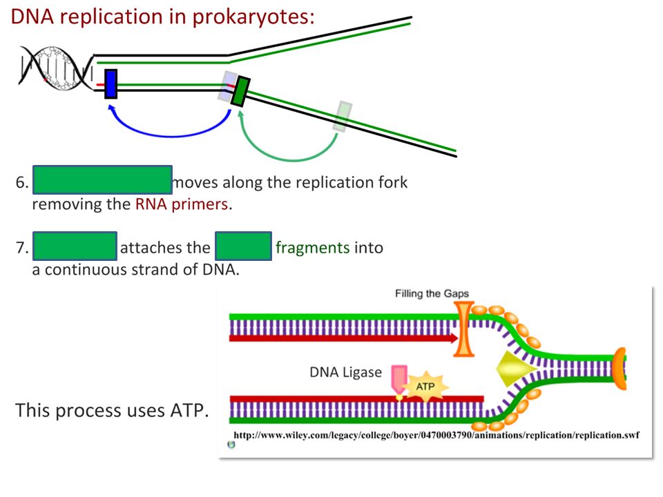
Explain the process of DNA replication in prokaryotes, including the role of enzymes Origin of replication (bubble; prok has one origin, euk has several) Helicase: uncoils the double helix RNA primase: @ replication fork; short sequence of RNA (5-10 n’tides) DNA polymerase III: adds DNA n’tides in 5’ to 3’ direction N’tide is actually a deoxynucleoside triphosphate (dNTP) 2 P groups lost during bonding energy for bonding DNA polymerase I: removes primer from 5’ end and replaces it with DNA n’tides
 Helicase: uncoils the double helix. RNA replication fork; short sequence of RNA (5-10 n’tides) DNA polymerase III: adds DNA n’tides in 5’ to 3’ direction. N’tide is actually a deoxynucleoside triphosphate (dNTP) 2 P groups lost during bonding energy for bonding. DNA polymerase I: removes primer from 5’ end and replaces it with DNA n’tides.")
24
ANTI-PARALLEL...OKAZAKI FRAGMENTS!
Explain the process of DNA replication in prokaryotes, including the role of enzymes ANTI-PARALLEL...OKAZAKI FRAGMENTS! Can only go 5’ to 3’ b/c DNA Pol III Leading strand Fast; toward rep fork in 5’ 3’ Needs primase, primer, DNA pol III only once Lagging strands Slower; Fragments away from rep fork, 5’ 3’ Each fragment needs primase, primer, DNA pol III DNA ligase attaches S-P backbones of Okazaki fragments to make 1 strand

26
IB Book, fig 7.7 page 200 Examiner’s Hint Draw the figure from memory
Annotate what’s happening at specific locations

27
7.2.3 State that DNA replication is initiated at many points in eukaryotic chromosomes.
1. replication begins at origin, strands separate b/c helicase breaks H bonds Replication fork at each end of bubble (DBL strand opens to expose 2 template strands) Bubble enlarges in both directions (bidirectional) & eventually fuse together Multiple bubbles replication faster for the BIG eukaryotic genomes
 Bubble enlarges in both directions (bidirectional) & eventually fuse together. Multiple bubbles replication faster for the BIG eukaryotic genomes.")
30
Primer

31
Primer

37
LIFE book

38
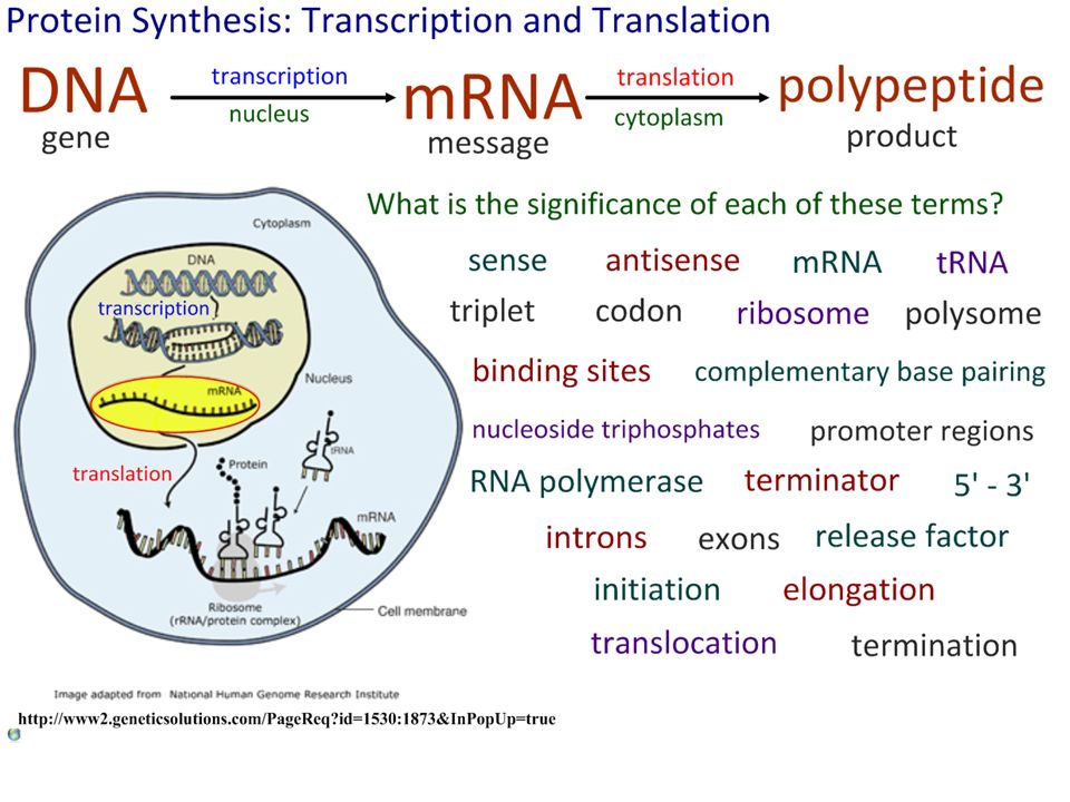
Happy …. Explain the process of transcription in eukaryotes. (8)
")
39
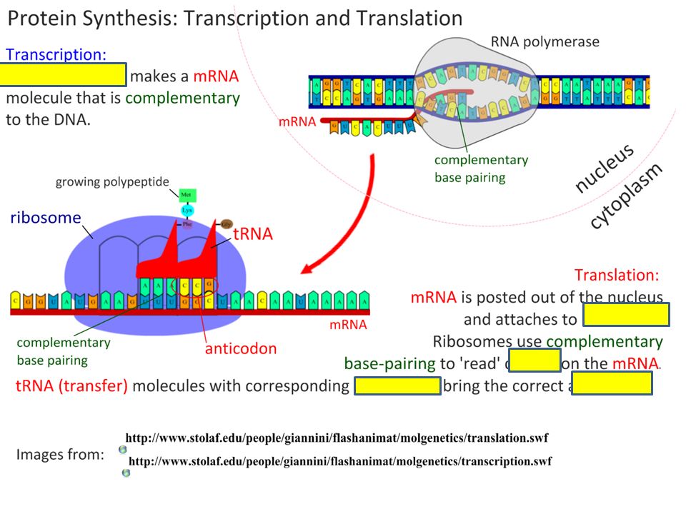
RNA polymerase controls transcription / is the enzyme used in transcription;
DNA is unwound by RNA polymerase; DNA is split into two strands; mRNA is made by transcription; promoter region (by start of gene) causes RNA polymerase to bind; anti-sense / template strand of DNA is transcribed; direction of transcription is ; free nucleotide triphosphates used; complementary base pairing between template strand and RNA nucleotides / bases; Accept this marking point if illustrated using a diagram RNA contains uracil instead of thymine; terminator (sequence) stops RNA polymerase / transcription; mRNA is released / RNA polymerase released; 8 max
 causes RNA polymerase to bind; anti-sense / template strand of DNA is transcribed; direction of transcription is ; free nucleotide triphosphates used; complementary base pairing between template strand and RNA nucleotides / bases; Accept this marking point if illustrated using a diagram. RNA contains uracil instead of thymine; terminator (sequence) stops RNA polymerase / transcription; mRNA is released / RNA polymerase released; 8 max.")
40
TUUUUUUUESDAY, 8/31!!! List three of the other molecules, apart from mRNA, required for transcription. 3 mks

41
DNA; RNA polymerase; (ribose) nucleotides / ribonucleotides / RNA nucleotides; transcription factors; nucleoside / ribonucleoside triphosphates; 3 max Any two of the following: A / C / G / U;

42
Transcription

43
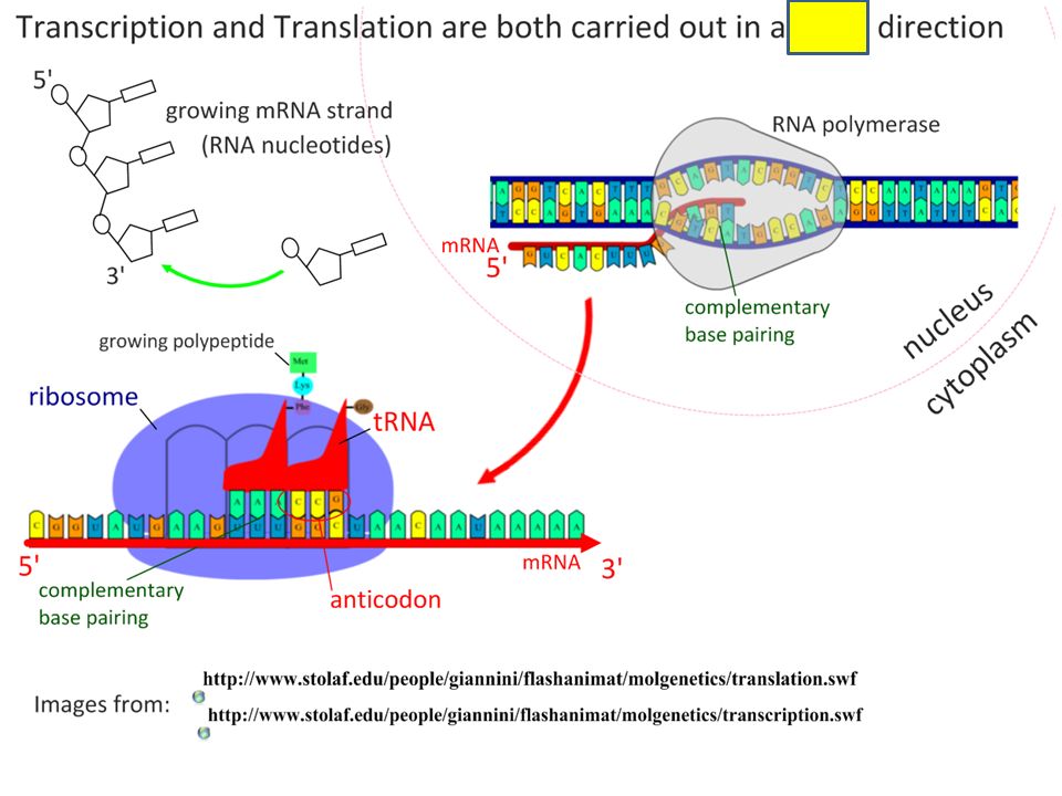
7.3.1 State that transcription is carried out in a 5¢ ® 3¢ direction.
SIMILAR to replication No helicase! RNA polymerase separates DNA strands binds to promoter region of DNA 5’ 3’ The 5¢ end of the free RNA nucleotide is added to the 3¢ end of the RNA molecule that is already synthesized.

44
7.3.2 Distinguish between the sense & antisense strands of DNA.
sense strand = coding strand, carries the genetic code has the same base sequence as mRNA with uracil instead of thymine antisense strand = template strand is transcribed, has the same base sequence as the tRNA Promoter region determines which is antisense Is always the same strand for a particular gene Can differ for different genes

45
Front of RNA polym unwinds the DNA helix
Explain the process of transcription in prokaryotes, including the role of the promoter region, RNA polymerase, nucleoside triphosphates terminator. Front of RNA polym unwinds the DNA helix Adds nucleoside triphosphates to produce mRNA (2 P groups released), nucleotides added to 3’ end of growing strand Anti-sense is template Back of RNA polym rewinds DNA strands Terminator: DNA sequence, transcribed, causes RNA polym to detach & transcription stops
, nucleotides added to 3’ end of growing strand. Anti-sense is template. Back of RNA polym rewinds DNA strands. Terminator: DNA sequence, transcribed, causes RNA polym to detach & transcription stops.")
47
7.3.4 State that eukaryotic RNA needs the removal of introns to form mature mRNA.
No introns in prokaryotic mRNA Eukaryotes—must be removed to get functional mRNA

55
THURSDAY, 9/2 The information needed to make polypeptides is carried in the mRNA from the nucleus to the ribosomes of eukaryotic cells. This information is decoded during translation. The diagram below represents the process of translation. State the name of the next amino acid which will attach to the polypeptide. (1) Explain how the amino acid was attached to the tRNA. (3)
 Explain how the amino acid was attached to the tRNA. (3)")
56
Alanine / Ala 1 an activating enzyme attaches amino acid to the tRNA; specific enzyme for specific tRNA; recognizes tRNA by its shape / chemical properties; energy (ATP) is needed; amino acid attached at end; amino acid attached at CCA; 3 max
 is needed; amino acid attached at end; amino acid attached at CCA; 3 max.")
57
Happy Thursday! 9/2/2010 Explain the process of translation.
(Total 9 marks)
")
58
consists of initiation, elongation and termination;
mRNA translated in a 5' to 3' direction; binding of ribosome to mRNA; small sub-unit then large; first / initiator tRNA binds to start codon / to small subunit of ribosome; AUG is the start codon; second tRNA binds to ribosome; large subunit moves down mRNA after a second tRNA binds; amino acid / polypeptide on first tRNA is transferred / bonded to amino acid on second tRNA; peptide bonds between amino acids / peptidyl transferase; requires GTP; movement of ribosome / small subunit of ribosome down the mRNA; loss of tRNA and new tRNA binds; reach a stop codon / termination; polypeptide released; tRNA activating enzymes link correct amino acid to each tRNA; (activated) tRNA has an anticodon and the corresponding amino acid attached; [9]
 tRNA has an anticodon and the corresponding amino acid attached; [9]")
59
Translation

60
H bonds form in 4 areas, create “clover”
Explain that each tRNA molecule is recognized by a tRNA-activating enzyme that binds a specific amino acid to the tRNA, using ATP for energy. 3’ end is free, has CCA Site of a.a. attachment H bonds form in 4 areas, create “clover” 1 loop of clover has anticodon (unique to each tRNA) Each a.a. has a specific tRNA-activating enzyme (aminoacyl-tRNA synthetase; 20 of them) Active site fits only 1 a.a. & its tRNA, rxn requires ATP “activated” amino acid, tRNA takes it to ribosome The shape of tRNA and CCA at the 3’ end should be included.
 Each a.a. has a specific tRNA-activating enzyme (aminoacyl-tRNA synthetase; 20 of them) Active site fits only 1 a.a. & its tRNA, rxn requires ATP. activated amino acid, tRNA takes it to ribosome. The shape of tRNA and CCA at the 3’ end should be included.")
61
mRNA binding site – in cavity b/w 2 subunits
Outline the structure of ribosomes, including protein and RNA composition, large and small subunits, three tRNA binding sites, mRNA binding sites. Lg & sm subunits rRNA (2/3 of its mass) and many RNA proteins Prokaryotic – smaller than eukaryotic 3 tRNA binding sites A site: holds tRNA carrying the next amino acid to be added to the polypeptide chain P site: holds the tRNA carrying the growing polypeptide chain E site: site from which tRNA that has lost its amino acid is discharged mRNA binding site – in cavity b/w 2 subunits
 and many RNA proteins. Prokaryotic – smaller than eukaryotic. 3 tRNA binding sites. A site: holds tRNA carrying the next amino acid to be added to the polypeptide chain. P site: holds the tRNA carrying the growing polypeptide chain. E site: site from which tRNA that has lost its amino acid is discharged. mRNA binding site – in cavity b/w 2 subunits.")
63
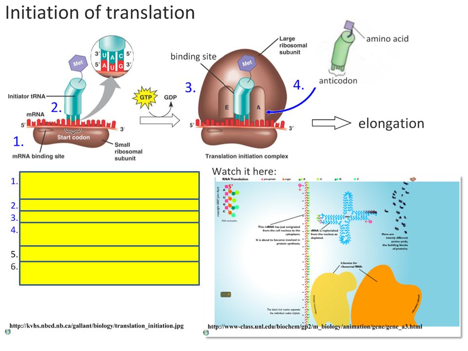
7.4.3 State that translation consists of initiation, elongation, translocation, termination.
Start codon AUG on 5’ end of all mRNA Activated amino acid (methionine + tRNA w/anticodon UAC) attaches to mRNA & small ribosomal subunit Small subunit travels down mRNA to start codon (AUG) Starts translation H bonds b/w initiator tRNA & start codon Large ribosomal subunit attaches Elongation Translocation Termination
 attaches to mRNA & small ribosomal subunit. Small subunit travels down mRNA to start codon (AUG) Starts translation. H bonds b/w initiator tRNA & start codon. Large ribosomal subunit attaches. Elongation. Translocation. Termination.")
64
7.4.3 State that translation consists of initiation, elongation, translocation, termination.
tRNAs bring amino acids to mRNA-ribosomal complex in order specified by codons on mRNA Elongation factors (ptns) bind tRNAs to exposed A site tRNA P site next Ribosomes catalyze peptide bonds forming b/w amino acids (condensation) Translocation Termination
 bind tRNAs to exposed A site. tRNA P site next. Ribosomes catalyze peptide bonds forming b/w amino acids (condensation) Translocation. Termination.")
65
7.4.3 State that translation consists of initiation, elongation, translocation, termination.
During elongation phase tRNAs move down mRNA Binds with A site Its amino acid bonds to polypeptide (attached to A site) Moves to P site Transfers polypeptide to new tRNA in A site Empty tRNA to E site, released 5’ to 3’ direction (ribosome moves along mRNA TOWARD 3’ end—start codon was on 5’ end) Termination
 Moves to P site. Transfers polypeptide to new tRNA in A site. Empty tRNA to E site, released. 5’ to 3’ direction (ribosome moves along mRNA TOWARD 3’ end—start codon was on 5’ end) Termination.")
66
7.4.3 State that translation consists of initiation, elongation, translocation, termination.
1 of 3 stop A site Release factor (protein) fills A site (doesn’t have amino acid) Catalyzes hydrolysis of bond linking P site’s tRNA to polypeptide All released
 fills A site (doesn’t have amino acid) Catalyzes hydrolysis of bond linking P site’s tRNA to polypeptide. All released.")
67
7.4.4 State that translation occurs in a 5¢ ® 3¢ direction.
During translation, the ribosome moves along the mRNA towards the 3¢ end. The start codon is nearer to the 5¢ end.

68
7.4.5 Draw and label the structure of a peptide bond between two amino acids.

69
Explain the process of translation, including ribosomes, polysomes, start codons and stop codons.

70
Polysomes: string of ribosomes all translating the same mRNA
common

71
State that free ribosomes synthesize proteins for use primarily within the cell, and that bound ribosomes synthesize proteins primarily for secretion or for lysosomes. “bound” to what??? ER!

Similar presentations