Download presentation
Presentation is loading. Please wait.

1
外傷及感染之放射線影像檢查 Radiographic Interpretation of Trauma and Infection

2
內容綱要 顎顏面外傷之影像檢查 技術 顎顏面外傷分類 判讀 感染的影像檢查 軟硬組織感染

3
影像檢查在顎顏面外傷的應用 Plays a critical role
Identify the location and orientation of fractures Indicate the degree of separation or displacement

4
顎顏面外傷常用 放射線影像檢查技術 -I Routine view bones 2. Lateral view of facial bone
1. Posterior-anterior (PA) view of facial bones 2. Lateral view of facial bone 3. Panorex 4. Water’s ( Occipitomental ) Suspect mid-face fracture
 view of facial. bones. 2. Lateral view of facial bone. 3. Panorex. 4. Water’s ( Occipitomental ) Suspect mid-face fracture.")
5
顎顏面外傷常用 放射線影像檢查技術-II 5. Periapical view 6. Occlusal view
Occlusal view of nasal bone Occlusal view of mandible 7. Submental-vertex view 8. Lateral view of nasal bone

6
顎顏面外傷常用 放射線影像檢查技術-III
9. Tomography 10. Towne’s view 11. PA view of mandible 12. Oblique view of mandible

7
1. PA view Skull Jaws

8
2. Lateral view Skull Jaws Neck

9
Mandible, maxilla, dentition,
3. Panex Mandible, maxilla, dentition, 70 % of mid-face fracture

10
4. Water’ view (Occipito-mental )
Maxillary fracture Orbital fracture Frontal bone / sinus

11
5. Periapical view Tooth and alveolar injury

12
6. Occlusal view - Mandible
Fracture line , direction Axial section

13
6. Occlusal view - Maxilla

14
7. Submental vertex view Zygomatic arch fracture Coronoid fracture

15
8. Lateral nasal view

16
9. Tomography Blow –out fracture TMJ fracture 185 180

17
175 170

18
165 160

19
10. Modified Town’s view Condylar fracture Mandibular angle fracture

20
11.PA symphysis view

21
12. Oblique lateral view Was replaced by Panoex
Used when patient can not sit or stand

22
CT scan

23
Reconstructive 3-D CT scan

24
顎顏面外傷之影像檢查判讀 General interpretation of fracture line
Condyle and other mandibular fracture Middle facial fracture Cavity: sinus, orbital Dento-alveolar fracture

25
General interpretation of fracture line
Displacement ( deviation, dislocation ) Step, gap, overlapping Discontinuity Asymmetry Comminuted Malocclusion Cavity: (air-fluid level ) *** Degree and direction
 Step, gap, overlapping. Discontinuity. Asymmetry. Comminuted. Malocclusion. Cavity: (air-fluid level ) *** Degree and direction.")
26
Mandibular fracture

27
Angle and symphysis fracture
Malocclusion Step Angle and symphysis fracture

28
Condyle and symphysis fracture
Overlap , discontinuity and displacement Asymmetry Comminuted

29
Coronoid and ramus fracture
Gap, Discontinuity Step , Displacement

30
Condyle Deviation Displacement Dislocation Displacement Undisplaced

31
condyle fracture Displacement

32
Bilateral condyle fracture
Dislocation

33
Bone gap

34
Split fracture Coronoid fracture

35
Chin horizontal fracture

36
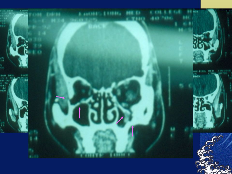
Mid-facial trauma Cavity: sinus, hernia of orbital soft tissue
Tomography of orbital fracture CT Air-emphysema Middle fracture ( Le Fort I, II, III ) ZMC fracture Basilar skull fracture: air-fluid level in sphenoid sinus
 ZMC fracture. Basilar skull fracture: air-fluid level in sphenoid sinus.")
37
Le fort I fracture

38
Air-fluid level

40
Le Fort II fracture

41
Le Fort III fracture

42
Blow-out fracture

44
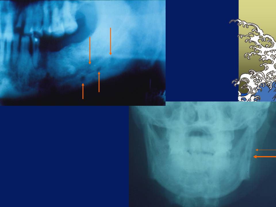
Zygomatic arch fracture

45
Zygomatico-maxillary complex ( ZMC) fracture
 fracture")
46
Classification of dentoalveolar injuries
A. Tooth structures B. Supporting structures 1. Crown craze or crack 2. Crown fracture 1) Enamel 2) Enamel-Dentin 3) Enamel-Dentin- Pulp 3. Crown and root fracture 1) Pulp involvement 2) No pulp involvement
 Enamel. 2) Enamel-Dentin. 3) Enamel-Dentin- Pulp. 3. Crown and root fracture. 1) Pulp involvement. 2) No pulp involvement.")
47
Classification of dentoalveolar injuries
4. Root fracture 1) Apical third 2) Middle third 3) Cervical third Shift to another angle
 Apical third. 2) Middle third. 3) Cervical third. Shift to another angle.")
48
B. Supporting Structure
1. Sensitivity ( concussion ) * percussion pain * no displacement * no mobility * Image widening of PDL space 2. Subluxation * loosening, no displacement, * Image portion of PDL widening
 * percussion pain. * no displacement. * no mobility. * Image widening of PDL space. 2. Subluxation. * loosening, no displacement, * Image portion of PDL widening.")
49
Classification of dentoalveolar injuries
3. Tooth displacement 1) Intrusion 2) Extrusion 3) Labial displacement 4) Lingual displacement 5) Lateral displacement 4. Avulsion 5. Alveolar process fracture
 Intrusion. 2) Extrusion. 3) Labial displacement. 4) Lingual displacement. 5) Lateral displacement. 4. Avulsion. 5. Alveolar process fracture.")
50
感染的影像檢查技術 Plain film radiography CT scan MRI Nuclear bone scans
Tomography Ultrasonography

51
齒源性感染的常用影像檢查 Plain film radiography 根尖片 : 對於根尖及早期病變的顯示最佳
咬合片 : (Axial) Garrie’s osteomyelitis 全口片 (panoex) 有張口困難的病人, 同時對牙齒的情況做,骨頭的破壞檢查,
 Garrie’s osteomyelitis. 全口片 (panoex) 有張口困難的病人, 同時對牙齒的情況做,骨頭的破壞檢查,")
52
CT Scan Space infection Neck: Air way, pharynx Sinus Orbit
Intracranial abscess Soft tissue

53
MRI Noninvasion, no radiation, high soft tissue resolution﹐high sensitivity and specificity 對骨的細部變化 space infection, presence of pus, cavitation TMJ abscess

54
選擇的要領 Plain film : 一般診斷及治療反應後的追蹤
CT / MRI : Extension into soft tissue , air way Bone scan: Response to treatment

55
Image finding and Bone changes
Difficult to visualize by conventional techniques in early stage Until substantial mineral removed % After infection :5 -14 days

56
感染部位與描述名稱 Margin: well or poor demarcation / defined
Lesion: radiolucent / radiopaque Periapical changes: PDL , trabeculae . Cavity (sinus) : cloudy, air-fluid level… Osteomyelitis: periosteal reaction﹐moth eaten , rarefaction, …. Sinus tract ( fistula )
 : cloudy, air-fluid level… Osteomyelitis: periosteal reaction﹐moth eaten , rarefaction, …. Sinus tract ( fistula )")
57
1. Periapical Infection ( acute / chronic ) Widening of PDL
Lamina dura discontinuity Trabeculae destruction Chronic Periapical abscess Periapical granuloma Fistula Root resorption

58
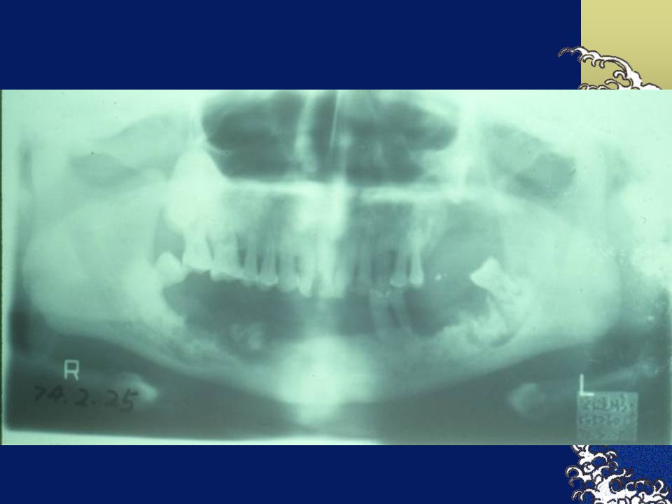
2. Osteomyelitis (骨髓炎 ) Acute suppurative osteomyelitis
Trabeculae: fuzzy, diffuse destruction﹐ Radiolucent area Poor demarcation Irregular border

59
Chronic suppurative osteomyelitis
Necrotic Bone Moth eaten* Radiolucent area with poor demarcation Necrotic bone Sequestrum Radiopaque with peripheral rediolucent area Rarefaction

60
Moth eaten

62
Sequestrum Radiopaque with peripheral radiolucent area

64
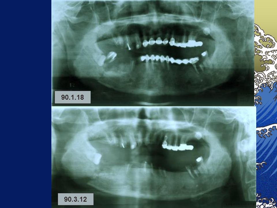
Pathologic fracture

65
Expansion Rarefaction

66
Sclerosing Osteomyelitis
Focal type Increasing density ( disposition of the bone ) rarefaction Periapical area Thickening of PDL Bone scar
 rarefaction. Periapical area. Thickening of PDL. Bone scar.")
67
Sclerosing Osteomyelitis
Diffuse type Border between normal and sclerosis .. poor defined Cotton wool appearance。

69
Garre’s proliferative periostitis (Osteomyelitis)
Subperiosteal reaction: onion skinning﹐ Duplication of the cortical layer of bone

70
ORN ( Osteo-Radio Necrosis )
No remodeling

72
Bone necrosis due to Arsenic
Tooth germ, nerve damage…

73
軟組織感染 Infections involving soft tissues are not readily
Demonstrated by many imaging techniques Gas producing organism

74
Infratemporal space Submasseteric space

75
Air way

77
MRI of TMJ space abscess

78
Sinusitis Cloudy Air-Fluid level

79
下 課

Similar presentations


 Oral Digital Image Artifacts 口腔數位影像失誤 陳玉昆副教授 : 高雄醫學大學 口腔病理科 07-3121101~2755>")


