Download presentation
Presentation is loading. Please wait.

1
Case 1 Extrahepatic Biliary Obstruction
Bondoc-Borja, J.- Borja, P.-Buenavente- Bustamante-Buti-Cabanag-Calaquian-Calayan

2
Case 1 month PTA: 58-year old male CC: progressive jaundice HPI:
2 months PTA: experienced vague abdominal pain and anorexia 1 month PTA: progressive yellowish discoloration of the sclera tea-colored urine, pruritus, and acholic stools weight loss of around 20%

3
Past Personal History Past Medical History
known hypertensive for the past 10 years; no history of hepatitis no history of diabetes on captopril and metoprolol heavy smoker( 1 pack a day for the last 3 years); occasional alcoholic beverage drinker
; occasional alcoholic beverage drinker.")
4
Physical Examination HEENT: icteric sclerae; no palpable cervical lymph nodes Heart/Lungs: unremarkable Abdomen: globular with a vague ballotable mass at the RUQ, smooth, not tender and moves with respiration, (-) fluid wave. Rectal exam: acholic stools
 fluid wave. Rectal exam: acholic stools.")
5
Diagnostic evaluation
CBC – normal; Creatinine: 2 mg/dl Alkaline phosphatase: 500 u/L; Total protein: 6.5 g/dl; albumin: 3.5g/dl; globulin: 2.5g/dl Total bilirubin: 10 mg/dl; Direct bilirubin: 8 mg/dl; Indirect bilirubin: 2 mg/dl CA 19-9: 350 units/ml Chest x-ray: normal Ultrasound: distended gallbladder with no stones; CBD 2.5 cm; dilated intrahepatic ducts; enlarged head of the pancreas; normal liver CEA ng/ml (normal to 5 ), CA 19-9 u/ml ( normal to 37 ). The result from June 2007 ( after surgery ) was CEA ng/nl, CA more than 12000u/ml.
, CA u/ml ( normal to 37 ). The result from June 2007 ( after surgery ) was CEA ng/nl, CA more than 12000u/ml.")
6
Diagnostic Evaluation
ERCP CT scan

7
Diagnostic Evaluation
MRI Endoscopic ultrasound

8
Salient Features Subjective Objective 58 y/o, male
Icteric sclerae Globular abdomen, with a vague ballotable mass at the RUQ smooth, not tender and moves with respiration Rectal exam: acholic stools Labs: Alkaline phosphatase: 500 u/L; Total bilirubin: 10 mg/dl Direct bilirubin: 8 mg/dl CA 19-9: 350 units/ml Imaging: Ultrasound: distended gallbladder with no stones; CBD 2.5 cm; dilated intrahepatic ducts; enlarged head of the pancreas; normal liver 58 y/o, male CC: progressive jaundice Vague abdominal pain and anorexia Progressive yellowish discoloration of the sclera, tea-colored urine, pruritus, and acholic stools Weight loss of 20% heavy smoker (3-pack years)occasional alcoholic beverage drinker; (+)HPN The normal range is 20 to 140 IU/L-alp Anything higher than 37 U/ml is considered abnormal. The higher the number, the more advanced the disease may be.-ca 19-9
occasional alcoholic beverage drinker; (+)HPN. The normal range is 20 to 140 IU/L-alp. Anything higher than 37 U/ml is considered abnormal. The higher the number, the more advanced the disease may be.-ca")
9
Biliary Obstruction secondary to Pancreatic Head Carcinoma
Diagnosis: Biliary Obstruction secondary to Pancreatic Head Carcinoma

10
Pancreatic Head Carcinoma
Cancer of the pancreas is the 5th leading cause of cancer death in the US Risk factor consistently linked to pancreatic cancer is smoking; smoking increases the risk of developing pancreatic cancer by at least 2- fold Other risk factors: long-standing diabetes, chronic pancreatitis, family history

11
Pancreatic Head Carcinoma
Head 80%, body 15%, tail 5% •Types •Ductal adenocarcinoma- most common •Intraductal papillary mucinous carcinoma •Mucinous cystadenocarcinoma Peak age incidence: years old

12
Pancreatic Head Carcinoma
Signs and symptoms •Jaundice, pruritus •Anorexia, weight loss •Back pain •Late-onset diabetes •Vomiting due to duodenal obstruction •Palpable GB (Courvoisier’s sign) •Virchow’s node, Sister Joseph’s sign
 •Virchow’s node, Sister Joseph’s sign.")
13
Differential diagnoses

14
CHOLEDOCHOLITHIASIS

15
Choledocholithiasis found in 6 to 12% of patients with stones in the gallbladder incidence increases with age secondary common bile duct stones majority of ductal stones in Western countries are formed within the gallbladder cystic duct common bile duct usually cholesterol stones primary stones form in the bile ducts usually of the brown pigment type more commonly seen in Asian populations associated with biliary stasis and infection causes of biliary stasis biliary stricture papillary stenosis Tumors other (secondary) stones
 stones.")
16
vague ballotable mass at RUQ
Choledocholithiasis Patient > 60 yrs. Old Female Abdominal pain Colicky, moderate in severity, located in the RUQ, intermittent, transient, and recurrent jaundice Icteric sclerae nausea vomiting Tea-colored urine Acholic stools RUQ tenderness 58-year old male vague abdominal pain progressive jaundice icteric sclerae anorexia tea-colored urine pruritus acholic stools 20% weight loss globular abdomen vague ballotable mass at RUQ smooth, not tender and moves with respiration Jaundice occurs when the CBD becomes obstructed and conjugated bilirubin enters the bloodstream.

17
MIRIZZI SYNDROME

18
Mirizzi Syndrome Rare cause of extrahepatic biliary obstruction caused by gallstone impaction in either Hartmann's pouch of the gall bladder or the cystic duct Chronic and/or acute inflammatory changes lead to contraction of the gallbladder secondary stenosis of the CHD Large impacted stones lead to cholecystocholedochal fistula formation Impaction of a large gallstone (or multiple small gallstones) in the Hartmann pouch or cystic duct results in the Mirizzi syndrome in 2 ways: (1) chronic and/or acute inflammatory changes lead to contraction of the gallbladder, which then fuses with and causes secondary stenosis of the CHD, or (2) large impacted stones lead to cholecystocholedochal fistula formation secondary to direct pressure necrosis of the adjacent duct walls. Increasingly, these phenomena are seen not as distinct and separate steps but as part of a continuum
 in the Hartmann pouch or cystic duct results in the Mirizzi syndrome in 2 ways: (1) chronic and/or acute inflammatory changes lead to contraction of the gallbladder, which then fuses with and causes secondary stenosis of the CHD, or (2) large impacted stones lead to cholecystocholedochal fistula formation secondary to direct pressure necrosis of the adjacent duct walls. Increasingly, these phenomena are seen not as distinct and separate steps but as part of a continuum.")
19
Mirizzi Syndrome Classification To aid in surgical management
groups to aid in the surgical treatment of patients: Type I - No fistula present Type IA - Presence of the cystic duct Type IB - Obliteration of the cystic duct Types II-IV - Fistula present Type II - Defect smaller than 33% of the CBD diameter Type III - Defect 33-66% of the CBD diameter Type IV - Defect larger than 66% of the CBD diameter

20
elderly; any patient with cholelithiasis at risk
Mirrizi Patient elderly; any patient with cholelithiasis at risk Male = Female No consistent or unique clinical features that distinguish it from other more common forms of obstructive jaundice painless jaundice or with cholangitis with cholecystitis or pancreatitis 58-year old male vague abdominal pain progressive jaundice icteric sclerae anorexia tea-colored urine pruritus acholic stools 20% weight loss globular abdomen vague ballotable mass at RUQ smooth, not tender and moves with respiration

21
CHRONIC PANCREATITIS

22
CHRONIC PANCREATITIS long-standing inflammation of the pancreas that results in irreversible deterioration of pancreatic structure and function. Chronic inflammation, fibrosis, progressive destruction of both exocrine and eventually endocrine tissue

23
CHRONIC PANCREATITIS CHRONIC PANCREATITIS PATIENT
Age: 46 ±13 years old Sex: male > female Abdominal pain Mid abdominal/epigastric Constant or intermittent Exacerbated by eating Weight Loss Malabsorption Steatorrhea Acholic stools Tender fullness or mass palpated in epigastrium Jaundice Complication 58-year old male vague abdominal pain progressive jaundice icteric sclerae anorexia tea-colored urine pruritus acholic stools 20% weight loss globular abdomen vague ballotable mass at RUQ smooth, not tender and moves with respiration

24
CHRONIC PANCREATITIS CHRONIC PANCREATITIS PATIENT

25
CHOLANGIOCARCINOMA

26
CHOLANGIOCARCINOMA mucin-producing adenocarcinomas that arise from the bile ducts grouped by their anatomic site of origin as intrahepatic, hilar (central) and peripheral (distal) Several predisposing factors: primary sclerosing cholangitis liver fluke in Asians: Opisthorchis viverrini and Clonorchis sinensis. chronic biliary inflammation and injury with alcoholic liver disease, choledocholithiasis, choledochal cysts and Caroli's disease.
 and peripheral (distal) Several predisposing factors: primary sclerosing cholangitis. liver fluke in Asians: Opisthorchis viverrini and Clonorchis sinensis. chronic biliary inflammation and injury. with alcoholic liver disease, choledocholithiasis, choledochal cysts and Caroli s disease.")
27
CHOLANGIOCARCINOMA Elderly: 60’s-70’s M:F ratio is 1:2.5
painless jaundice pruritus weight loss acholic stools Abdominal pain 58-year old male vague abdominal pain progressive jaundice icteric sclerae anorexia tea-colored urine pruritus acholic stools 20% weight loss globular abdomen vague ballotable mass at RUQ smooth, not tender and moves with respiration CHOLANGIOCARCIMA PATIENT

28
CHOLANGIOCARCINOMA CHOLANGIOCARCINOMA PATIENT
57-year-old man with mass-forming intrahepatic cholangiocarcinomas. CT scan shows irregularly shaped mass with peripheral enhancement. Note three small peritumoral satellite nodules (arrow). PATIENT
. PATIENT.")
29
CARCINOMA OF THE AMPULLA OF VATER

30
Carcinoma of the Ampulla of Vater
Arises within 2 cm of the distal CBD 90% an adenocarcinoma May invovle locoregional lymph nodes Liver is the most frequent site for metastases This tumor arises within 2 cm of the distal end of the common bile duct, and is mainly (90%) an adenocarcinoma. Locoregional lymph nodes are commonly involved (50%), and the liver is the most frequent site for metastases. The commonest clinical presentation is jaundice, and many patients also have pruritus, weight loss, and epigastric pain. Initial evaluation is performed with an abdominal ultrasound to assess vascular involvement, biliary dilatation, and liver lesions. This is followed by a CT scan, or MRI and especially MRCP. The most effective therapy is resection by pylorus-sparing pancreaticoduodenectomy, an aggressive procedure resulting in better survival rates than local resection. Survival rates are ~25% at 5 years in operable patients with involved lymph nodes and ~50% in patients without involved nodes. Unlike CCC, ~80% of patients are thought to be resectable at diagnosis. Adjuvant chemotherapy or radiotherapy has not been shown to be useful in enhancing survival. For metastatic tumors, chemotherapy is currently experimental.
 an adenocarcinoma. Locoregional lymph nodes are commonly involved (50%), and the liver is the most frequent site for metastases. The commonest clinical presentation is jaundice, and many patients also have pruritus, weight loss, and epigastric pain. Initial evaluation is performed with an abdominal ultrasound to assess vascular involvement, biliary dilatation, and liver lesions. This is followed by a CT scan, or MRI and especially MRCP. The most effective therapy is resection by pylorus-sparing pancreaticoduodenectomy, an aggressive procedure resulting in better survival rates than local resection. Survival rates are ~25% at 5 years in operable patients with involved lymph nodes and ~50% in patients without involved nodes. Unlike CCC, ~80% of patients are thought to be resectable at diagnosis. Adjuvant chemotherapy or radiotherapy has not been shown to be useful in enhancing survival. For metastatic tumors, chemotherapy is currently experimental.")
31
AMPULLARY CARCINOMA AMPULLARY CARCINOMA PATIENT 58-year old male
Jaundice (pruritus) Weight loss Epigastric pain Acholic Stools Diarrhea Nausea Vomitting (+)Courvoisier ‘s Fever 58-year old male vague abdominal pain progressive jaundice icteric sclerae anorexia tea-colored urine pruritus acholic stools 20% weight loss globular abdomen vague ballotable mass at RUQ smooth, not tender and moves with respiration Diarrhea, a common but not universal symptom, might be associated with an absence of lipase within the gut because of pancreatic duct obstruction. Physical examination sometimes discloses a Courvoisier gallbladder (ie, a distended, palpable gallbladder in a patient with jaundice). Fever can be present, particularly when the biliary tract has been explored previously (eg, after common duct exploration for stones). AMPULLARY CARCINOMA PATIENT
 Weight loss. Epigastric pain. Acholic Stools. Diarrhea. Nausea. Vomitting. (+)Courvoisier ‘s. Fever. 58-year old. male. vague abdominal pain. progressive jaundice. icteric sclerae. anorexia. tea-colored urine. pruritus. acholic stools. 20% weight loss. globular abdomen. vague ballotable mass at RUQ. smooth, not tender and moves with respiration. Diarrhea, a common but not universal symptom, might be associated with an absence of lipase within the gut because of pancreatic duct obstruction. Physical examination sometimes discloses a Courvoisier gallbladder (ie, a distended, palpable gallbladder in a patient with jaundice). Fever can be present, particularly when the biliary tract has been explored previously (eg, after common duct exploration for stones). AMPULLARY CARCINOMA. PATIENT.")
32
Abdominal US: Ampullary Carcinoma
Para-sagittal scan of the right upper quadrant Description This image shows a dilated extrahepatic common bile duct. The common duct and the main pancreatic duct are seen coursing together towards the duodenum.

33
CT Scan: Ampullary Carcinoma
DOUBLE DUCT SIGN: dilated CBD and pancreatic duct Also seen: Metastasis in the liver

34
DIAGNOSTIC EVALUATION

35
Physical exam Blood tests
presents clinically with non-specific signs and symptoms such as pain, jaundice (yellowing of the skin) and weight loss Blood tests CA 19-9 (carbohydrate antigen 19-9) is the mainstay tumor marker and is ordered when pancreatic cancer is suspected
 and weight loss. Blood tests. CA 19-9 (carbohydrate antigen 19-9) is the mainstay tumor marker and is ordered when pancreatic cancer is suspected.")
36
TISSUE EXAMINATION Tissue for microscopic examination can be obtained by Fine needle biopsy Tissue needle cone biopsy Excisional biopsy (at the time of laparotomy)
")
37
Angiography useful to determine if the vessels around the pancreas are involved by the tumor

38
IMAGING TECHNIQUES CAT scan Endoscopic ultrasound (EUS)
Endoscopic retrograde cholangiopancreatography (ERCP) PTC (percutaneous transhepatic cholangiography)
 PTC (percutaneous transhepatic cholangiography)")
39
Histopathology “Gold Standard”
80% are adenocarcinomas of the ductal epithelium Only 2% of tumors of the exocrine pancreas are benign

40
MANAGEMENT

41
SURGICAL RESECTION resectable, unresectable, or borderline resectable.
Only potentially curative treatment for patients with pancreatic cancer The resectability of malignant pancreatic tumors needs to be established Pancreatic masses are characterized resectable, unresectable, or borderline resectable. Treatment Surgical resection is the only potentially curative treatment for patients with pancreatic cancer, although many patients are not candidates for resection. RESECTABLE LESIONS About 15 to 20 percent of patients with pancreatic adenocarcinoma have resectable disease at the time of diagnosis.12 The classic Whipple procedure (Figure 3) involves removal of the head and uncinate process of the pancreas, duodenum, proximal 6 in (15 cm) of jejunum, gallbladder, common bile duct, and distal stomach, with anastomosis of the common hepatic duct and the remaining pancreas and stomach to the jejunum.32 The perioperative mortality rate of patients undergoing this procedure has improved significantly over the past three decades. Surgical teams performing more than 16 procedures per year report significantly lower perioperative mortality rates than centers with less experience (3.8 versus 7.5 to 17.6 percent).33 Pyloruspreserving pancreaticoduodenostomy appears to offer the same long-term survival benefits as the standard Whipple procedure with shorter operative time and reduced blood loss, decreasing the need for blood transfusions.34 Risks associated with both procedures include delayed gastric emptying, pancreatic fistula, anastomotic leaks, wound infection, intraabdominal abscess, hemorrhage, diabetes, and pancreatic exocrine insufficiency.34 Distal pancreatectomy is performed in patients with resectable cancer in the body or tail of the pancreas. The spleen usually is removed as well. The resectability rate for body and tail lesions is less than one half of that for head lesions35 because diagnosis usually occurs late in the disease process after local invasion has occurred. Five-year survival for resection of body or tail lesions is similar to that of resection for pancreatic head lesions.35 Five-year survival rates after surgical resection range from 10 to 30 percent.36–41 Negative prognostic factors include poorly differentiated histology, positive resection margins, lymph node involvement, and a tumor larger than 0.8 in (2 cm).36–38
 involves removal of the head and uncinate process of the pancreas, duodenum, proximal 6 in (15 cm) of jejunum, gallbladder, common bile duct, and distal stomach, with anastomosis of the common hepatic duct and the remaining pancreas and stomach to the jejunum.32 The perioperative mortality rate of patients undergoing this procedure has improved significantly over the past three decades. Surgical teams performing more than 16 procedures per year report significantly lower perioperative mortality rates than centers with less experience (3.8 versus 7.5 to 17.6 percent).33. Pyloruspreserving pancreaticoduodenostomy appears to offer the same long-term survival benefits as the standard Whipple procedure with shorter operative time and reduced blood loss, decreasing the need for blood transfusions.34 Risks associated with both procedures include delayed gastric emptying, pancreatic fistula, anastomotic leaks, wound infection, intraabdominal abscess, hemorrhage, diabetes, and pancreatic exocrine insufficiency.34 Distal pancreatectomy is performed in patients with resectable cancer in the body or tail of the pancreas. The spleen usually is removed as well. The resectability rate for body and tail lesions is less than one half of that for head lesions35 because diagnosis usually occurs late in the disease process after local invasion has occurred. Five-year survival for resection of body or tail lesions is similar to that of resection for pancreatic head lesions.35 Five-year survival rates after surgical resection range from 10 to 30 percent.36–41 Negative prognostic factors include poorly differentiated histology, positive resection margins, lymph node involvement, and a tumor larger than 0.8 in (2 cm).36–38.")
42
SURGICAL RESECTION Pancreaticoduodenectomy (whipple procedure)
Distal pancreatectomy Total pancreatectomy

43
PANCREATICODUODENECTOMY (WHIPPLE PROCEDURE)
Removal of the head and uncinate process of the pancreas, duodenum, proximal 6 in (15 cm) of jejunum, gallbladder, common bile duct, and distal stomach With anastomosis of the common hepatic duct and the remaining pancreas and stomach to the jejunum All share a common blood supply Pancreaticoduodenectomy (Whipple procedure) Patients who will most likely benefit from this procedure have a tumor located in the head of the pancreas or the periampullary region. The Whipple procedure is not strictly the surgical approach for pancreatic head tumors. Pancreatic ductal tumors, cholangiocarcinoma (bile duct cancer), and duodenal masses will all require this resection. The operation traditionally involves the following: removal of the pancreatic head, duodenum, gallbladder, and the antrum of the stomach with surgical drainage of the distal pancreatic duct and biliary system, usually accomplished through anastomosis to the jejunum. The primary reason for removing such a large quantity of intraabdominal structures is that they all share a common blood supply. Pancreaticoduodenectomy has been shown to have an overall mortality rate of 6.6%.38 Many forms of morbidity are associated with the operation. One of these is delayed gastric emptying. This occurs in approximately 25% of patients. This condition may require nasogastric decompression and will lead to a longer hospital stay.39 Other morbidities include pancreatic anastomotic leak. This can be treated with adequate drainage. Postoperative abscesses are not uncommon. It is unclear whether preoperative biliary drainage leads to increased rates of postoperative infection.40 The standard Whipple operation may be altered in order to include a pylorus-sparing procedure. This modification was previously incorporated to increased nutritional strength in these patients as the increased-gastric emptying associated with antrectomy caused nutritional deficiencies. Although many believe that delayed gastric emptying is worsened by this modification, studies have proven both resections to be equivalent in that regard. Another source of controversy is the extent of lymphadenectomy that is necessary in a Whipple operation. In an elegant study, Pawlik et al found the ratio of positive nodes to total nodes removed was an important prognostic factor.41 This was even more significant than margin positivity.42
 of jejunum, gallbladder, common bile duct, and distal stomach. With anastomosis of the common hepatic duct and the remaining pancreas and stomach to the jejunum. All share a common blood supply. Pancreaticoduodenectomy (Whipple procedure) Patients who will most likely benefit from this procedure have a tumor located in the head of the pancreas or the periampullary region. The Whipple procedure is not strictly the surgical approach for pancreatic head tumors. Pancreatic ductal tumors, cholangiocarcinoma (bile duct cancer), and duodenal masses will all require this resection. The operation traditionally involves the following: removal of the pancreatic head, duodenum, gallbladder, and the antrum of the stomach with surgical drainage of the distal pancreatic duct and biliary system, usually accomplished through anastomosis to the jejunum. The primary reason for removing such a large quantity of intraabdominal structures is that they all share a common blood supply. Pancreaticoduodenectomy has been shown to have an overall mortality rate of 6.6%.38 Many forms of morbidity are associated with the operation. One of these is delayed gastric emptying. This occurs in approximately 25% of patients. This condition may require nasogastric decompression and will lead to a longer hospital stay.39 Other morbidities include pancreatic anastomotic leak. This can be treated with adequate drainage. Postoperative abscesses are not uncommon. It is unclear whether preoperative biliary drainage leads to increased rates of postoperative infection.40. The standard Whipple operation may be altered in order to include a pylorus-sparing procedure. This modification was previously incorporated to increased nutritional strength in these patients as the increased-gastric emptying associated with antrectomy caused nutritional deficiencies. Although many believe that delayed gastric emptying is worsened by this modification, studies have proven both resections to be equivalent in that regard. Another source of controversy is the extent of lymphadenectomy that is necessary in a Whipple operation. In an elegant study, Pawlik et al found the ratio of positive nodes to total nodes removed was an important prognostic factor.41 This was even more significant than margin positivity.42.")
44
PANCREATICODUODENECTOMY (WHIPPLE PROCEDURE)
The Whipple procedure. Before the procedure(A). After the procedure; note the anastomosis of the hepatic duct and the remaining pancreas and stomach to the jejunum(B).
. After the procedure; note the anastomosis of the hepatic duct and the remaining pancreas and stomach to the jejunum(B).")
45
PANCREATICODUODENECTOMY (WHIPPLE PROCEDURE)
Patients who will most likely benefit from this procedure have a tumor located in the head of the pancreas or the periampullary region

46
DISTAL PANCREATECTOMY
May be an effective procedure for tumors located in the body and tail of the pancreas Isolation of the distal portion of the pancreas containing the tumor Resection of that segment Oversewing of the distal pancreatic duct Distal pancreatectomy This procedure possesses a lower mortality rate than the standard Whipple procedure at 3.5%, but its use in curative resection remain limited.38 Essentially, a distal pancreatectomy may be an effective procedure for tumors located in the body and tail of the pancreas. Unfortunately, masses located in this area present later than the periampullary tumors and hence have a higher unresectability rate. The procedure involves isolation of the distal portion of the pancreas containing the tumor followed by resection of that segment, with oversewing of the distal pancreatic duct. The main complications for distal pancreatectomy involve pancreatic stump leak, hemorrhage, or endocrine insufficiency.43 Once again, the best treatment for the pancreatic leak is adequate drainage.

47
TOTAL PANCREATECTOMY Tumor involves the neck of the pancreas.
Either the tumor originates from the neck or is growing into the neck Total pancreatectomy Although this procedure is the least commonly done with the highest associated mortality at 8.3%, it may still remain a valuable instrument in the surgical cure of pancreatic cancer.38 The indication is cases in which the tumor involves the neck of the pancreas. This can either be a situation in which the tumor originates from the neck or is growing into the neck. These patients obviously get insulin-dependent diabetes. In some cases, the diabetes can be hard to control. Despite this, the morbidity of a total pancreatectomy is comparable to that of a Whipple procedure.44

48
Metastatic Lesions Single- and multiple-agent chemotherapeutic regimens gemcitabine vs. fluorouracil first-line therapy 12-month survival advantage improves or stabilizes pain, performance status, and weight Clinical trial (gene therapy) METASTATIC LESIONS Researchers have studied many single- and multiple-agent chemotherapeutic regimens for patients with metastatic disease, and more studies are ongoing; however, few studies have shown survival or clinical benefit. The use of gemcitabine as first-line therapy has a 12-month survival advantage and improves or stabilizes pain, performance status, and weight compared with fluorouracil monotherapy.42 Although the combination of leucovorin and fluorouracil is effective as adjuvant chemotherapy in resectable disease, it does not seem to be any more effective than fluorouracil monotherapy for treatment of unresectable disease.43–44 Palliative Chemotherapy and Radiation In patients with unresectable pancreatic cancer, gemcitabine results in symptomatic improvement, improved pain control and performance status, and weight gain. 289 However, survival is improved by only 1 to 2 months. Although these results may warrant treatment in patients who understand the benefits and risks, the lack of significant survival advantage should encourage physicians to refer motivated patients for experimental protocols such as gene therapy, since it is only through continued clinical research that more meaningful treatments for pancreatic cancer will be developed.
 METASTATIC LESIONS. Researchers have studied many single- and multiple-agent chemotherapeutic regimens for patients with metastatic disease, and more studies are ongoing; however, few studies have shown survival or clinical benefit. The use of gemcitabine as first-line therapy has a 12-month survival advantage and improves or stabilizes pain, performance status, and weight compared with fluorouracil monotherapy.42 Although the combination of leucovorin and fluorouracil is effective as adjuvant chemotherapy in resectable disease, it does not seem to be any more effective than fluorouracil monotherapy for treatment of unresectable disease.43–44. Palliative Chemotherapy and Radiation. In patients with unresectable pancreatic cancer, gemcitabine results in symptomatic improvement, improved pain control and performance status, and weight gain. 289 However, survival is improved by only 1 to 2 months. Although these results may warrant treatment in patients who understand the benefits and risks, the lack of significant survival advantage should encourage physicians to refer motivated patients for experimental protocols such as gene therapy, since it is only through continued clinical research that more meaningful treatments for pancreatic cancer will be developed.")
49
Locally Advanced Lesions
External beam and intraoperative radiation therapy ↓ local progression neither affects survival or metastasis Radiation therapy alone – not effective Combined radiation therapy and fluorouracil-based chemotherapy vs. radiation therapy alone 40 vs. 10% survival after 1 year, NNT = 3 LOCALLY ADVANCED LESIONS External beam and intraoperative radiation therapy decrease local progression in patients with unresectable, locally advanced disease, but neither affects survival or metastasis.45 Therefore, radiation therapy alone does not effectively treat patients with locally advanced pancreatic cancer outside of palliation. Combined radiation therapy and fluorouracil-based chemotherapy offer significant survival improvement compared with radiation therapy alone (40 versus 10 percent survival after one year, number needed to treat = 3) and are routinely used unless a patient is enrolled in an investigational study of another treatment regimen.12,45–47 Radiation with gemcitabine increases toxicity rates but does not significantly impact survival compared with radiation and fluorouracil.48 Regardless of stage, the potential benefits of therapy for pancreatic cancer must be balanced against the significant side effects, costs, and quality-of-life factors.
 and are routinely used unless a patient is enrolled in an investigational study of another treatment regimen.12,45–47 Radiation with gemcitabine increases toxicity rates but does not significantly impact survival compared with radiation and fluorouracil.48 Regardless of stage, the potential benefits of therapy for pancreatic cancer must be balanced against the significant side effects, costs, and quality-of-life factors.")
50
Palliative Care Pain Jaundice Duodenal obstruction
3 clinical problems in advanced pancreatic CA: Pain Jaundice Duodenal obstruction ** cachexia, malabsorption In general, there are three clinical problems in advanced pancreatic cancer that require palliation: pain, jaundice, and duodenal obstruction. The mainstay of pain control is oral narcotics. Sustained-release preparations of morphine sulfate are frequently used. Invasion of retroperitoneal nerve trunks accounts for the severe pain experienced by patients with advanced pancreatic cancer. A celiac plexus nerve block can control pain effectively for a period of months, although the procedure sometimes needs to be repeated. At the time of initial exploration for pancreatic cancer, it is a good practice to think about pain control and consider performing an intraoperative celiac block regardless of whether a resection is performed. The procedure is performed by injecting 50% alcohol directly into the tissues along the sides of the aorta just cephalad and posterior to the origin of the celiac trunk. This can be accomplished quite easily with either open surgery or laparoscopic surgery, and does not prolong the operation by more than a few minutes. If necessary, the procedure can be repeated postoperatively, either percutaneously or with use of endoscopic ultrasound guidance. Jaundice is present in the majority of patients with pancreatic cancer, and the most troublesome aspect for the patient is the accompanying pruritus. Biliary obstruction may also lead to cholangitis, coagulopathy, digestive symptoms, and hepatocellular failure. In the past, surgeons traditionally performed a biliary bypass when unresectable disease was found at laparotomy. As many patients today already have a bile duct stent in place by the time of operation, it is not clear that operative biliary bypass is required. If an operative bypass is performed, choledochojejunostomy is the preferred approach. Although an easy procedure to perform, choledochoduodenostomy is felt to be unwise because of the proximity of the duodenum to tumor. Some have discouraged the use of the gallbladder for biliary bypass; 287 however, it is suitable as long as the cystic duct clearly enters the common duct well above the tumor. Duodenal obstruction is usually a late event in pancreatic cancer and occurs in only about 20% of patients. 288 Therefore, in the absence of signs or symptoms of obstruction, such as nausea or vomiting, or a tumor that is already encroaching on the duodenum at the time of surgery, the routine use of prophylactic gastrojejunostomy when exploration reveals unresectable tumor is controversial. Although anastomotic leaks are uncommon, gastrojejunostomy is sometimes associated with delayed gastric emptying, the very symptom the procedure is designed to treat. Whether performing both a biliary and enteric bypass or just a biliary bypass, the jejunum is brought anterior to the colon rather than retrocolic, where the tumor potentially would invade the bowel sooner. Some surgeons use a loop of jejunum with a jejunojejunostomy to divert the enteric stream away from the biliary-enteric anastomosis. Others use a Roux-en-Y limb with the gastrojejunostomy located 50 cm downstream from the hepaticojejunostomy (Fig ). Potential advantages of the defunctionalized Roux-en-Y limb include the ease with which it will reach up to the hepatic hilum, probable decreased risk of cholangitis, and easier management of biliary anastomotic leaks. If a gastrojejunostomy is performed, it should be placed dependently and posterior along the greater curvature to improve gastric emptying, and a vagotomy should not be performed. If patients are explored laparoscopically and found to have unresectable disease, palliation of jaundice can be achieved in a minimally-invasive fashion with ERCP and placement of a coated, expandable metallic endoscopic biliary stent (Fig ). Endoscopic stents are definitely not as durable as a surgical bypass. Recurrent obstruction and cholangitis is more common with stents and results in inferior palliation. However, this minimally-invasive approach is associated with considerably less initial morbidity and mortality than surgical bypass. Newer, expandable metallic Wallstents demonstrate improved patency and provide better palliation than plastic stents. If an initial diagnostic laparoscopy reveals a contraindication to the Whipple procedure, such as liver metastases, it is not appropriate to perform a laparotomy simply to create a biliary bypass. In such a patient it is better to place an endoscopic stent. In contrast, if a laparotomy has already been performed as part of the assessment of resectability and the Whipple procedure is not possible, a surgical bypass is usually performed. However, if the patient has a functioning endoscopic stent already in place, it may be reasonable to forego surgical bypass.
. Potential advantages of the defunctionalized Roux-en-Y limb include the ease with which it will reach up to the hepatic hilum, probable decreased risk of cholangitis, and easier management of biliary anastomotic leaks. If a gastrojejunostomy is performed, it should be placed dependently and posterior along the greater curvature to improve gastric emptying, and a vagotomy should not be performed. If patients are explored laparoscopically and found to have unresectable disease, palliation of jaundice can be achieved in a minimally-invasive fashion with ERCP and placement of a coated, expandable metallic endoscopic biliary stent (Fig ). Endoscopic stents are definitely not as durable as a surgical bypass. Recurrent obstruction and cholangitis is more common with stents and results in inferior palliation. However, this minimally-invasive approach is associated with considerably less initial morbidity and mortality than surgical bypass. Newer, expandable metallic Wallstents demonstrate improved patency and provide better palliation than plastic stents. If an initial diagnostic laparoscopy reveals a contraindication to the Whipple procedure, such as liver metastases, it is not appropriate to perform a laparotomy simply to create a biliary bypass. In such a patient it is better to place an endoscopic stent. In contrast, if a laparotomy has already been performed as part of the assessment of resectability and the Whipple procedure is not possible, a surgical bypass is usually performed. However, if the patient has a functioning endoscopic stent already in place, it may be reasonable to forego surgical bypass.")
51
Palliative Care: PAIN Oral narcotics – mainstay
SR preparations of morphine sulfate Celiac plexus neurolysis i.e. chemical splanchnicectomy of the celiac plexus with alcohol. injecting 50% alcohol directly into the tissues along the sides of the aorta just cephalad and posterior to the origin of the celiac trunk. intraoperatively, percutaneously, or endoscopic ultrasonography. effective minimal risk of the potentially serious complications The mainstay of pain control is oral narcotics. Sustained-release preparations of morphine sulfate are frequently used. Invasion of retroperitoneal nerve trunks accounts for the severe pain experienced by patients with advanced pancreatic cancer. A celiac plexus nerve block can control pain effectively for a period of months, although the procedure sometimes needs to be repeated. At the time of initial exploration for pancreatic cancer, it is a good practice to think about pain control and consider performing an intraoperative celiac block regardless of whether a resection is performed. The procedure is performed by injecting 50% alcohol directly into the tissues along the sides of the aorta just cephalad and posterior to the origin of the celiac trunk. This can be accomplished quite easily with either open surgery or laparoscopic surgery, and does not prolong the operation by more than a few minutes. If necessary, the procedure can be repeated postoperatively, either percutaneously or with use of endoscopic ultrasound guidance. Pain from pancreatic cancer can be managed with opioid analgesics, radiation therapy, chemotherapy, or celiac plexus neurolysis (i.e., chemical splanchnicectomy of the celiac plexus with alcohol). Celiac plexus neurolysis eases pain without the side effects of opioids and can be administered intraoperatively, percutaneously, or by endoscopic ultrasonography. Endoscopic ultrasonography–guided neurolysis is effective and has minimal risk of the potentially serious complications associated with the surgical or percutaneous approaches.49
. Celiac plexus neurolysis eases pain without the side effects of opioids and can be administered intraoperatively, percutaneously, or by endoscopic ultrasonography. Endoscopic ultrasonography–guided neurolysis is effective and has minimal risk of the potentially serious complications associated with the surgical or percutaneous approaches.49.")
52
Palliative Care: JAUNDICE
Choledochojejunostomy surgical formation of a communication between the common bile duct and the jejunum Cholecystojejunostomy surgical formation of a communication between the gallbladder and the jejunum. ** can be performed with gastrojejunostomy -- The surgical formation of a communication between the common bile duct and the jejunum. Jaundice is present in the majority of patients with pancreatic cancer, and the most troublesome aspect for the patient is the accompanying pruritus. Biliary obstruction may also lead to cholangitis, coagulopathy, digestive symptoms, and hepatocellular failure. In the past, surgeons traditionally performed a biliary bypass when unresectable disease was found at laparotomy. As many patients today already have a bile duct stent in place by the time of operation, it is not clear that operative biliary bypass is required. If an operative bypass is performed, choledochojejunostomy is the preferred approach. Although an easy procedure to perform, choledochoduodenostomy is felt to be unwise because of the proximity of the duodenum to tumor. Some have discouraged the use of the gallbladder for biliary bypass; 287 however, it is suitable as long as the cystic duct clearly enters the common duct well above the tumor. Duodenal obstruction is usually a late event in pancreatic cancer and occurs in only about 20% of patients. 288 Therefore, in the absence of signs or symptoms of obstruction, such as nausea or vomiting, or a tumor that is already encroaching on the duodenum at the time of surgery, the routine use of prophylactic gastrojejunostomy when exploration reveals unresectable tumor is controversial. Although anastomotic leaks are uncommon, gastrojejunostomy is sometimes associated with delayed gastric emptying, the very symptom the procedure is designed to treat. Biliary decompression for palliation of jaundice can be achieved surgically through choledochojejunostomy or cholecystojejunostomy. These procedures can be performed at the same time as gastrojejunostomy, which can relieve gastric outlet or duodenal obstruction. Biliary decompression also can be achieved endoscopically using expandable wire stents. Endoscopic placement of metal stents has a much lower risk than with surgery and less stent occlusion than with plastic stent use.12,50 This method relieves obstructive symptoms in 97 percent of patients and has morbidity and mortality rates of 12 and 3 percent, respectively. Complications include bleeding, infection, and pancreatitis.50 Similarly, metal stent placement can effectively manage duodenal obstruction in 81 percent of patients. Metal stents cost less and require a shorter hospital stay than surgical treatment.51

54
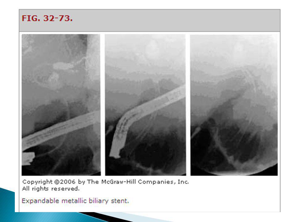
Palliative Care: JAUNDICE
Expandable wire stents: endoscopically Lower risk vs. surgery not as durable as a surgical bypass Complications: bleeding, infection, and pancreatitis; recurrent obstruction & cholangitis effectively manage duodenal obstruction in 81% of patients Metal stents cost less and require a shorter hospital stay than surgical treatment Biliary decompression also can be achieved endoscopically using expandable wire stents. Endoscopic placement of metal stents has a much lower risk than with surgery and less stent occlusion than with plastic stent use.12,50 This method relieves obstructive symptoms in 97 percent of patients and has morbidity and mortality rates of 12 and 3 percent, respectively. Complications include bleeding, infection, and pancreatitis.50 Similarly, metal stent placement can effectively manage duodenal obstruction in 81 percent of patients. Metal stents cost less and require a shorter hospital stay than surgical treatment.51 Endoscopic stents are definitely not as durable as a surgical bypass. Recurrent obstruction and cholangitis is more common with stents and results in inferior palliation. However, this minimally-invasive approach is associated with considerably less initial morbidity and mortality than surgical bypass. Newer, expandable metallic Wallstents demonstrate improved patency and provide better palliation than plastic stents.

56
Palliative Care: DUODENAL OBSTRUCTION
Gastrojejunostomy GI surgery procedure in which the duodenum is excised or bypassed and the stomach is end-to- end anastomosed to the jejunum relieves gastric outlet or duodenal obstruction sometimes associated with delayed gastric emptying Palliative Surgery and Endoscopy For the 85 to 90% of patients with pancreatic cancer who have disease that precludes surgical resection, appropriate and effective palliative treatment is critical to the quality of their remaining life. Because of the poor prognosis of the disease, it is not appropriate to employ invasive and toxic regimens in patients with extremely advanced disease and poor performance status. When patients do desire antineoplastic therapy, it is important to encourage them to enroll in clinical trials so that therapeutic advances can be made. Duodenal obstruction is usually a late event in pancreatic cancer and occurs in only about 20% of patients. 288 Therefore, in the absence of signs or symptoms of obstruction, such as nausea or vomiting, or a tumor that is already encroaching on the duodenum at the time of surgery, the routine use of prophylactic gastrojejunostomy when exploration reveals unresectable tumor is controversial. Although anastomotic leaks are uncommon, gastrojejunostomy is sometimes associated with delayed gastric emptying, the very symptom the procedure is designed to treat. Biliary decompression for palliation of jaundice can be achieved surgically through choledochojejunostomy or cholecystojejunostomy. These procedures can be performed at the same time as gastrojejunostomy, which can relieve gastric outlet or duodenal obstruction. If a gastrojejunostomy is performed, it should be placed dependently and posterior along the greater curvature to improve gastric emptying, and a vagotomy should not be performed. If patients are explored laparoscopically and found to have unresectable disease, palliation of jaundice can be achieved in a minimally-invasive fashion with ERCP and placement of a coated, expandable metallic endoscopic biliary stent (Fig ). Endoscopic stents are definitely not as durable as a surgical bypass. Recurrent obstruction and cholangitis is more common with stents and results in inferior palliation. However, this minimally-invasive approach is associated with considerably less initial morbidity and mortality than surgical bypass. Newer, expandable metallic Wallstents demonstrate improved patency and provide better palliation than plastic stents. If an initial diagnostic laparoscopy reveals a contraindication to the Whipple procedure, such as liver metastases, it is not appropriate to perform a laparotomy simply to create a biliary bypass. In such a patient it is better to place an endoscopic stent. In contrast, if a laparotomy has already been performed as part of the assessment of resectability and the Whipple procedure is not possible, a surgical bypass is usually performed. However, if the patient has a functioning endoscopic stent already in place, it may be reasonable to forego surgical bypass.
. Endoscopic stents are definitely not as durable as a surgical bypass. Recurrent obstruction and cholangitis is more common with stents and results in inferior palliation. However, this minimally-invasive approach is associated with considerably less initial morbidity and mortality than surgical bypass. Newer, expandable metallic Wallstents demonstrate improved patency and provide better palliation than plastic stents. If an initial diagnostic laparoscopy reveals a contraindication to the Whipple procedure, such as liver metastases, it is not appropriate to perform a laparotomy simply to create a biliary bypass. In such a patient it is better to place an endoscopic stent. In contrast, if a laparotomy has already been performed as part of the assessment of resectability and the Whipple procedure is not possible, a surgical bypass is usually performed. However, if the patient has a functioning endoscopic stent already in place, it may be reasonable to forego surgical bypass.")
57
Gastrojejunostomy Billroth I gastroduodenostomy
This procedure creates a direct anastomosis between the stomach and duodenum. It is the most physiologic procedure and is therefore the operation of choice. Several factors may preclude its use, including previous subtotal gastrectomy or extensive scarring around the duodenum. In these situations, the surgeon may be unable to gain enough mobility on the stomach and duodenum to create an anastomosis without excessive tension. Billroth's operation II, is an operation in which the lower part of the stomach (antrum) is removed and a loop of small bowel (jejunum) is brought up and joined to it in a (side-to-side manner) for drainage (gastrojejunostomy).[ Roux-en-Y gastrojejunostomy For a Roux-en-Y gastrojejunostomy, the jejunum is divided several centimeters distal to the ligament of Treitz. The proximal portion of the distal jejunal segment is anastomosed to the stomach. The distal end of the Roux limb is anastomosed to the distal jejunal segment. This jejunojejunostomy is created approximately 40 cm downstream from the gastrojejunostomy in order to minimize the possibility of developing alkaline (bile) reflux gastritis. Van Stiegmann and Goff described a variant of this operation in which the jejunum is not divided.51 This is the so-called uncut Roux-en-Y gastrojejunostomy. The procedure was developed to avoid Roux stasis syndrome, which was thought to be caused by interruption of jejunal intestinal pacesetting potentials. In an uncut Roux procedure, a loop gastrojejunostomy is fashioned. The afferent limb of this loop is occluded—but not divided—by a staple line. A jejunojejunostomy is made between the afferent and efferent jejunal limbs just proximal to the occluding staple line. This operation has not gained wide acceptance, partially because of the problem of dehiscence of the occluding staple line, as reported by Mulholland and colleagues.52
 is removed and a loop of small bowel (jejunum) is brought up and joined to it in a (side-to-side manner) for drainage (gastrojejunostomy).[ Roux-en-Y gastrojejunostomy. For a Roux-en-Y gastrojejunostomy, the jejunum is divided several centimeters distal to the ligament of Treitz. The proximal portion of the distal jejunal segment is anastomosed to the stomach. The distal end of the Roux limb is anastomosed to the distal jejunal segment. This jejunojejunostomy is created approximately 40 cm downstream from the gastrojejunostomy in order to minimize the possibility of developing alkaline (bile) reflux gastritis. Van Stiegmann and Goff described a variant of this operation in which the jejunum is not divided.51 This is the so-called uncut Roux-en-Y gastrojejunostomy. The procedure was developed to avoid Roux stasis syndrome, which was thought to be caused by interruption of jejunal intestinal pacesetting potentials. In an uncut Roux procedure, a loop gastrojejunostomy is fashioned. The afferent limb of this loop is occluded—but not divided—by a staple line. A jejunojejunostomy is made between the afferent and efferent jejunal limbs just proximal to the occluding staple line. This operation has not gained wide acceptance, partially because of the problem of dehiscence of the occluding staple line, as reported by Mulholland and colleagues.52.")
58
Palliative Care: CACHEXIA, MALABSORPTION
Pancreatic enzyme replacement Exocrine pancreatic insufficiency and subsequent malabsorption 30,000 IU of pancrelipase before, during, and after a meal, with ↑ titration as needed Appetite stimulants, high-calorie diet or nutritional supplements PALLIATIVE CARE Palliative treatment of patients with pancreatic cancer is important, and involving hospice early is appropriate. Patients should be monitored closely for depression and treated when it arises. Other complications that require palliative intervention include pain; gastric outlet or duodenal obstruction; and bile duct obstruction and subsequent jaundice, cachexia, and malabsorption caused by exocrine pancreatic insufficiency. Exocrine pancreatic insufficiency and subsequent malabsorption should be treated with pancreatic enzyme replacement (30,000 IU) of pancrelipase before, during, and after a meal, with increased titration as needed. Weight loss unrelated to malabsorption generally is multifactorial and may be treated with appetite stimulants (e.g., megestrol [Megace], dronabinol [Marinol], corticosteroids) and a high-calorie diet or nutritional supplements. Pain from pancreatic cancer can be managed with opioid analgesics, radiation therapy, chemotherapy, or celiac plexus neurolysis (i.e., chemical splanchnicectomy of the celiac plexus with alcohol). Celiac plexus neurolysis eases pain without the side effects of opioids and can be administered intraoperatively, percutaneously, or by endoscopic ultrasonography. Endoscopic ultrasonography–guided neurolysis is effective and has minimal risk of the potentially serious complications associated with the surgical or percutaneous approaches.49 Biliary decompression for palliation of jaundice can be achieved surgically through choledochojejunostomy or cholecystojejunostomy. These procedures can be performed at the same time as gastrojejunostomy, which can relieve gastric outlet or duodenal obstruction. Biliary decompression also can be achieved endoscopically using expandable wire stents. Endoscopic placement of metal stents has a much lower risk than with surgery and less stent occlusion than with plastic stent use.12,50 This method relieves obstructive symptoms in 97 percent of patients and has morbidity and mortality rates of 12 and 3 percent, respectively. Complications include bleeding, infection, and pancreatitis.50 Similarly, metal stent placement can effectively manage duodenal obstruction in 81 percent of patients. Metal stents cost less and require a shorter hospital stay than surgical treatment.51
 of pancrelipase before, during, and after a meal, with increased titration as needed. Weight loss unrelated to malabsorption generally is multifactorial and may be treated with appetite stimulants (e.g., megestrol [Megace], dronabinol [Marinol], corticosteroids) and a high-calorie diet or nutritional supplements. Pain from pancreatic cancer can be managed with opioid analgesics, radiation therapy, chemotherapy, or celiac plexus neurolysis (i.e., chemical splanchnicectomy of the celiac plexus with alcohol). Celiac plexus neurolysis eases pain without the side effects of opioids and can be administered intraoperatively, percutaneously, or by endoscopic ultrasonography. Endoscopic ultrasonography–guided neurolysis is effective and has minimal risk of the potentially serious complications associated with the surgical or percutaneous approaches.49. Biliary decompression for palliation of jaundice can be achieved surgically through choledochojejunostomy or cholecystojejunostomy. These procedures can be performed at the same time as gastrojejunostomy, which can relieve gastric outlet or duodenal obstruction. Biliary decompression also can be achieved endoscopically using expandable wire stents. Endoscopic placement of metal stents has a much lower risk than with surgery and less stent occlusion than with plastic stent use.12,50 This method relieves obstructive symptoms in 97 percent of patients and has morbidity and mortality rates of 12 and 3 percent, respectively. Complications include bleeding, infection, and pancreatitis.50 Similarly, metal stent placement can effectively manage duodenal obstruction in 81 percent of patients. Metal stents cost less and require a shorter hospital stay than surgical treatment.51.")
59
Thank You

Similar presentations


, body (20%), tail (10%) Median age 72 years (65-84 y/o) Most tumors are diagnosed.>")


